Ⅰ. 서 론
, . .
, . Ki Tae Suk[1]
.
. Takaki[2]
Min Leabu[3] (interstitial
cells of Cajal)
, ,
.
, ,
.
.
.
.
. 2
, (cross-correlation) Ophir[4]
. Chen[5]
TOF(time of flight) .
이균정1, 최우혁1, 유재원1, 서종범1, 최서형2, 신태민1
1
Development of a Non-invasive Ultrasonic Measurement System for tissue elasticity
G. J. Lee1, W. H. choi1, J. W. Yu1, J. B. Seo1, S. H. Choi2, T. M. Shin1
1Department of Biomedical Engineering, Yonsei University
2Hana traditional Oriental medicine hospital (Received January 22, 2009. Accepted December 2, 2009)
Diseases caused by indurate tissues of the internal organs are liver cirrhosis and abdominal sclerosis. The cause of chronic gastro-intestinal disease is a digestive system disorder and a defecation disorder. They impede peristaltic movement and digestive system with the symptom that indurate tissues. The purpose of the present study was to determine the disease grade quantitatively by measuring an indurated standard of tissues and organs.
For the measurement of elasticity, we designed the system that measure the displacement of the substance and approved pressure using ultrasound transducer. For verification of developed system, we compared elasticity as results of experiment between the developed system and public elasticity measurement machine at individual plastic phantoms made by plastic hardener and softener. Elasticity of the plastic phantoms is averagely 0.007MPa lower measured by developed system than Micro-indenter, and less than 10% errors. Comparing with economical value and accuracy between developed system and Micro-indenter, the system is significant of measurement for tissue elasticity.
Thus, it is possible to measure a elasticity at human tissue and organ. A chronic gastro-disease as well as grade could be decided objective validity using this system.
indurate tissues, elasticity, ultrasound echo, pressure, measurement
Corresponding Author : 신태민
(220-842)강원도 원주시 흥업면 매지리 첨단의료기기테크노타워 202호 Tel : +82- 33-760-2807 / Fax : +82-33-763-1953 E-mail : [email protected]
(speckle) (speckle tracking)
, O'Donnell[6]
Lubinski[7] .
,
.
.
(plastic phantom)
, .
Ⅱ. 이론적 배경
, .
.
.
.
.
. ,
.
( (1)).
.
. phase sensitive cross-correlation
, [8].
,
(2) . 1
. (2) τ , T
.
Lubinski
[7] FHWM(full half width maximum)
. (auto-
correlation) , . (2)
(Maximum Cross Correlation
Coefficient) (3) .
(τ) .
[7].
(1)
.
Zhang[8]
(4) . (4)
.
(υ) 0.5 .
Indentation
- (4) k
Wellman[9] (5) K . (5)
.
K ,
[8].
.
.
(1)
(2)
Amplitude
15
10
5
0
550 600 650 700 750 800 850 900 950 1000 Sample number
r2(n)
×10-3
r1(n)
그림 1. 압력 인가 전 (r1(n))과 후 초음파 신호(r2(n))를 나타냄 Fig. 1. Appear befor and after the approved pressure echo-signal
(3)
′
(4)
(5)
Ⅲ. 시스템 설계
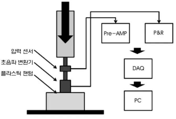
A. 시스템 시스템 구성시스템 시스템 구성구성구성
. , ,
.
, .
. ( 2).
,
. , .
.
PC .
2 .
(( ) ) 5MHz ,
7.5mm . (Load Cell Model34, Honeywell)
(N) . 0.5N ,
0 45N .
(PR5073, Panametric-NDT, USA) . 59dB DAQ(PCI5122, NI:national
instrument) .
, LabVIEW9.2 ,
.
B. 신호 신호 신호 신호 처리부처리부처리부처리부
50MHz
1~4cm 2000 .
LabVIEW .
(demodulation) (base
band signal) . (time
gain compensation) n
n-1
. 3
, .
. n
n-1 ,
.
. .
peak hoping (hanning
window) [6].
.
Ⅳ. 시스템 검증을 위한 팬텀 실험
.
5:5
[10].
Micro-indenter
.
압력 센서 초음파 변환기 플라스틱 팬텀
P&R
DAQ
PC Pre-AMP
그림 2. 초음파 탄성도 측정 시스템의 구성도
Fig. 2. The blocks of the ultrasound elasticity measurement system
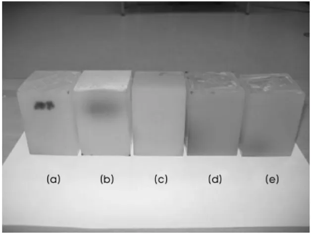
A. 플라스틱 플라스틱 팬텀의 플라스틱 플라스틱 팬텀의 팬텀의 제작팬텀의 제작제작제작
3 .
1:9, 2:8,
3:7, 4:6, 5:5 .
(Scatterer) .
Amberlite ,
0.5% . 50mm×50mm×70mm
. 50%
.
, SNR ,
[10].
B. 실험 실험 실험 실험 방법방법방법방법
Micro-indenter .
Indentation ,
- .
1) Micro Indenter
Micro-Indenter(model
5848, Instron) .
4 .
Micro Indenter
(Bluehill) 0~2 N
. 0~20 N
, 10 .
1
.
2)
. .
0~20N .
. 5
. ,
0~20N
0N , 1
10 .
그림 4. Micro Indenter를 이용한 팬텀의 탄성도 측정 Fig. 4. Elasticity measurement of the phantom with Micro Indenter
(a) (b) (c) (d) (e)
그림 3.제작된 플라스틱 팬텀 (1:9(a), 2:8(b), 3:7(c), 4:6(d), 5:5(e)) Fig. 3. Designed plastic phantoms : plastic softener ratio
그림 5.설계된 초음파 탄성도 측정 시스템으로 팬텀 탄성도 측정
Fig. 5. Measurement of the elasticity on phantom with designed ultrasound elasticity measurement system
Ⅳ. 결 과
Micro-Indenter
. 6
Micro-Indenter -
.
. 1:9
7 .
.
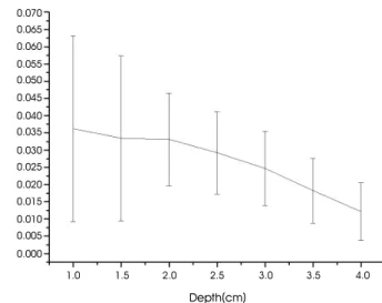
10 .
0.5cm .
8 1:9
0.5cm .
10 1 .
(4) (stress)
. - 9
. Micro-Indenter
, 10
Stress(Pa)
10000
8000
6000
4000
2000
0
0.00 0.04 0.08 0.12 0.16 0.20 0.24
Strain
10% (a) 20% (b) 30% (c) 40% (d) 50% (e)
Displacement(mm)
0.070 0.065 0.060 0.055 0.050 0.045 0.040 0.035 0.030 0.025 0.020 0.015 0.010 0.005 0.000
1.0 1.5 2.0 2.5 3.0 3.5 4.0
Depth(cm)
그림 6. Micro Indenter의 응력-변형률 선도 Fig. 6. Stress-Strain Diagram of the Micro Indenter
그림 8. 연화제 비가 1:9인 팬텀에서 깊이에 따른 평균 변위와 표준 편차 Fig. 8. Averaged displacement and standard deviation with depth of the
phantom as softener ratio 10%
0.0002
Displacement(cm)
0.0000
-0.0002
-0.0004
-0.0006 -0.0008
-0.0010
-0.0012
-0.0014
0 5 10 15 20 25
Pressure(N)
100000
80000
60000
40000
20000
0
Stress(Pa)
0.00 0.04 0.08 0.12 0.16 0.20 0.24 0.28 0.32 Strain
10% (a) 20% (b) 30% (c) 40% (d) 50% (e)
그림 7. 인가 압력값에 대한 평균 변위 Fig. 7. Average displacement with the input pressure
그림 9.설계된 시스템의 응력-변형률 선도 Fig. 9. Stress-Strain Diagram of the designed system
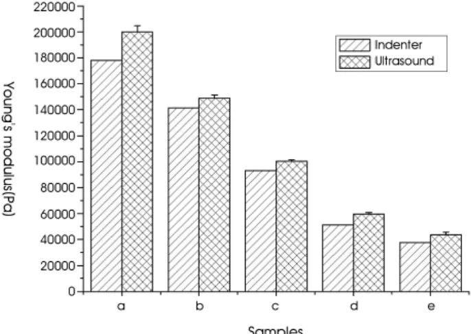
. Micro- Indenter
5% 16% . (a)
0.007MPa .
Ⅴ. 고 찰
, [ 6].
.
, 0.9
.
1 (c) 4cm .
.
10 Micro Indenter
Indentation
0.5 . Wallman[9]
K
Micro Indenter .
6 (a), (b), (c)
, (d), (e) .
, 50%
.
. .
, .
,
, .
.
.
Ⅵ. 결 론
.
, Pulser&Receiver
.
. Micro Indenter
.
0.007MPa
. ,
1cm 1.5cm 2cm 2.5cm 3cm 3.5cm 4cm
(a) 36.2 33.4 33.1 29.2 24.6 18.2 12.1
(b) 44.0 37.4 40.8 35.0 30.5 21.2 11.5
(c) 50.8 52.5 47.6 41.1 34.5 25.6 34.2
(d) 97.0 92.6 84.8 73.2 59.0 48.0 16.9
(e) 98.9 94.0 87.8 76.2 61.5 47.3 25.1
표 1. 각 팬텀에서 깊이에 따른 평균 변위(μm)
Table 1. Average displacement with depth of each phantom
220000
Young's modulus(Pa)
200000 180000 160000 140000 120000 100000 80000 60000 40000 20000 0
a b c d e
Samples
Indenter Ultrasound
그림 10.각 팬텀에서 Indenter와 초음파 탄성도 측정 시스템를 이용한 탄성도 측정 값(Pa)
Fig. 10. Elasticity measurement value used Micro Indenter and designed system at each phantoms
.
. Micro Indenter
, .
.
LabView
, .
.
참고문헌
[1] Ki Tae Suk, Dae Wook Lim, Moon Young Kim, Dong Hun Park, Kyu Hong Kim, Jung Min Kim, Jae Woo Kim, Hyun Soo Kim, Sang Ok Kwon, Soon Koo Baik, Sung Jin Park, “Thickening of the Gastric Wall on Transabdominal Sonography: A Sign of Gastric Cancer”, Journal of Clinical Ultrasound, Vol. 36 No. 8, pp462 - 466, 2007.
[2] Takaki M., “Gut pacemaker cells: The interstitial cells of Cajal (ICC)”, J Smooth Muscle Res. J Smooth Muscle Res.,Vol. 39, No.
5, pp137-161, 2003.
[3] K. W. Min, and M. Leabu,”interstitial cells of cajal(ICC) and gastrointestinal stromal tumor(GIST): facts, speculations, and myths,”J, Cell Mol Med.,vol. 10,pp.995-1013,2006.
[4] Ophir, J., CCspedcs, I., Ponnekanti, H., Yazdi, Y., et al..,
“Elastography: a quantitative method for imaging the elasticity of biological tissues,” Ultrasonic Imaging, Vol. 14, pp. I1 1-134, 1991.
[5] Chen EJ, Novakofski J, Jenkins WK, O’Brien WD Jr. “Young’s modulus measurement of soft tissues with application to elasticity imaging,” IEEE trans. Ultrasonics, Ferroelectrics and Frequency control. 1996;43(1):191-194.
[6] M. O'Donnell, A. R.Skovoroda, B. M. Shapo, and S. Y.
Emelianov,” Internal displacement and strain imaging using ultrasonic speckle tracking,” IEEE Trans. Ultrason., Ferroelect., Freq. Contr., vol. 41, pp. 314-325, May 1994.
[7] Mark A. Lubinski “Speckle Tracking Methods for Ultrasonic Elasticity Imaging Using Short-Time Correlation”, IEEE Trans.
Ultrason., Ferroelect., Freq. Contr. vol. 46, NO. 1, January 1999.
[8] Zhang, M. Zhang, YP and Mak AFT. “Estimating the effective Young's modulus of soft tissues form indentation tests-nonlinear finite element analysis of effects of friction and large deformation” Med. Eng. Phys. Vol. 19, No.6 pp512-517, 1997.
[9] P. Wellman, R. Howe, E. Dalton, and K. A. Kern, “Breast tissue stiffness in compression is correlated to histological diagnosis,”
Tech. Rep., Harvard BioRobotics Laboratory, Harvard University, Cambridge, Mass, USA, 1999.
[10] , , , , “
” Journal of Biomedical Engineering Research, 29 , 4 pp302-306, 2008.