The Geranium nepalense has been used traditionally for treatment of various diseases. However, the molecular studies on the effect of Geranium nepalense have not been carried out. In the present study, Quercetin, quercitrin, and afzelin were isolated from the methanol extract of Geranium nepalense were tested for their anti-inflammatory effect. The anti-inflammatory effect of the compounds was studied in lipopolysaccharide(lps)-treated mouse macrophage cells, RAW 264.7.
RAW 264.7 cells were pre-incubated with isolated compounds(0, 5, 10, 20, 40, 50 ㎍/㎖) for 4h and treated with 1 ㎍/㎖ lps for 18h, and then the anti-inflammatory effects of compounds were determined. The results are as follows: Quercetin at various concentration inhibited the viability of Raw 264.7 from 7% to 45%, quercitrin from 25% to 80%, and afzelin from 13% to 52%.
현초에서 분리한 생리활성물질의 항염증효과 (단보)
김보미, 김의성1, 이영행, 유병수, 채규윤
원광대학교 자연과학대학 나노생명화학부, 1한국생명공학연구원 전북분원,
ABSTRACT
Antiinflammatory Effect of Bioactive Compounds from Geranium nepalense (Notes)
Bo-Mi Kim, Eui-Sung Kim1, Young-Hang Lee, Byung-Soo Yu, Kyu-Yun Chai Division of Nanobiochemistry, College of Natural Sciences, Wonkwang University
1Eco-Friendly Biomaterial Research Center, Korea Research Institute of Bioscience and Biotechnology
16)
•Corresponding author : Kyu-Yun Chai,
•Department of Bionanochemistry, School of Natural Science, Wonkwang University, Iksan 570-749, South Korea.
•Tel : +82-63-850-6230 E-mail : [email protected]
•접수 : 2010/ 10/ 27 수정 : 2010/ 11/ 02 채택 : 2010/ 11/ 26
Isolated compounds showed a significant decrease in i NOS (inducible nitric oxide synthase) and COX-2 (cyclooxygenase-2). These results suggest that these compounds can be used as stable anti-inflammatory materials.
Key word : Quercetin, quercitrin, afzelin, Geranium nepalense, nitric oxide, prostaglandin E2, inducible nitric oxide synthase, cyclooxygenase-2.
Ⅰ. 서 론
Geranium nepalense subsp. thunbergii
)는 이질 풀이라 불리는 식물의 한의학적 명칭으로 꽃이 필 무렵 풀 전체를 채취해서 말린 것을 의미한다. 노 관초 라고도 하며 산과 들에서 자란다. 한국, 일 본, 타이완 등지에서 자생하는 다년생 초본으로 쥐손이풀과에 속하며, 꽃은 8~9월에 피고, 홍색, 홍자색, 백색 등을 띤다. 많은 양의 탄닌과 케르세 틴이 들어 있어, 소염, 지혈, 수렴, 살균작용이 있 으며, 민간에서는 적리, 역리, 변비, 통경, 대하증, 방광염, 피부병, 창종, 위궤양, 지이, 대장카타르, 이질, 십이지장궤양 등에 약재로 사용하였으며, 한 방에서는 지사제로 쓰인다1). 이질풀을 달여 마시 면, 어떠한 설사병도 말끔히 낫기 때문에 일본에 서는 이를 5대 민간 영약으로 부른다. 꽃이 필 무 렵 풀 전체를 채취해서 말린 것을 현초라고 하는 데, 탄닌(tannin)ㆍ푸로신(furosin) 등이 들어 있 다. 지사제, 정장제, 항진균성 효능이 있어 위장복 통, 변비, 종기, 감기, 피부병 등을 치료하는 효과 가 있다. 민간요법에서는 종기와 피부병 등에 외 용한다2). 현초 추출물이 활성 산소종을 소거함으 로써 그리고 활성 산소종에 대항하여 세포막을 보 호함으로써 생체계, 특히 태양 자외선에 노출된 피부에서 항산화제로서 작용할 수 있음을 가리키며, 현초 성분에 대한 분석과 ethyl actate 분획의 당 제거 실험 후 얻어진 aglycone 분획의 큰 elastase 저해활성 으로부터 주름개선 기능성 화장품 원료 로서도 응용 가능성이 있음을 보고하였다3).
본 연구에서는 현초에서 분리한 quercetin, quercitrin, afzelin를 이용하여, 염증억제 효과에 관 한 연구 결과를 얻었기에 보고합니다.
Ⅱ. 실험재료 및 방법
1. 실험재료
본 실험에서 사용한 현초는 원광대학교 한의학 대학 부속한방병원에서 구입하여, 외부형태를 비 교 조사하고 확인 후 사용하였다. 실험에 사용된 현초는 원광대학교 자연과학대학 천연물실험실에 보관되어있다.
2. 시약 및 기기
현초의 추출 및 open columm 분리에서 사용된 용매는 제 1급 시약을 사용하였으며, 물질의 분리 에는 25 TLC plastic sheets (20 × 20 cm) silica gel 60 F254 (MERCK), 25 TLC plates 5 × 10 cm RP-18 F254S(MERCK), silica gel 60 (0.015~0.140 mm, MERCK), silica gel 60 (0.040~0.063 mm MERCK), octadecyl-functioalized silica gel (Aldrich),
lipophilic sephadex LH-20 (SIGMA) 등 충진제와 Phenomenex 사의 Luna 5u C18 (2) (250 x 21.20 mm5 micron) HPLC column 과 YMC 사의 Hydrosphere C18 (250 x 20 mm I. D. S-5 m, 12 mm) HPLC column, EYEL4 rotary vacuum evaporator, vision workstation perseptive bio-systems HPLC 등을 사용하였다. 순수한 물질의 확인은 ESI-MS를 얻 기위해, Micromass Quatro LC를 사용하였다. NMR spectrum은 JEOL Eclipse 500 FT-NMR Spectrometer (500 MHz)를 사용하였으며, NMR 용매는 methanol-
d
4, dimethyl-sulfoxide-d
6,,
chloroform-d
, 내부 표준물 질은 tetramethylsilane (TMS)를 사용하였다.생리활성에 대한 측정을 위해, MTT (2-[4,5- dimethylthiazol-2-yl]-2,5-diphenyl tetrazolium bromide) 와 DMSO (dimethyl sulfoxide), LPS (lipopoly- sacchride)는 Sigma사에서 구입하였으며, Antibiotic antimyotic solution 과 FBS (fetal bovine serum), RPMI 1640 은Gibco/Invitrogen에서 구입하였다.
Anti-
i
NOS, anti-COX-2, anti-tumor necrosis factor -alpha (TNF-) antibody는 Santa Cruz Biotechnology INC. (California, USA)로부터 구입하였으며, nitrocellulose membrane (NC membrane)은 Amersharm Pharmacia 사로부터 구입하여 사용하였다. 용매로는 methanol (MeOH), ethanol (EtOH), chloroform (CHCl3), ethyl acetate (EA),n
-butanol (n
-BuOH),n
-hexane, 정제하지 않고 사용하였다. 추출물들의 농축에는 원심분리기 (Hanil Industrial Company, Hm 150Ⅳ)와 진공 회전식 농축기를 사용하였으며, 흡광도 측정에는 UV-Visible Spectrophotometer (Molecular Devices Spectra Max 190)와UV-Visible S-2100 (Scinco Co. Ltd.)을 사용하였다.
3. 생리활성물질의 분리
건조된 현초 2.0 kg을 상온에서 메탄올 (18.0 L)에 일주일 동안 침출시킨 후, 여과하여 얻은 추 출물을 감압 농축하여 메탄올 추출물 (641.0 g, 32.05%)을 얻었다. 메탄올 추출물은 3차 증류수에
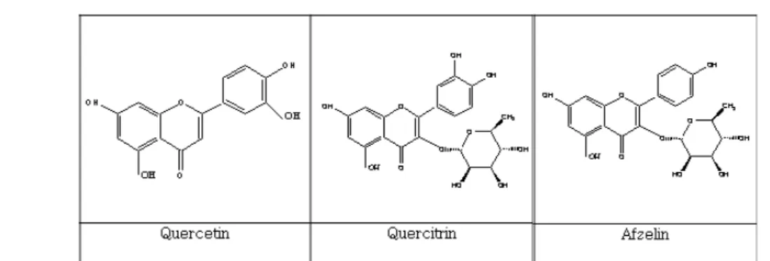
포화시킨 후, 용매의 극성에 따라 계통 분획하였 다. 노르말 헥산으로 3회 추출하여, 노르말 헥산 추출물 (108.00 g, 16.85%)을 얻었으며, 같은 방법 으로 에틸 아세테이트 추출물 (80.76 g, 12.60%), 노르말 부탄올 추출물 (109.15 g, 17.03%) 그리고 물 추출물 (332.00 g, 51.79%)을 얻었다. 에틸 아 세테이트 추출물 (5.0 g)을 메탄올 (5.0 mL)에 녹 인 후, silica 60 (200 g)로 충진 된 flash column 에 넣어, 에틸 아세테이트 : 포름산 : 초산 : 물 (200 : 1 : 1 : 3) 조건으로 분리하였다. 분획 1 (200.0 mg), 분획 2 (420.0 mg), 분획 3 (370.0 mg), 분획 4 (1,330.0 mg), 분획 5 (560.0 mg), 분 획 6 (900.0 mg)을 얻었다(Sche. 1). 그 중 생리활 성이 있는 분획 1 을Sephadex LH-20을 이용하여, 메탄올 : 물 (4 : 6)와 메탄올의 용매조건으로 분 리하여 노란색 고체 quercetin (Fig. 8)(13.7 mg)을 얻었다. 분획 4를 역상 HPLC 을 이용하여, 10 - 55 % acetonitrile/H2O의 용매조건으로 60분 동안 분리하여, quercitrin(Fig. 8) (머무름시간, 46.556분, 15.3 mg)4-8)과 afzelin(Fig. 8) (머무름시간 49.543 분, 27.1 mg)9-12)을 얻었다(Fig. 7).
4. 세포배양
생쥐의 대식세포주 (Murine macrophage Raw 264.7 cell line)를 10% FBS (56 ℃ heat inactivated), 1 % L-glutamine, nonessential amino acids 및1
% antibiotic/antimycotic solution (100 U/mL of penicillin, 25 g /mL of amphotericin D, and 100 g/mL of streptomycin), 2 % sodium bicarbonate 등이 포함된 RPMI 1640 배지에 넣고 37℃, 5 % CO2incubator에서 24시간 배양하였다.
5. 세포 생존율 측정
Quercetin, quercitrin 및 afzelin에 대한 RAW 264.7 세포주의 세포 생존율을 MTT 방법을 이용하여 분석하였다. RAW 264.7 세포주를 96-Well plate 에 2.0 × 10⁵ cells/mL 되도록 seeding한 다음,
IFN-γ/LPS 및 약제를 24시간 처리하고 새로운 배지에 50.0 µg/mL이 되도록, MTT를 첨가하여 2 - 4시간 동안 반응시켰다. MTT와 생존세포로부터 생성된 보라색 불용성 formazan을 DMSO로 용해 하여, 540 nm 파장에서 흡광도를 측정하고 대조군 과 비교하여 백분율 (%)로 표시하였다.
6. Nitric oxide(NO) 의 농도의 측정
Quercetin, quercitrin 및 afzelin을 DMSO에 녹 여 저장용액을 만들었으며, sarcoma 생성된 NO의 양은 세포 배양액 중에 존재하는 NO2-의 형태로 서 griess 시약(1 % sulfanilamide, 0.1 % N-(1- naphthyl)ethylenediamine dihydrochloride in 2.5
% phosphoric acid solution)을 이용하여 측정하였 다. 이것을 희석하여 적정용액을 만든 후 LPS를 1.0 µg/mL처리하여, 18시간 동안 배양하였다. 배양 액 중 100 L를 취하고, 동량의 griess시약을 넣고, 실온에서 10분간 방치한 다음, ELISA plate reader 를 이용하여 540 nm 에서 흡광도를 측정하였다.
NO2- 는 sodium nitrite를 표준용액으로 측정하였 으며, 각 측정치는 5 - 8 µM 의 NO2- 을 함유하 는 cell-free 배양액의 흡광도를 이용하여 보정하였다.
7. Western Blotting
Quercetin, quercitrin 및 afzelin과 LPS에 의해 자 극된 세포를 PBS로 세척한 다음, cell lysis buffer (50 mM Tris, pH 8.0, 110 mM NaCl, 5 mM EDTA, 1% Triton X-100, PMSF 100 µg/mL)로 용혈시킨 다 음, 원심분리 하여 단백질 용액을 얻었다. Bradford 분석 방법을 이용하여 단백질을 정량한 다음, 동 량의 sample buffer (125 mM Tris pH 6.8, 4 % SDS, 20 % glycerol, 10 % mercaptoethanol)를 혼 합한 다음 95℃에서 5분 동안 가열하여 단백질 변 성을 유도하였다. 변성된 단백질을 12% acrylamide gel에서 전기영동을 수행한 다음, nitrocellulose membrane (Amersharm Pharmacia)으로 전위시키고, 5 % skim milk/ TBS-T (20 mL)로 상온에서 1시간
동안 반응시켜 비특이적인 항체반응을 억제시켰 다. COX-2와
i
NOS 및 ß-actin에 대한 1차 항체 (primary antibody) 를 3 % skim milk/1 x TBS-T 에서 1 : 1,000 으로 희석하여, membrane과 상온 에서 1시간 30분 동안 반응시키고 세척한 다음, anti-mouse, rabbit 혹은 goat IgG conjugated Horseradish peroxidase 2차 항체 (secondary antibody) 를1% skim milk/ TBS-T에서 1 : 5,000으로 희석 하여, membrane과 상온에서 1시간 동안 반응시킨 다. TBS-T로 세척한 다음, ECL reagent kit (ECL, Amersham, Buckinghamshire, England)로 발색시킨 다음, X-ray film에 감광하는 방법으로 분석하였다.8. Cyclooxygenase-2(COX-2) 농도의 측정
Quercetin, quercitrin 및 afzelin을 농도별로 RAW 264.7 세포에 1시간 전처리한 다음, LPS로 자극하 고 24시간 후에 배양액을 회수하여 방치한 다음, ELISA(enzyme linked immunosorbent assay, R&D System)를 이용하여 분석하였다. 모든 분석과정은 R&D System에서 제공된 방법에 준하여 측정하 였다.9. 통계분석
대조군과 각 시료들로부터 얻은 실험 결과는 mean ± S.D. 값으로 표시하였으며, 각 실험 결과 로부터 ANOVA (analysis of variance)를 구한 후 Duncan’s multiple range test를 이용하여 각 군의 평균간의 유의성을 검정하였다. 일반적으로, p 값 이 0.005 이하인 것만 통계적으로 유의성이 있는 것으로 검증하였다.
10. 실험결과 및 고찰
식물체에 플라보노이드는 광범위하게 분포되어 있으며, 구조적 특이성 즉, pyrone고리 자체의 산 소, 카보닐기, 페놀성 히드록실기에 기인하여, 생 체내에 강한 친화력으로 인하여 약리작용이 있는 것으로 알려져 있다13). 식물체에 많이 함유된 플라
보노이드는 A, B, C, 세고리로 나누며, A고리 proton이 B고리나 C고리 proton보다 고자장에서 나타난다. A고리의 oxygenated pattern 가운데서 가장 많이 나타나는 5,7-dihydroxy group에서는 6 번 proton이 8번proton보다 고자장에서 나타난다14).
11. Quercetin, quercitrin 및 afzelin의 세포 생
존률에 미치는 영향
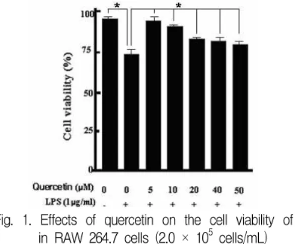
Quercetin, quercitrin 및 afzelin을540 nm파장에 서 흡광도를 측정한 결과, 대조군을 100%로 하였 을 경우, LPS로 자극한 대조군은 75%의 세포 생 존율을 나타냈으며, 생리활성물질과 LPS를 처리 한 군은 생리활성물질들이 가장 높은 농도인 50.0 μM을 처리한 군에서 80% 이상의 세포 생존율이 측정되었다. 이러한 연구결과는 전보에서 보고한 것처럼15), quercetin, quercitrin 및 afzelin은 5.0 - 50.0 μM에서는 세포 생존율에 영향을 미치지 않 는 플라보노이드 계열의 화합물임을 확인되었다 (Fig. 1, 2과 3)16).
Fig. 1. Effects of quercetin on the cell viability of in RAW 264.7 cells (2.0 × 10⁵ cells/mL) Cells were incubated with or without 1 g/mL LPS for 24 hrs in the presence or absence of quercetin at indicated concentrations. The cell viability was determined by the MTT method. One of three blots is shown and each bar represents the mean ± S.D. from three separate experiments. Significantly different from the control values. *p<0.005
Fig. 2. Effects of quercitrin on the cell viability of in RAW 264.7 cells (2.0 × 10⁵ cells/mL) Cells were incubated with or without 1 g/mL LPS for 24 hrs in the presence or absence of quercitrin at indicated concentrations. The cell viability was determined by the MTT method. One of three blots is shown and each bar represents the mean ± S.D. from three separate experiments. Significantly different from the control values. *p<0.005
Fig. 3. Effects of afzelin on the cell viability of in RAW 264.7 cells (2.0 × 10⁵ cells/mL) Cells were incubated with or without 1 g/mL LPS for 24 hrs in the presence or absence of afzelin at indicated concentrations. The cell viability was determined by the MTT method. One of three blots is shown and each bar represents the mean ± S.D. from three separate experiments. Significantly different from the control values. *p<0.005
12. Quercetin, quercitrin 및 afzelin에 대한 nitric oxide의 농도 측정
Quercetin, quercitrin 및 afzelin이 RAW 264.7 세포주에서 LPS의 유도에 의한 NO 생성에 미치 는 영향에 대해 알아보기 위하여, RAW 264.7 세 포주 (1 x 10⁵ cells/mL)를 5시간 후에 5.0 - 50.0 M 생리활성물질의 농도로 2시간 동안 전처리한 다음, 1.0 mg/mL LPS를 처리하여 18시간 배양하 였다. 그 결과 생리활성물질이 처리되지않고 LPS 로 자극한 대조군은 23.6 M의 아질산염을 생성시 킨 반면, 5.0 - 50.0 M 생리활성물질을 처리한 실 험군은 약 3.0 - 15.0 M의 아질산염을 생성시켰다.
이러한 연구결과에 의하면, quercetin, quercitrin 및 afzelin는 LPS로 활성화된 RAW 264.7 세포에서 NO 생성을 억제하는 효과가 정공피 메탄올 추출물에서 분리된 prunetin, sakuranetin, 및 dihydroquercetin보 다 효능이 좋은 것으로 관찰되었다(Fig. 4, 5와 6)15,16).
13. Quercetin, quercitrin 및 afzelin에 대한 iNOS 의 농도 측정
Quercetin, quercitrin 및 afzelin과 1.0 g/mL LPS가 처리된 RAW 264.7 세포를 용해시킨 후, Bradford법으로 분석한 결과에 의하면, 농도 의존 적으로 감소되는 현상을 관찰할 수가 있었으며, 이들 생리활성물질은 대조군과 비교할 때에 50.0 M에서 발생하는 NO의 30% 이상 감소하는 것으 로 나타났다. 그러나 정공피 메탄올 추출물에서 분리된 prunetin, sakuranetin, 및 dihydroquercetin 은 30 M에서 발생하는 NO의 50%이상 감소하는 것으로 관찰되었다. 이러한 연구결과는 quercetin, quercitrin 및 afzelin이 항염증제로서 활용할 가치 가 있는 것으로 생각된다(Fig. 4, 5와 6)15,16).
14. Quercetin, quercitrin 및 afzelin에 대한
COX-2의 농도 측정
PGE2 생성에 직접적으로 영향을 미치는 COX-2 의 발현을 western immunoblot analysis 방법과 ELISA 방법으로 조사하였다. COX-2 단백질 발현 이 농도 의존적으로 억제되는 사실을 증명하였으며, LPS로 자극된 대조군을 기준으로 했을 때, 50 M 생 리활성물질을 처리하였을 때, 50%의 감소율이 관찰 되었다. 이러한 연구결과는 정공피 메탄올 추출물에 서 분리된 prunetin, sakuranetin, 및 dihydroquercetin 은 30 M에서 발생하는 NO의 50%이상 감소하는 것으로 관찰되었다. 플라보노이드 화합물인 quercetin, quercitrin 및 afzelin이 항염증제로서 활용할 가치 가 있는 것으로 생각된다(Fig. 4, 5와 6)15,16).
15. 생리활성 물질이 단백질 발현에 미치는
영향
대식세포가 interferon-γ (INF-γ)또는 LPS의 자극에 의해서 larginine으로부터 NOS에 의한 대 사과정을 통해 NO가 다량 생산되면, 염증반응으 로 유발되는 조직손상, 돌연변이 및 신경조직의 손상 등을 일으켜 생체에 유해한 작용을 나타내는 것으로 알려져 있다. NO가 생성되는 대사과정에 관여하는 사이토카인으로 알려진 iNOS와 NOS의 생성에 관여하는 조효소로 COX-2의 발현을 관측 하였다. 또한, 모든 세포가 일정하게 가지고 단백 질로 알려진 β-actin 을 측정함으로써, 각 농도별 단백질의 양이 일정함을 확인하였다. 자극원만 처 리한 대조군의 사이토카인 발현 정도를 기준으로 하여, 생리활성 물질이 사이토카인을 억제하는 정 도를 확인하였다. 현초에서 분리한 생리활성물질 을 5.0 - 50.0 μM의 농도로 세포주에 처리하고, 이 로부터 단백질을 추출하여 전기영동장치를 이용한 단백질 분리과정을 통해 얻은 띠의 두께를 분석한 결과, 생리활성물질이 β-actin의 발현에 영향을 미 치지 않고, 농도 의존적으로
i
NOS 와 COX-2 의 생성을 억제함을 확인하였다(Fig. 4, 5와 6)15,16).(A)
(B)
Fig. 4. Effects of quercetin on NO production and
i
NOS protein expression in RAW 264.7 Cells.(A) cells (2.0 × 10⁵/ ml) were incubated 24 h for Nitric Oxide assay or (B) 18 h for iNOS Western blot with medium, Nitric Oxide concentration was determined by Griess reagent For the determination of intracellular iNOS protein, Western immunoblot analysis was carried out as described in materials and methods.
(A)
(B)
Fig. 5. Effects of quercitrin on NO production and
i
NOS protein expression in RAW 264.7 Cells.(A) cells (2.0 × 10⁵/ ml) were incubated 24 h for Nitric Oxide assay or (B) 18 h for iNOS Western blot with
medium, Nitric Oxide concentration was determined by Griess reagent For the determination of intracellular iNOS protein, Western immunoblot analysis was carried out as described in materials and methods.
(A)
(B)
Fig. 6. Effects of afzelin on NO production and
i
NOS protein expression in RAW 264.7 Cells.(A) cells (2.0 × 10⁵/ ml) were incubated 24 h for Nitric Oxide assay or (B) 18 h for iNOS Western blot with medium, Nitric Oxide concentration was determined by Griess reagent For the determination of intracellular iNOS protein, Western immunoblot analysis was carried out as described in materials and methods.
Fig. 7. The HPLC chromatograms extracts of fraction ethyl acetate in hydrosphere C18 HPLC column (250 × 20 mm ID, particle S-5 ㎛).
HPLC conditions:
Linear gradient: 0 min: 10% acetinitrile in water ; 60 min - 55 % acetinitrile in water
Detection: 254, 280, 350, 370 nm, Flow rate: 3 mL/ min
Fig. 8 The structure of quercetin, quercitrin, afzelin.
Scheme 1. The pathway on the extraction and fraction of
Geranium nepalense subsp
.thunbergii
.Ⅲ. 결 론
현초의 메탄올 추출물로부터 분리한 quercetin 와quercitrin 그리고, afzelin 화합물이 면역반응에 미치는 영향에 대한 연구 결과에 의하면, 면역반 응에 의한 염증 대사 과정에 관여하는 것으로 알 려진 대식세포 중 쥐의 대식세포 RAW 264.7 세포 주를 사용하여 활성화된 대식세포로부터 생성되는 사이토카인 및 NO의 생성, iNOS의 발현, COX-2 의 발현와 PGE2 생성이 농도 의존적으로 저해하
는 것을 확인하였다. 이러한 연구결과에 의하면, quercetin와 quercitrin 그리고, afzelin 화합물이 항 균 치료제나 염증 억제제로서 관련 질환을 예방하 거나 치료할 가능성을 시사하고 있다.
감사의 글
본 연구는 2009년도 원광대학교 교비연구비 지 원에 의하여 이루어졌으며, 이에 감사드립니다.
참고문헌
1. 신민교. 임상본초학, 남산당. 1986:337.
2. Park, S.N. and Lee, K.H.. Antioxidative activities and antiaging effects of
Geranium nepalense
Extracts.J. Soc. Cosm. Scient. Kor.
2008;34:25.3. Park, J.C., Yu, Y.B. and Lee, J.H.: Isolation and structure elucidation of flavonoid glycosides from
Lindera obtasiloba BL
.J. Korean Soc.
Food. Nutr
, 1996;25:76.4. Choi, J.S., Young, H.S., Park, J.C., Choi, J.H.
and Woo, W.S.: Flavonids from the leaves of
Rhododendron brachycarpum
,Arch pharm Res
, 1986;9:233.5. Zhong, X.N., Otsuka, H., Ida, T., Hirata, E., Takushi, A. and Takeda, Y.: Three flavonol glycosides from leaves of myrsine.
Phytochem.
1997;46:943.
6. Beck, P.O., Dijoux, M.G., Cartier, G. and Mariotte, A.M.: Quercitrin 3'-sulphater from leaves of
Leea Guinenis, Phytochem.
1998;47:1171.7. Choi, Y.H., Kim, J.H. and Kim, M.J.:
Antioxidative compounds in leaves of
Castanea crenata S. et Z. Korean J. Medicinal Crop.
Sci.
2000;8:373.8. Dinya, D. and Hetenyi, E. Y.: Topics in flavonoid chemistry and biochemistry, Elservier Scientific Publishing Comp, 1975:589.
9. Mabry, T. J., Markharn, K. R. and Thomas, M. B. The systematic identification of flavonoids, Springer-Verlag, 1970:253.
10. Park, H.J., Young, H.S., Park, K.Y., Rhe, S.H., Chung, H.Y. and Choi, J.S.: Flavonoids from the whole plants of
Orostachys japonicus, Arch. Pharm. Res.
1991;14:167.11. Hur, J.M., Park, J.C. and Hwang, Y.H.:
Aromatic acid and flavonoids from the leaves of
Zanthoxylum piperitum
,Nat. Prod. Sci.,
2001;71:23.12. Dinya, D. and Hetenyi, E.Y.: Topics in Flavonoid Chemistry and Biochemistry, Elservier Scientific Publishing Comp, 1975:
589.
13. Mabry, T.J., Markharn, K.R. and Thomas, M.B.: The Systematic Identification of Flavonoids, Springer-Verlag, 1970:253.
14. Yun, Y.G., Chai, K.Y., Lee, K.K, Bhatt, L.R. and Baek, S.H.: Studies on the Antiinflammatory compounds from
Sorbus commixta. J. Pharm.
Soc. Korea
, 2009;53:351.15. Rho, J.R., Jun, C.S., Ha, Y.A., Yoo, M.J., Cui, M.X., Baek, S.H., Lim, J.A, Lee, Y.H. and Chai. K.Y.: Isolation and characterization of a new alkaloid from the seed of