Korean J Gastroenterol Vol. 73 No. 1, 35-38 https://doi.org/10.4166/kjg.2019.73.1.35 pISSN 1598-9992 eISSN 2233-6869
CASE REPORT
Korean J Gastroenterol, Vol. 73 No. 1, January 2019 www.kjg.or.kr
토혈을 동반하지 않은 대동맥식도누공의 내시경적 진단 1예
이종윤, 장진석, 김동균, 차재황, 최원종
동아대학교병원 소화기내과
Endoscopic Diagnosis of Aortoesophageal Fistula Not Presenting Hematemesis
Jong Yoon Lee, Jin Seok Jang, Dong Kyun Kim, Jae Hwang Cha and Won Jong Choi Department of Gastroenterology, Dong-A University Hospital, Busan, Korea
Aortoesophageal fistula (AEF) is an extremely rare but lethal cause of massive gastrointestinal hemorrhage. Characteristic symptoms are mid-thoracic pain, sentinel minor hemorrhage, and massive hemorrhage after a symptom-free interval. Prompt diagnosis and immediate treatments are necessary to reduce mortality. However, AEF is difficult to diagnose because it is uncommon and often leads to death with massive bleeding before proper evaluation. We report a case of endoscopic diagnosis of AEF that did not present with hematemesis; it was treated with thoracic endovascular aortic repair (TEVAR) and surgery. A 71-year-old female presented to the emergency department with epigastric discomfort. Endoscopy demonstrated a submucosal tumor-like protrusion and pulsating mass with blood clots. Contrast-enhanced chest CT confirmed AEF due to descending thoracic aortic aneurysm. The patient immedi- ately underwent TEVAR to prevent massive bleeding and subsequently underwent surgery. Endoscopists should consider AEF if they see a submucosal tumor-like mass with a central ulcerative lesion or a pulsating protrusion covered with blood clots in mid-esophagus during an endoscopy. (Korean J Gastroenterol 2019;73:35-38)
Key Words: Aortoesophageal fistula; Gastrointestinal hemorrhage; Aortic aneurysm; Endoscopy
Received May 10, 2018. Revised May 29, 2018. Accepted June 28, 2018.
CC This is an open access article distributed under the terms of the Creative Commons Attribution Non-Commercial License (http://creativecommons.org/licenses/
by-nc/4.0) which permits unrestricted non-commercial use, distribution, and reproduction in any medium, provided the original work is properly cited.
Copyright © 2019. Korean Society of Gastroenterology.
교신저자: 장진석, 49201, 부산시 서구 대신공원로 26, 동아대학교병원 소화기내과
Correspondence to: Jin Seok Jang, Department of Gastroenterology, Dong-A University Hospital, 26 Daesingongwon-ro, Seo-gu, Busan 49201, Korea. Tel:
+82-51-240-5042, Fax: +82-51-242-5852, E-mail: [email protected], ORCID: https://orcid.org/0000-0001-8067-4598 Financial support: None. Conflict of interest: None.
서 론
대동맥식도누공은 드문 질환으로서 상부위장관의 대량 출혈 을 일으켜 치명률이 높은 질환이다.1,2 병인은 식도와 대동맥 사이에 비정상적 교통에 의한 것으로 원발성과 이차성으로 나 눌 수 있다. 원발성은 흉부 대동맥류, 식도암 등에 의하며, 이차 성은 이전 흉부 수술이나 이물질 삼킴, 외상 등에 의하여 발생할 수 있다. 특징적인 증상은 Chiari3에 의하여 처음 보고된 흉부 통증, 대량 출혈이 나타나기 전 보초 출혈(sentinel hemor- rhage), 무증상 기간 후 나타나는 대량 출혈을 포함하는 이른바 Chiari 징후이다.진단은 내시경과 흉부 전산화단층촬영의 특 징적인 소견을 토대로 해야 하지만 환자의 대부분이 정확한
진단을 받기 전에 사망한다.4,5저자들은 토혈의 증상 없이 상복 부 불편감을 호소하는 환자를 내시경을 통하여 대동맥식도누공 으로 진단하고 흉부 전산화단층촬영을 통하여 하행 흉부 대동 맥류에 의한 원발성 대동맥식도누공으로 확인하여 혈관 내 스 텐트 삽입술(thoracic endovascular aortic repair)과 이후 수 술적 치료를 통하여 성공적으로 치료한 1예를 경험하였기에 문헌고찰과 함께 보고하는 바이다.
36
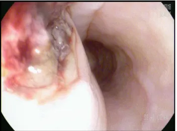
이종윤 등. 대동맥식도누공의 내시경적 진단 1예The Korean Journal of Gastroenterology Fig. 1. Endoscopic finding. Submucosal mass-like protrusion and
pulsating mass with blood clots is found at 30 cm below the incisors.
Fig. 2. Contrast-enhanced chest CT. (A) Esophageal lumen is narrow and esophageal wall is thickening (arrow). (B) Peripheral enhancing loculated cavity filled with fluid and multiple air-bubbles in posterior mediastinum (arrow).
증 례
71세 여자가 2주간의 상복부 불편감을 주소로 내원하였다.
수일 전부터 흑색변이 있었다고 하며 직장수지 검사에서도 흑 색변 소견을 보였다. 코위삽입관을 통한 위세척에서는 응고된 혈액이 소량 섞여 있었다. 고혈압으로 10년 전부터 약물 복용 중이었고, 7년 전부터 당뇨병으로 경구 혈당강하제를 복용 중 이었다. 음주력과 흡연력은 없었다. 내원 당시 계통 문진에서 주 호소 이외에 증상은 호소하지 않았다. 신체 검진에서 급성 병색을 보였으며, 결막은 다소 창백하였다. 공막의 황달 소견 을 보이지 않았고 간과 비장의 비대는 없었다. 혈압은 120/80
mmHg, 맥박 85회/분, 호흡수 20회/분, 체온 36.5℃였다. 흉 부 X선 촬영에는 이상 소견이 없었고 심전도 검사는 V1,2에 T파 역위가 있었다. 검사실 소견으로 말초혈액 검사는 백혈구 19,390/mm3, 혈색소 7.2 g/dL, 혈소판 232,000/mm3였고, 생 화학 검사에서는 AST 30 mg/dL, ALT 32 mg/dL, 총 단백 6.8 g/dL, 알부민 3.3 g/dL, 총 빌리루빈 0.5 mg/dL, 혈청 나트륨 134 mEq/L, 혈청 칼륨 4.4 mEq/L, 혈당 260 mg/dL로 다소 상승되었으며, BUN 49 mg/dL, creatinine 1.38 mg/dL, CRP는 8.96 mg/dL로 각각 상승되어 있었다. 상복부 불편감, 혈색소 감소 소견을 토대로 위 내시경을 시행하기로 하였다.
위 내시경 검사에서 내시경 삽입 30 cm 부위 중부 식도에 약 3 cm 크기와 1 cm 높이의 융기형 병변이 관찰되었다. 융 기된 표면은 정상 점막과 동일한 성상의 점막하 종괴의 형태 를 가지고 중심부에는 응고된 혈액으로 덮힌 궤양성 병변을 보였다(Fig. 1). 내시경 선단을 대었을 때, 박동이 느껴졌다.
식도 주변의 외부 압박에 의한 병변으로 판단하였고, 해부학 적 구조를 고려하여 식도에 인접한 대동맥에 의한 것임을 의 심하였다. 내시경을 더 진입시키지 않고 융기된 병변에 조직 검사나 궤양성 병변에 지혈술을 시행하지 않은 채 검사를 종 료하였다. 환자는 흉부 전산화단층촬영술을 시행받았다. 식도 내강이 협소하였고 하행 흉부 대동맥류가 관찰되었다. 대동맥의 직경은 5.5 cm, 대동맥류의 크기는 3 cm에 이르렀다. 하행 흉부 대동맥 주변에는 불규칙한 경계의 식도벽이 관찰되었고 후방 종격동에는 다발성 공기 음영이 관찰되었다(Fig. 2). 하행 흉부 대동맥류에 의한 대동맥식도누공과 동반된 종격동염으 로 진단하였다. 대량 출혈의 예방을 위하여 응급으로 혈관 내
A B
Lee JY, et al. Endoscopic Diagnosis of AEF
37
Vol. 73 No. 1, January 2019 Fig. 3. Thoracic endovascular aortic repair (TEVAR). (A) Saccular
aneurysm is visible. (B) TEVAR was performed in thoracic descending aorta.
Fig. 4. Endoscopic findings after thoracic endovascular aortic repair.
Ulcerative lesion covered with exudate is found at 30 cm below the incisors.
Fig. 5. Esophagography. Outpouching typed contrast leakage in esophagus is observed.
스텐트 삽입술을 우선적으로 시행하기로 하였다. 대동맥 조영 술에서 관찰되는 주머니(saccular) 모양의 동맥류에 직경 28 mm, 길이 80 mm의 스텐트를 삽입하였다(Fig. 3). 종격동염 과 이로 인한 백혈구 상승에 대하여 정맥 내 항생제 치료를 병행하였다. 입원 13일째 대동맥식도누공의 호전을 평가하기 위하여 위 내시경을 시행하였다. 융기된 병변은 호전을 보였 으나 여전히 기저 바닥에 삼출물로 덮힌 궤양성 병변이 관찰 되었다(Fig. 4). 입원 15일째 시행한 식도 조영 검사에서는 혈 관 내 스텐트 삽입에도 불구하고 누공이 지속되고 있음을 확
인하였다(Fig. 5). 입원 28일째 환자는 식도 절제술을 성공적 으로 시행받고 입원 44일만에 퇴원 후 현재 외래에서 추적 관찰 중이다.
고 찰
원발성 대동맥식도누공은 드물게 발생하는 원발성 대동맥 장관누공 중에서도 약 10%만을 차지하고,6연간 발생률은 천 만 명 당 15명으로 추정된다.7,8 Hollander와 Quick9의 보고 에 따르면 Chiari 징후 세 가지 중 상복부 혹은 흉부 통증은 59%, 대량 출혈 전에 나타나는 보초 출혈은 65%의 환자들에 서 나타났지만 대량 출혈을 포함하여 Chiari 징후 세 가지 모 두가 나타나는 경우는 45%에 불과한 것으로 보고하고 있다.
또한 대량 출혈이 동반되는 경우에는 급격한 경과로 사망에 이르기 때문에 진단하기 전에 사망하는 경우가 많다.4,5 이와 같은 이유로 임상의가 실제 대동맥식도누공 환자를 진료하게 되더라도 이를 정확히 진단해 내기란 매우 어렵다. 본 증례에 서는 상복부 불편감의 증상과 함께 빈혈과 흑색변으로 상부위 장관 출혈을 의심할 수 있었지만 토혈의 증상이 없었다. 혈관 내 스텐트 삽입술을 시행받은 72명의 대동맥식도누공 환자에 대한 Canaud 등10의 문헌고찰에 따르면 72명의 대동맥식도 누공 환자 중 62명이 토혈을 동반하였다고 보고하고 있어 본 증례의 임상 양상은 흔하지 않은 경우라 할 수 있다.
대동맥식도누공의 진단에는 전산화단층촬영이 위장관과 대동맥 사이의 관계를 잘 보여주기 때문에 가장 효과적이
다.11,12 흉부 전산화단층촬영에서 대동맥 주위에 공기 음영이
A B
38
이종윤 등. 대동맥식도누공의 내시경적 진단 1예The Korean Journal of Gastroenterology
나 인접한 식도 주변에 조영제가 누출되고 식도벽의 비후가 관찰된다면 대동맥식도누공을 진단할 수 있다. 하지만 상부위 장관 출혈의 흔한 원인이 소화성 궤양, Mallory-Weiss 증후 군, 식도 정맥류 출혈이기 때문에 전산화단층촬영 전에 상부 위장관 내시경을 우선적으로 시행하게 된다. 따라서 대동맥식 도누공의 특징적인 내시경 소견을 사전에 인지하고 있어야만 이를 의심하여 신속하게 전산화단층촬영을 통한 진단과 혈관 내 스텐트 삽입이나 수술적 치료를 할 수 있다. 대동맥식도누 공의 공통적인 내시경 소견은 대동맥의 압박에 의한 점막하 종괴 양상의 융기와 중심부 궤양이다. 융기된 병변에는 박동 이 느껴질 수 있고 누공이 관찰될 수도 있다.12,13 하지만 점막 하 종괴 양상의 융기 병변 중심부에 궤양이나 누공이 관찰되 지 않아 식도 폴립으로 오진된 보고도 있다.14 따라서 식도에 점막하 종괴 양상의 융기가 있다면 해부학적 구조를 고려하여 식도 주변에 인접한 대동맥의 외부 압박에 의한 것인지를 감 별하기 위하여 겸자를 대어 박동성을 확인할 필요가 있다. 만 약 융기된 병변을 종양으로 오인하여 조직 검사를 하거나 출 혈 부위에 지혈술을 시행할 경우 식도와 인접한 대동맥 벽에 침습적 행위를 하여 대량 출혈을 유발할 수 있으므로 대동맥 식도누공이 의심되는 경우에는 절대 시행해서는 안될 것이다.
대동맥식도누공의 치료는 수술적 치료가 원칙이지만 최근 응급 치료로 혈관 내 스텐트 삽입술로 치료하기도 한다.2,10 혈관 내 스텐트 삽입은 대량 출혈의 예방을 위하여 시행할 수 있으나 누공이 지속되는 경우, 심한 종격동염으로 세척이 필요한 경우는 수술적 치료가 병행되어야 한다.10본 증례에서 도 응급으로 혈관 내 스텐트 삽입술을 시행하여 대량 출혈을 예방하였으나 식도 조영술에서 누공이 지속되고 있음을 확인 하여 수술적 치료를 추가로 시행하였다.
대동맥식도누공은 낮은 발생률, 대량 출혈로 인한 급격한 경과 그리고 다른 상부위장관 출혈로 오인되어 상부위장관 내 시경을 우선적으로 시행하게 되는 등의 이유로 오진이나 치료 지연으로 인한 사망 가능성이 높다. 하지만 조기에 진단할 경 우 혈관 내 스텐트 삽입이나 수술로 성공적인 치료를 할 수 있기 때문에 소화기 내시경을 시행하는 임상의들은 대동맥식 도누공이 비록 드문 질환이라 하더라도 내시경적 특징을 충분 히 인지해야 한다. 저자들은 토혈의 증상 없이 상복부 불편감 을 호소하는 환자를 내시경을 통하여 대동맥식도누공으로 진 단하고 흉부 전산화단층촬영으로 하행 흉부 대동맥류에 의한
원발성 대동맥식도누공을 확인하여 이를 혈관 내 스텐트 삽입 술과 수술로 성공적으로 치료한 1예를 경험하였기에 이를 보 고하는 바이다.
REFERENCES
1. Bergqvist D. Arterioenteric fistula. Review of a vascular emergency.
Acta Chir Scand 1987;153:81-86.
2. Prokakis C, Koletsis E, Apostolakis E, Dedeilias P, Dougenis D.
Aortoesophageal fistulas due to thoracic aorta aneurysm: surgi- cal versus endovascular repair. Is there a role for combined aortic management? Med Sci Monit 2008;14:RA48-RA54.
3. Chiari H. Ueber Fremdkorpeverletzung des Oesophagus mit Aortenperforation. Ber Klin Wochenschr 1914;51:7-9.
4. Ikeda Y, Morita N, Kurihara H, Niimi M, Okinaga K. A primary aortoesophageal fistula due to esophageal carcinoma suc- cessfully treated with endoluminal aortic stent grafting. J Thorac Cardiovasc Surg 2006;131:486-487.
5. Shiraishi S, Watarida S, Matsubayashi K, Motoishi M, Satsu T.
Successful management of an aortoesophageal fistula resulting from an aneurysm of the thoracic aorta with a covered stent. J Cardiovasc Surg (Torino) 2002;43:95-98.
6. Contini S, Corrente V, Nervi G, Franzè A, Scarpignato C.
Dysphagia aortica: a neglected symptom of aortoesophageal fistula. Dig Liver Dis 2006;38:51-54.
7. Saers SJ, Scheltinga MR. Primary aortoenteric fistula. Br J Surg 2005;92:143-152.
8. Cutler JA, Mendeloff AI. Upper gastrointestinal bleeding. Nature and magnitude of the problem in the U.S. Dig Dis Sci 1981;26(7 Suppl):90S-96S.
9. Hollander JE, Quick G. Aortoesophageal fistula: a compre- hensive review of the literature. Am J Med 1991;91:279-287.
10. Canaud L, Ozdemir BA, Bee WW, Bahia S, Holt P, Thompson M.
Thoracic endovascular aortic repair in management of aortoeso- phageal fistulas. J Vasc Surg 2014;59:248-254.
11. Lee JT, Saroyan RM, Belzberg G, Pianim NA, Bongard FS. Primary aortoenteric fistula: computed tomographic diagnosis of an atypical presentation. Ann Vasc Surg 2001;15:251-254.
12. Yang Y, Hu D, Peng D. Primary aortoesophageal fistula: a fatal outcome. Am J Emerg Med 2018;36:343.e1-e343.e3.
13. Hsu WF, Lin CC, Chang KM, Lee TH. Primary aortoesophageal fis- tula: a rare but fatal cause of upper gastrointestinal bleeding. J Dig Dis 2013;14:676-678.
14. Jiao Y, Zong Y, Yu ZL, Yu YZ, Zhang ST. Aortoesophageal fistula:
a case misdiagnosed as esophageal polyp. World J Gastroenterol 2009;15:6007-6009.