J Korean Ophthalmol Soc 2014;55(1):66-72 pISSN: 0378-6471
eISSN: 2092-9374
http://dx.doi.org/10.3341/jkos.2014.55.1.66
= 증례보고 =
펨토초 레이저와 매뉴얼 트레파인을 이용한 심부표층각막이식의 초기 임상성적 비교
김근영⋅박진형⋅이용은⋅주천기 가톨릭대학교 의과대학 안과 및 시과학교실
목적: 펨토초레이저를 이용하여 심부표층각막이식술을 시행하여 그 임상결과를 매뉴얼 트레파인(manual trephine)을 이용한 심부표층 각막이식술과 비교하여 보고하고자 한다.
대상과 방법: 매뉴얼 트레파인(manual trephine)을 이용한 심부표층각막이식술을 받은 17안(17명), 펨토초레이저를 이용한 심부표층각 막이식술을 받은 13안(13명)에 대해 최대교정시력, 각막난시, 굴절난시, 각막내피세포수 등의 술후 임상성적을 비교하였다.
결과: 펨토초레이저를 이용한 심부표층각막이식술군에서 최대교정시력(logMAR)은 수술 후 2, 4, 6개월째에 각각 0.31 ± 0.17, 0.23
± 0.15, 0.18 ± 0.14였으며, 매뉴얼 트레파인(manual trephine)을 사용한 군에서는 각각 0.55 ± 0.41, 0.45 ± 0.28, 0.35 ± 0.22 (p=0.056, p=0.025, p=0.313)로 측정되었다. 자동굴절검사상 평균 각막난시는 펨토초레이저군에서 2, 4, 6개월째에 각각 5.35 ± 1.57, 4.24 ± 1.97, 3.65 ± 1.31, 매뉴얼 트레파인 사용군 8.32 ± 2.75, 6.80 ± 2.50, 4.54 ± 1.25 (p=0.031, p=0.041, p=0.370)로 측정되었 다. 술후 6개월째 두 군에 있어서 각막내피세포수는 두 군에서 유의한 차이를 보이지 않았다.
결론: 펨토초레이저를 이용한 심부표층각막이식술은 기존의 매뉴얼 트레파인을 이용한 심부표층각막이식술과 비교해 수술 후 초기 시력 회복과정이 빠르고, 난시의 유발량이 더 적어 이를 대체할 수 있는 수술방법이라 하겠다.
<대한안과학회지 2014;55(1):66-72>
■Received: 2013. 3. 18. ■ Revised: 2013. 10. 22.
■Accepted: 2013. 12. 7.
■Address reprint requests to Choun-Ki Joo, MD, PhD Department of Ophthalmology, Seoul St. Mary’s Hospital,
#222 Banpo-daero, Seocho-gu, Seoul 137-701, Korea Tel: 82-2-2258-1188, Fax: 82-2-599-7405
E-mail: [email protected]
심부표층각막이식은 각막의 앞층인 각막상피, 바닥막, 보 우만막, 간질을 제거하고, 공여안의 조직으로 대체시키는 방법으로 건강한 각막내피를 가진 각막혼탁 환자에서 전층 각막이식술을 대체하는 수술방법으로 제시되었다. 이 수술 방법의 장점은 수여자 자신의 각막내피를 보존하여 이식거 부반응의 발생 및 이식실패를 감소시키는 것이다. 반면 단 점으로는 기술적으로 어렵고, 술자가 데스메막이 노출될 때 까지 간질층을 점진적으로 박리해 나갈 경우 수술시간이 길어질 수 있다는 것이다.1
최근 대두된 펨토초레이저(Femtosecond laser)는 Nd:YAG 레이저와 비슷하나 초단파의 진동주기(ultra-short pulse duration)를 가지고 있어 더 작은 충격파(shock wave)와 기포를 발생시킨다.2이것은 레이저를 각막의 원하는 위치에 다양한 깊이로 정확하게 전달 가능하며 원형으로 조사하여 앞층판, 뒷층판, 수평절개를 만들게 된다. 각 층판의 크기를 조절하여 버섯모양, 중절모자(top-hat), 수직형(vertical),
Z-방형(Z-square) 등의 다양한 창상 모양의 각막을 원하 는 크기대로 신속하게 절개할 수 있으며 재현성이 있다는 장점이 있다. 또한 이러한 정확한 창상의 모양은 공여-수 여 각막의 접촉면을 증가시킴으로써 조기 봉합사 제거 및 난시의 감소로 빠른 시력회복을 유도하는 장점이 있다.3-5
이에 저자들은 본 연구에서 이러한 펨토초레이저를 이용한 심부표층각막이식술(IntraLase femtosecond laser-enabled deep anterior lamellar keratoplasty; IE-DALK, 펨토초레 이저군)을 시행하고 술 후 임상양상을 매뉴얼 트레파인 (manual trephine, Barron vacuum trephine)을 이용한 심 부표층각막이식술(Manual DALK; M-DALK, 매뉴얼군)을 시행한 환자들의 임상양상과 임상기록의 후향적 고찰을 통 하여 비교 분석하고자 하였다.
대상과 방법
1. 대상
2009년 5월부터 2011년 5월까지 본원에서 심부표층각 막이식술을 받은 33안(33명) 중 매뉴얼 트레파인을 이용 한 심부표층각막이식술을 받은 20안(20명) 또는 펨토초레 이저(IntraLase FS laser, IntraLase. Corp, USA)를 이용
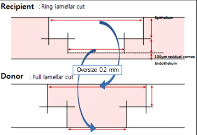
Figure 1. Settings on the FSL to create a partial-thickness
mushroom configuration in the recipient and doner cornea as- suming a minimum corneal pachymetry of 550 mm. Vertical cuts overlap 0.2 mm horizontally and 20 mm vertically with the horizontal lamellar cut at a 90-degree angulation to the cor- neal surface to ensure completely cut intersecting wound edges.The posterior side cut depth was calculated by subtracting 100 mm of residual cornea from the minimum corneal pachymetry (solid line, FSL cuts; dotted line, manual dissection by the sur- geon).
한 심부표층각막이식술을 받은 13안(13명, 원추각막 9안, 각막혼탁 4안)을 대상으로 하였으며 수술 후 효과 및 임상경과 를 비교하기 위해 의무기록을 후향적으로 조사하였다. 본 연구 는 병원 임상시험심사위원회(IRB 과제번호 KC12RISI0656) 의 심의를 통과하였다.
2. 수술방법
마취는 매뉴얼군(20안)에서는 전신마취가 16안, 구후마 취가 4안, 펨토초레이저군에서는 13안 모두에서 전신마취 가 시행되었다.
두 군 모두에서 각막간질과 데스메막 사이를 박리하기 위 해 Anwar and Teichmann가 제안한 “big bubble technique6”을 이용하여 최대깊이 층판박리(maximal depth lamellar dis- section) 방법을 사용하였다.
공여각막과 수여각막의 준비는 매뉴얼군에서는 개검기 로 안검을 벌린 후 헤스버그 바론 원형 절제기를 이용하여 수 여각막을 각막 두께의 2/3 깊이로 원형절개한 후, 30 guage 주사침을 간질에 삽입하여 공기를 주입해서 big-bubble 형 성한 후 간질과 데스메막 박리를 유도하였다. Beaver blade 로 원형 절개한 깊이의 절반 정도를 360도 층판 박리하여 제거하였다. 남아 있는 간질의 중심부에 Vannas scissors로 고랑을 만들고 Sinsky hook으로 데스메막 직전까지 고랑을 깊게 하였다. 주걱(spatula)을 이용하여 층판 분리술을 시
행하였고, 분리 정복의 방법으로 남아 있는 간질을 Vannas scissors를 이용하여 제거하였다. 잔여 섬유조직을 제거하 여 매끈한 면을 가진 데스메막을 완전히 노출시켜 이식수 여안의 준비를 완료하였다. 공여각막은 Trephine blade를 이용하여 원형절개한 후 smooth forcep으로 내피와 데스메 막을 비교적 저항 없이 쉽게 분리 제거하였고, 이렇게 준비 된 각막이식편을 수여안에 올려 놓고 10-0 nylon을 이용 하여 16개의 단속 봉합을 시행하였다.
펨토초레이저군에서는 외래굴절수술실에서 수여각막과 공여각막 모두 60-kHZ 펨토초레이저(Intrarase®, USA)를 이용하여 13안 모두에서 전통적인 수직 절개(straight cut) 에 비해 기계적 안정성(mechanical stability)이 우수하다 고 알려져 있는 버섯모양(mushroom configuration)으로 에너지 파(pulse)를 전달하도록 기계에 설정하여 절개를 시행하였고 수여각막의 경우 천공을 막기 위해 뒤쪽 부위 에 100 μm를 남겨놓았다. 각막간질과 데스메막 사이를 박 리하기 위해 매뉴얼군과 동일한 방법으로 분리를 시행하였 다(Fig. 1).
또한 수여각막과 같은 창상구조를 만들기 위해 공여각막 을 인공전방(artificial anterior chamber) 위에 올려놓은 후 평형염액(Balanced Salt solution: BSS)을 전방에 주입하 여 적당한 안압을 유지하였다. 각막중심부에 표시를 하고 레이저의 압평 렌즈 아래 흡입 없이 중심을 맞춘 후 계획된 각각의 창상 모양에 따라 수여각막과 같은 방식으로 앞층 판 및 뒤층판이 수여각막보다 0.2 mm 정도로 크게 레이저 로 각막절개를 시행하였다(Fig. 1). 그리고 공여각막을 수 여각막 위에 정확하게 위치시킨 뒤 매뉴얼군과 동일하게 10-0 nylon으로 16개의 단속 봉합을 시행하였다.
수술 후 1% prednisolone acetate (Pred forte®, Allergan, USA), 0.3% Gatifloxacin (Gatiflo®, Handok Pharmaceuticals, Korea)을 전자는 하루 2시간마다, 후자는 하루 4회 점안하 였다.
3. 측정변수
두 군의 모든 환자에서 수술 전 경과관찰 기간 동안 최대 교정시력, 세극등현미경검사 및 자동굴절계를 통한 굴절검 사, 각막두께 측정계(SP-3000, Tomey corporation, Japan) 로 각막두께, 수동각막곡률계(OM-4, Topcon, Japan)로 수직방향과 수평방향의 굴절값을 측정하였으며 골드만 압 평 안압계(GAT Haag-Streit, Switzerland)로 안압검사를 시행하였다. 또한 수술 전 빛간섭단층촬영(Visante anterior segment Optical coherence tomography, Carl Zeiss Meditech, Germany)을 이용하여 수여각막의 절개방법과 도안을 결정
Oversize 0.2 mm
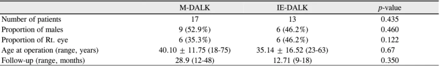
Table 1. Demographic data of the patients who underwent M-DALK and IE-DALK
M-DALK IE-DALK p-value
Number of patients 17 13 0.435
Proportion of males 9 (52.9%) 6 (46.2%) 0.460
Proportion of Rt. eye 6 (35.3%) 6 (46.2%) 0.122
Age at operation (range, years) 40.10 ± 11.75 (18-75) 35.14 ± 16.52 (23-63) 0.67
Follow-up (range, months) 28.9 (12-48) 12.71 (9-18) 0.350
Values are presented as mean ± SD.
Table 2. The indications of transplants in the two groups,
IntraLase-enabled DALK and manual DALK (p = 0.533)M-DALK IE-DALK
Keratoconus 10 (58.9%) 9 (69.2%)
Corneal opacity 7 (41.2%) 4 (30.8%)
BCVA (log MAR)
Preop Postop 2 months Postop 4 months Postop 6 months 2
1.5
1
0.5
0
p = 0.875
p = 0.060
p = 0.034* p = 0.332
M-DALK IE-DALK
Figure 2. Postoperative changes of log MAR mean best-spec-
tacle corrected visual acuity (BCVA) after manual trephine us- ing deep anterior lamellar keratoplasty (M-DALK) versus IntraLase- enabled deep anterior lamellar keratoplasty (IE- DALK); Postoperative BCVA improved gradually in both groups. BCVA in IE-DALK group was better than M-DALK group postoperatively, but the differences between two groups were statistically significant at 4 months (p = 0.034).The stat- istical analysis was performed using Mann-W hitney U test. A p-value less than 0.05 is statistically significant.하는데 참고하였다. 술 후 2개월, 4개월, 6개월째 최대교정 시력을 측정하였고, 굴절력 안정성 및 난시를 평가하기 위 해 자동굴절계를 통하여 굴절력 및 각막난시를 측정하였다.
술 후 방문 시마다 세극등현미경검사를 시행하고 술 후 합 병증의 발생 양상을 비교 분석하였으며 각막두께 검사를 시행하였고 6개월째 두 군을 비교하였다. 또 술 후 구조적 안정성을 평가하기 위해 빛간섭 단층촬영으로 공여각막과 수여각막의 접합부를 관찰하였으며 전안부 사진(anterior segment photo)을 촬영하였다.
4. 통계방법
통계학적 처리는 SPSS 17.0 for Window (SPSS Inc.)를 이용하였으며 연속변수에 대해서 두 군 간의 차이를 비교 하기 위해 Mann-Whitney U test를 이용하였다. p값이 0.05 미만일 경우 통계적으로 유의하다고 평가하였다.
결 과
매뉴얼 트레파인(매뉴얼군)과 펨토초레이저(펨토초레 이저군)를 이용한 심부표층각막이식술을 받은 17안 및 13안의 수술시 평균 연령은 40.10 ±11.75 (18-75)세와 35.14 ± 16.52 (23-63)세, 평균 추적관찰기간은 28.9 (12-48)개월과 12.7 (9-18)개월이었다(Table 1).
각막이식을 하게 된 적응증을 각 군별로 보여주고 있으며 원추각막이 두 군 모두에서 가장 높은 비율을 차지하고 있었 으나 두 군간의 유의한 차는 없었다(p=0.533, Table 2).
두 군에서 각각 술 후 최대교정시력의 변화를 술 후 2개 월, 4개월, 6개월로 나누어 살펴본 결과 지속적으로 펨토초레 이저군에서 높은 시력을 유지하였다. 술 후 4개월째 logMAR 최대교정시력은 펨토초레이저군에서 0.23 ±0.15, 매뉴얼군 에서는 0.45 ±0.28로 전자에서 유의하게 좋았다(p=0.034).
술 후 2, 6개월째 logMAR 최대교정시력은 펨토초레이저군 에서 0.31 ± 0.17, 0.18 ± 0.14, 매뉴얼군에서 0.55 ± 0.41, 0.35 ±0.22로 펨토초레이저군에서 시력이 좋았으나 통 계학적으로 유의한 차이는 없었다(p=0.060, p=0.332)(Fig. 2)
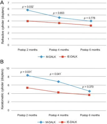
수술 후 2, 4, 6개월째 자동굴절검사 결과 굴절난시 (refractive astigmatism)와 평균각막난시(keratometric astigmatism)는 지속적으로 펨토초레이저군에서 미세각 막절삭기 사용군 보다 낮았으나 굴절난시(refractive as- tigmatism)의 유의한 차이는 술 후 2개월째, 각막난시 (keratometric astigmatism)는 술 후 2, 4개월째 비교에 서만 나타났다. 자동굴절검사에 의한 굴절난시는 2개월 째 펨토초레이저군에서 3.96 ± 0.55D, 매뉴얼군에서 5.80 ±2.35였다(p=0.032). 술 후 2, 4개월째 평균각막 난시 측정값은 펨토초레이저군에서 5.35 ±1.57, 4.24 ± 1.97였고, 매뉴얼군은 8.32 ±2.75, 6.80 ±2.50으로 낮 은 수치를 보였으며 통계적으로 유의하였다(p=0.031, 0.041)(Fig. 3).
M-DALK IE-DALK
Postop 2 months Postop 4 months Postop 6 months
Refractive cylinder (diopters)
7 6 5 4 3 2 1 0
M-DALK IE-DALK
Postop 2 months Postop 4 months Postop 6 months Keratometric cylinder (diopters) 10
8 6 4 2 0
p = 0.032*
p = 0.653
p = 0.778
p = 0.370 p = 0.041*
p = 0.031*
Figure 3. The cylinder measured using autorefractor in both
groups of M-DALK and IE-DALK at 2, 4 and 6 months postoperatively. The refractive cylinder showed lower value in IE-DALK during follow up periods, and the difference was statistically significant at 2 months postoperatively (p = 0.032) (A). The keratometric cylinder measured using manual keratometer showed lower value in IE-DALK than M-DALK, and the difference was statistically significant at 2 months (p = 0.031), and 4 months (p = 0.041) postoperatively (B). The statistical analysis was performed using Mann-Whitney U test.A p-value less than 0.05 is statistically significant.
p = 0.865
535.75 530.14
M-DALK IE-DALK
700 600 500 400 300 200 100 µic)m(s eskn thCalneorl ctraen 0
Central corneal thickness
Figure 4. Difference of postoperative central corneal thickness
between M-DALK (535.75 ± 54.0) and IE-DALK (530.14 ± 31.2) group (p = 0.865, Mann Whitney test). Pachymetry was performed at 6 months postoperatively (both group).p = 0.880
2361.6 2559.0
M-DALK IE-DALK
3000 2500 2000 1500 1000 500
2si)m/mllsce (tyenEl dell cliaheotnd 0
Specular microscopy
Figure 5. Difference of postoperative endothelial cell density
between M-DALK (2361.60 ± 320.5) and IE-DALK (2559.00± 531.75) group (p = 0.880, Mann W hitney test). Specular microscopy was performed at 6 months postoperatively (both group).
술 후 6개월째 측정한 중심각막두께는 펨토초레이저군에 서 530.14 ±51.81 μm, 매뉴얼군에서 535.75 ±54.0 μm로 통계적으로 유의한 차이를 보이지 않았다(p=0.865)(Fig. 4)
술 후 6개월째 측정한 각막내피세포수는 펨토초레이저군
에서 2559.0 ±531.7cells/mm2, 매뉴얼군에서 2361.6 ± 320.5 cells/mm2로 펨토초레이저군에서 높게 측정되었으나 통계적으로 유의한 차이는 보이지 않았다(p=0.880)(Fig. 5).
두 군 모두에서 술 후 6개월 동안 거부반응과 같은 합병 증은 발생하지 않았다.
고 찰
심부표층각막이식술은 1985년 Achila7에 의해 처음 보고 된 수술법으로 비교적 각막내피가 건강한 환자에서 병적인 각막기질을 제거, 건강한 공여각막으로 대체함으로써,8 각 막내피층의 거부반응을 줄일 수 있으며 뿐만 아니라, 수술 중 맥락막 출혈, 술후 각막내피세포부전, 봉합관련 난시의 감소와 조기발사로 인한 빠른 시력회복 등 다양한 이점이 있다.9-11이러한 장점으로 인하여 심부표층각막이식술은 원 추각막,9-15 각막혼탁,9,14,16-18 각막이영양증,9,14,17,18 굴절수술 후 각막확장증19등 다양한 질환에서 이용되고 있다(Fig. 6, 7).
심부표층각막이식술에 있어서 가장 중요한 수술 절차는 앞층판의 박리이다. 또한 술후 난시와 굴절이상을 유발할 수 있는 불규칙하거나 중심을 이탈한 절개를 피하기 위해 숙련된 원형절제기술(trephination technique) 역시 매우 중 요하다.20미세각막절삭기를 이용한 심부표층각막이식술의 경 우창상 변연이 매끄럽지 못해 공여 각막의 이탈(slippage) 을 유발할 수 있고 불규칙한 각막표면 등의 단점을 가지고 있다. 더불어 진행된 원추각막과 같은 가파른 각막의 경우 효과가 떨어지는 단점이 있다.10각막이식에 있어 펨토초레 이저의 사용은 특히 앞층판 각막이식술에 있어 새로운 전 기를 마련하였는데 여러 연구자들이 각막 층판 수술에 있 어 펨토초레이저를 사용함으로써 공여각막과 수여각막의 절제에 있어 저주파에너지의 사용과 spot 스폿 분리 설정 (spot separation setting)을 통한 높은 질과 정확성, 재현
A
B
Figure 6. Anterior segment photographs of the patients 2 months after surgery show the clear central cornea with well attached pe-
ripheral flange in IE-DALK (A), and mild edematous cornea with well attached graft in M-DALK (B).Figure 7. Six months postoperatively, Visante optical coherence tomography of IE-DALK (mushroom shape) (A), demonstrating
the perfect match of the recipient to the donor. In contrast, conventional M-DALK (B) may show the lack of precise match of cut between the two pieces of tissues and protrusions such as hills.성에 대해 보고했다.21,22
Buratto and Böhm19은 펨토초레이저를 사용하여 개개의 각막질환에 맞게 세분화된각막 절편을 만들어 공여각막과 수여각막 사이의 접촉면적을 증가시켜 궁극적으로 빠르고 더 좋은 상처 회복 및 봉합사로 인한 난시를 줄일 수 있었 다고 보고했다. 본 연구의 경우에도 최대교정시력의 변화를 술 후 2개월, 4개월, 6개월로 나누어 살펴본 결과 지속적으 로 펨토초레이저군에서 높은 시력을 유지하였다. 술 후 4개 월째 logMAR 최대교정시력은 펨토초레이저군에서 0.23 ± 0.15, 매뉴얼군에서는 0.45 ± 0.28로 전자에서 유의하게 좋았다(p=0.034). 술 후 2, 6개월째 logMAR 최대교정시 력은 펨토초레이저군에서 0.31 ±0.17, 0.18 ±0.14, 매뉴 얼군에서 0.55 ±0.41, 0.35 ± 0.22로 펨토초레이저군에 서 시력이 좋았으나 통계학적으로 유의한 차이는 없었다 (p=0.060, p=0.332). 또한 굴절난시와 각막난시의 경우 미세각막절삭기 사용군과 비교할 때 펨토초레이저군에서 더 낮은 수치를 보였으며 전자의 경우 술후 2개월(p=0.028), 후자의 경우 술후 2, 4개월(p=0.022, 0.045)에서 통계적
으로 의미 있는 결과를 보여주었다(Figs. 6, 7).
각막이식술에서 또 다른 관심사 중의 하나는 수술 후 지 속적인 각막내피세포밀도의 감소에 관한 것으로 Bourne23 은 전층각막이식술 후 약 10년간에 걸쳐서 생리적인 내피 세포밀도의 감소율보다 빠르게(0.6%/년), 최대 50%까지 감소한다고 보고하고 있다. 반면 van Dooren et al24은 심부 표층각막이식술을 받은 눈에서 술 후 처음 6개월에 약 11%의 내피세포밀도의 감소가 있었으나 이후로는 생리적 인 감소율을 보였다고 보고하고 있다. 본 연구에서는 술전 자료가 없어 각막내피세포수 감소율에 대한 분석은 하지 못하였고 술 후 6개월째 양쪽 군에 있어 각막내피세포수를 비 교하였는데 펨토초레이저군에서 2559.0 ±531.7 cells/mm2, 매뉴얼군에서 2361.6 ±320.5 cells/mm2로 다소 높게 측 정되었으나 통계적으로 유의한 차이는 보이지 않았다.
심부표층각막이식술에 있어 매뉴얼로 층판 박리 도중 데 스메막의 미세천공이 발생할 수 있는데 기존 연구 결과를 보면 0-39.2%16,18까지의 발생률을 보여 주었으며 본 연구 의 경우 6.1% (전체 33안 중 2안)의 발생률을 보여 주었으
A B
A B
며 이는 매뉴얼군에서만 나타났으며, 이는 데스메막을 포함 한 반흔이나, 잠재적인 각막수종이 존재했을 것으로 추정된 다. 술 후 거부반응은 두 군 모두에서 술 후 6개월 이내에는 나타나지 않았다. 이는 기존의 연구결과인 전층각막이식술 의 술 후 발생하는 각막내피층의 거부반응20-30%25와 비 교할 때 매우 낮다고 보일 수도 있으나 6개월이라는 단기간 의 시간만을 추적 관찰하였기 때문에 추후 지속적인 경과 관찰을 해야 정확한 비교가 가능하다.
이 연구의 제한점으로는 첫째, 대상의 수가 적었고 둘째, 1년 이상의 장기간의 결과를 분석하지 않은 점, 셋째, 펨토 초레이저군의 공여각막 및 수여각막의 모양이 버섯모양으 로 제한되었다는 점, 넷째 발사시기에 따른 난시 변화의 분 석을 못한 점 등을 들 수 있으나, 국내에서 최초로 매뉴얼 트레파인과 펨토초레이저를 이용한 심부표층각막이식술을 비교한 논문이라는 점에서 의의가 있다.
결론적으로 본 연구에서 시행한 펨토초레이저를 이용한 심부표층각막이식술(IE-DALK)은 기존의 일반적인 매뉴 얼 트레파인을 이용한 심부표층각막이식술(M-DALK)과 비교해 수술 후 초기 시력 회복과정이 빠르고, 난시의 유발 량이 더 적어 이를 대체할 수 있는 수술방법이라 하겠다.
REFERENCES
1) Tsubota K, Kaido M, Monden Y, et al. A new surgical technique for deep lamellar keratoplasty with single running suture adjustment.
Am J Ophthalmol 1998;126:1-8.
2) Stern D, Schoenlein RW, Puliafito CA, et al. Corneal ablation by nanosecond, picosecond, and femtosecond lasers at 532 and 625 nm. Arch Ophthalmol 1989;107:587-92.
3) Farid M, Kim M, Steinert RF. Results of penetrating keratoplasty performed with a femtosecond laser zigzag incision initial report.
Ophthalmology 2007;114:2208-12.
4) Ignacio TS, Nguyen TB, Chuck RS, et al. Top hat wound config- uration for penetrating keratoplasty using the femtosecond laser: a laboratory model. Cornea 2006;25:336-40.
5) Malta JB, Soong HK, Shtein R, et al. Femtosecond laser-assisted keratoplasty: laboratory studies in eye bank eyes. Curr Eye Res 2009;34:18-25.
6) Anwar M, Teichmann KD. Deep lamellar keratoplasty: surgical techniques for anterior lamellar keratoplasty with and without bar- ing of Descemet's membrane. Cornea 2002;21:374-83.
7) Archila EA. Deep lamellar keratoplasty dissection of host tissue with intrastromal air injection. Cornea 1984-1985;3:217-8.
8) Benson WH, Goosey CB, Prager TC, Goosey JD. Visual improve- ment as a function of time after lamellar keratoplasty for keratoconus.
Am J Ophthalmol 1993;116:207-11.
9) Panda A, Bageshwar LM, Ray M, et al. Deep lamellar keratoplasty versus penetrating keratoplasty for corneal lesions. Cornea 1999;
18:172-5.
10) Tan DT, Mehta JS. Future directions in lamellar corneal transplantation.
Cornea 2007;26(9 Suppl 1):S21-8.
11) Watson SL, Ramsay A, Dart JKG, et al. Comparison of deep la- mellar keratoplasty and penetrating keratoplasty in patients with keratoconus. Ophthalmology 2004;111:1676-82.
12) Amayem AF, Anwar M. Fluid lamellar keratoplasty in keratoconus.
Ophthalmology 2000;107:76-9.
13) Funnell CL, Ball J, Noble BA. Comparative cohort study of the outcomes of deep lamellar keratoplasty and penetrating kerato- plasty for keratoconus. Eye (Lond) 2006;20:527-32.
14) Trimarchi F, Poppi E, Klersy C, Piacentini C. Deep lamellar keratoplasty. Ophthalmologica 2001;215:389-93.
15) Yoo SH, Kymionis GD, Koreishi A, et al. Femtosecond laser-as- sisted sutureless anterior lamellar keratoplasty. Ophthalmology 2008;115:1303-7.
16) Mosca L, Fasciani R, Tamburelli C, et al. Femtosecond laser-as- sisted lamellar keratoplasty: early results. Cornea 2008;27:668-72.
17) Noble BA, Agrawal A, Collins C, et al. Deep Anterior Lamellar Keratoplasty (DALK): visual outcome and complications for a het- erogeneous group of corneal pathologies. Cornea 2007;26:59-64.
18) Sugita J, Kondo J. Deep lamellar keratoplasty with complete re- moval of pathological stroma for vision improvement. Br J Ophthalmol 1997;81:184-8.
19) Buratto L, Böhm E. The use of the femtosecond laser in penetrating keratoplasty. Am J Ophthalmol 2007;143:737-42.
20) Bahar I, Kaiserman I, McAllum P, Rootman D. Femtosecond la- ser-assisted penetrating keratoplasty: stability evaluation of differ- ent wound configurations. Cornea 2008;27:209-11.
21) Sarayba MA, Ignacio TS, Tran DB, Binder PS. A 60 kHz IntraLase femtosecond laser creates a smoother LASIK stromal bed surface compared to a Zyoptix XP mechanical microkeratome in human donor eyes. J Refract Surg 2007;23:331-7.
22) Alió JL, Piñero DP. Very high-frequency digital ultrasound meas- urement of the LASIK flap thickness profile using the IntraLase femtosecond laser and M2 and Carriazo-Pendular microkeratomes.
J Refract Surg 2008;24:12-23.
23) Bourne WM. Cellular changes in transplanted human corneas.
Cornea 2001;20:560-9.
24) van Dooren BT, Mulder PG, Nieuwendaal CP, et al. Endothelial cell density after deep anterior lamellar keratoplasty (Melles tech- nique). Am J Ophthalmol 2004;137:397-400.
25) Arentsen JJ. Corneal transplant allograft reaction: possible predis- posing factors. Trans Am Ophthalmol Soc 1983;81:361-402.
=ABSTRACT=
Comparison of Early Clinical Result of Deep Anterior Lamellar Keratoplasty Using FSlaser Versus Manual Trephine
Eun Yeong Kim, MD, Jin-Heung Park, MD, Yong Eun Lee, MD, Choun-Ki Joo, MD, PhD
Department of Ophthalmology and Visual Science, The Catholic University of Korea School of Medicine, Seoul, Korea
Purpose: To compare outcomes of femtosecond laser-enabled deep anterior lamellar keratoplasty (IE-DALK) versus man- ual trephine using deep anterior lamellar keratoplasty (Manual DALK, M-DALK).
Methods: Seventeen eyes from 17 patients underwent manual deep anterior lamellar keratoplasty, and femtosecond la- ser-enabled deep anterior lamellar keratoplasty was performed in 13 eyes of 13 patients. Postoperative clinical outcomes such as best corrected visual acuity, refractive astigmatism, keratometric astigmatism, endothelial cell density were com- pared between the two groups.
Results: The mean log MAR best spectacle-corrected visual acuity (BSCVA) was 0.31 ± 0.17, 0.23 ± 0.15, 0.18 ± 0.14 in the IE-DALK group, and 0.55 ± 0.41, 0.45 ± 0.28, 0.35 ± 0.22 (p = 0.056, p = 0.025, p = 0.313) in the M-DALK group at post- operative 2, 4, and 6 months respectively. The mean keratometric cylinder was 5.35 ± 1.57, 4.24 ± 1.97, 3.65 ± 1.31 in the IE-DALK, 8.32 ± 2.75, 6.80 ± 2.50, 4.54 ± 1.25 (p = 0.031, p = 0.041, p = 0.370) in the M-DALK group at postoperative 2, 4, and 6 months respectively. Endothelial cell counts in the two groups did not differ significantly at postoperative 6 months.
Conclusions: The IE-DALK showed better results in BSCVA and keratometric astigmatism in the early postoperative peri- od, in comparison with the M-DALK, and can be considered as a valuable method in deep anterior lamellar keratoplasty.
J Korean Ophthalmol Soc 2014;55(1):66-72
Key Words: Deep anterior lamellar keratoplasty, Endothelial cell density, Femtosecond laser
Address reprint requests to Choun-Ki Joo, MD, PhD Department of Ophthalmology, Seoul St. Mary’s Hospital
#222 Banpo-daero, Seocho-gu, Seoul 137-701, Korea
Tel: 82-2-2258-1188, Fax: 82-2-599-7405, E-mail: [email protected]