접수일 : 2016 년 12 월 5 일 , 게재승인일 : 2017 년 3 월 22 일 책임저자 : 김형섭 , 경기도 고양시 일산동구 일산로 100
10444, 국민건강보험 일산병원 재활의학과
Tel: 031-900-0137, Fax: 031-900-0343 E-mail: [email protected]
외상성 뇌손상 환자에서 성인 척수견인증후군에 의한 요천추 신경근병증
증례 보고
국민건강보험 일산병원 재활의학과
장찬웅ㆍ김종문ㆍ김형섭
전체 글
증례 보고
장찬웅ㆍ김종문ㆍ김형섭
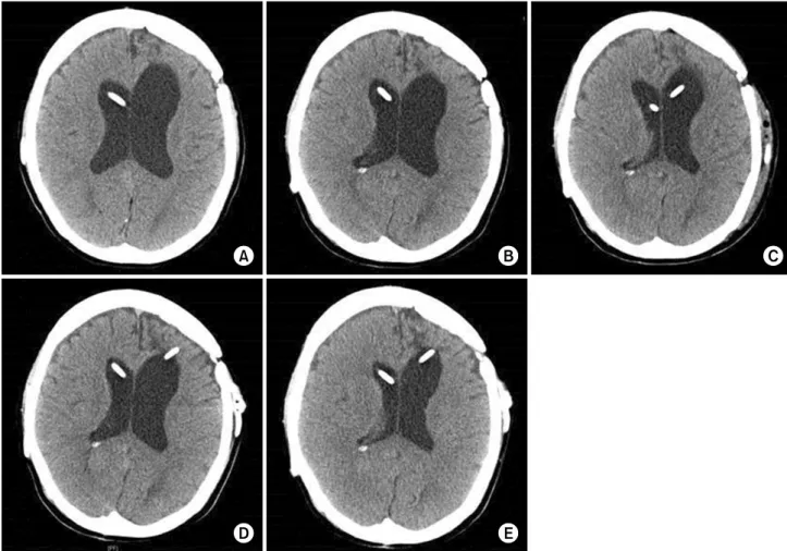
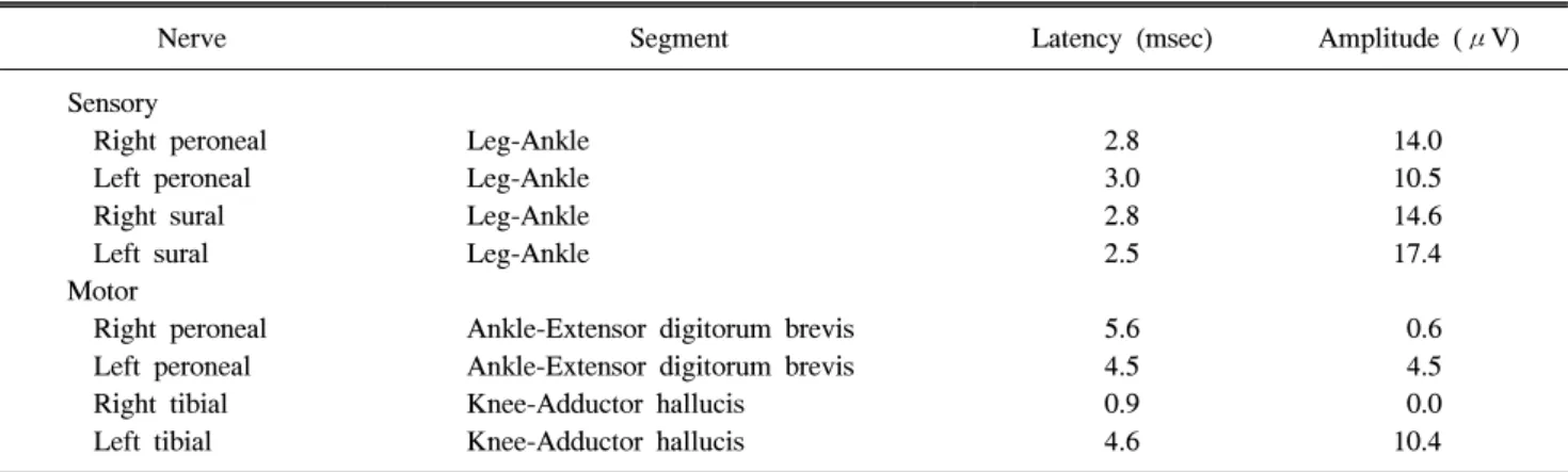
수치
관련 문서
GDP impact of COVID-19 spread, public health response, and economic policies. Virus spread and public
(1.1 points) According to the passage, which of the following is NOT mentioned as the nature of pain.. ① Pain is not tied to any referent in
W You’re going to have back and neck pain, if you sit like that for a long time4. When you’re sitting, it’s very important to keep the back straight, knees bent, and head
In conclusion, the sling stabilization exercise program for middle-aged women with non-specific low back pain was found to be effective in improving
Abstract: This study was aimed to investigate the adhesion control standards of pain relieving patch (PRP) drugs and to survey it′s adverse effects on the skin of patients
Depending on Tennis Forehand Stroke with elbow pain and without elbow pain Kinematic Analysis of
Comparison of pain scale between the conventional and 2-step needle insertion technique according to the injection area ··· 11... The combination of pain scale and
Sports Massage Using Thumb Pressure on the Effects on Subjective Neck
