Stimulation of eNOS-Ser617 Phosphorylation by Fluid Shear Stress in Endothelial Cells
Yong Chool Boo*
Department of Molecular Medicine, Kyungpook National University School of Medicine, Daegu 700-422, Korea Received October 13, 2005; Accepted November 11, 2005
Nitric oxide (NO) produced from endothelial cells plays a critical role in vascular physiology. The regulation of endothelial NO synthase (eNOS) involves various mechanisms including multiple Ser/Thr phosphorylations. Recently, eNOS-Ser617 was newly recognized to be phosphorylated in response to humoral factors including vascular endothelial growth factor. However, it remains unknown whether and how eNOS-Ser617 phosphorylation is stimulated by shear stress, the primary stimulus of endothelial NO production. This issue was explored in the present study using cultured bovine aortic endothelial cells (BAECs). Over-expression of a constitutively active protein kinase B (Akt) mutant in BAECs increased Ser617 phosphorylation while constitutively active protein kinase A mutant had no effect. When BAECs were subjected to an arterial level of laminar shear stress, eNOS-Ser617 phosphorylation was clearly increased in a time-dependent manner. Shear stress also stimulated Akt phosphorylation at Thr308, one of the key regulatory sites. The time courses of eNOS-Ser617 and Akt- Thr308 phosphorylations appeared to be very similar. These results suggested that eNOS-Ser617 phosphorylation, mediated by Akt, is a physiological response to the mechanical shear stress, involved in the regulation of NO production in endothelial cells.
Key words: shear stress, endothelial nitric oxide synthase (eNOS), phosphorylation, protein kinase B (Akt), protein kinase A (PKA)
Endothelial cells are constantly subjected to shear stress generated by blood flow. The importance of shear stress for vascular health has been recognized by the preferential development of atherosclerosis at the branched and curved points of arteries which experience disturbed rather than laminar flow.1,2) The regions of arteries experiencing laminar shear stress due to orderly blood flow are generally protected from lesion formation. Currently, it is appreciated that nitric oxide (NO) produced from endothelium plays a critical role in mediating the athero-protective effect of laminar shear stress while reactive oxygen species are implicated in the inflammatory reactions caused by oscillatory shear stress.3,4)
Endothelial NO production is stimulated by shear stress, cyclic strain and various humoral factors including vascular endothelial growth factor (VEGF).5-7) Endothelial nitric oxide synthase (eNOS) is regulated at the level of protein expression and by the acute modulation of activity through multiple mechanisms including an association in multiprotein complexes, protein-protein interactions, sub-cellular locations and post- translational modifications.8,9) Acute changes in shear stress
induce a Ca2+/CaM-dependent production of NO, followed by Ca2+-independent steady NO production.5) The precise mechanism of steady NO production is not entirely clear, yet binding of Hsp90 to eNOS and eNOS phosphorylation appear to play a role.4,9,10)
Over the past decade, we have come to appreciate the importance of eNOS phosphorylation at various sites. Currently, five Ser/Thr residues (Ser116, Thr497, Ser617, Ser635 and Ser1179) are known to be phosphorylated by numerous protein kinases including protein kinase B (Akt), protein kinase A (PKA), CaM kinase II, AMP-activated kinase and protein kinase C in response to physiological stimuli.9) We have shown that shear stress stimulates eNOS phosphorylation at Ser1179 and Ser635 without significant effect on Ser116 and Thr497.11,12) While S1179 phosphorylation in eNOS plays a critical role in response to both shear stress and humoral ligands such as VEGF, S635 phosphorylation renders it active under basal conditions, perhaps contributing to the basal NO production at a low level.13)
Recent studies have found that eNOS-S617 is phosphorylated in response to humoral factors such as ATP, bradykinin and VEGF.14) However it remains unknown if shear stress, the primary stimulus of endothelial NO production, stimulates the eNOS-S617 phosphorylation. Furthermore, the protein kinase responsible for the phosphorylation in cells remains to be identified. The present study was aimed to fill in these missing parts that are essential to understand the signaling events involved in shear-stimulated NO production.
*Corresponding author
Phone: +82-53-420-4946; Fax, +82-53-426-4944 E-mail: [email protected]
Abbreviations: NO, nitric oxide; VEGF, vascular endothelial growth factor; eNOS, endothelial nitric oxide synthase; CaM, calmodulin;
Hsp90, heat shock protein 90; Akt, protein kinase B; PKA, protein kinase A; VEGFR2, vascular endothelial growth factor receptor 2;
PECAM-1, platelet endothelial cell adhesion molecule-1
Materials and Methods
Cell culture. Bovine aortic endothelial cells (BAEC) harvested from descending thoracic aortas were maintained (37°C, 5% CO2) in a growth medium [Dulbecco’s modified Eagle’s medium (DMEM) containing 1 g/l glucose (GIBCO) and 20% fetal bovine serum (FBS, Atlanta Biologicals) without antibiotics].11)
Adenoviral infections. BAEC at ~90% confluency were infected with a recombinant adenovirus in serum-free DMEM for 1 h and incubated for 48 h in a growth medium. The recombinant adenovirus expressing a constitutively active Akt mutant generated by fusing a myristoylation signal to its amino terminus (Ad-Akt-Myr)11, 15) and the control adenovirus encoding β-galactosidase (Ad-β-Gal) were used in the present study.
Transfections. BAECs were transfected with a plasmid construct by using lipofectamin (Invitrogen). Briefly, cells grown to ~90% confluency in 100 mm culture dishes were washed with and kept in 5 ml Opti-MEM (Gibco). Plasmid construct (5µg DNA) was mixed with lipofectamin (10µg) in 500µl Opti-MEM, and the mixture was kept at room temperature for 30 min. Cells were treated with the mixture for 5 h at 37oC. The transfected cells were further incubated for 1 day. PKA-Cqr construct expresses a constitutively active mutant of mouse PKA catalytic subunit Cα which contains double mutations (His87Gln and Trp196Arg).13,16)
Shear stress studies. A confluent monolayer of BAEC grown in a 100-mm dish was exposed to non-pulsatile, laminar shear stress in a shear medium (phenol red-free DMEM containing 0.5% FBS and 25 mM HEPES, pH 7.4) by rotating a Teflon cone (0.5o cone angle) as described previously.11,12) Cells were exposed to an arterial level of shear stress (15 dyn/cm2).
Western blotting. Cells were washed in ice-cold phosphate-buffered saline (PBS) and lysed in 0.75 ml lysis buffer A (20 mM Tris · HCl, pH 7.6, 150 mM NaCl, 1 mM EDTA, 1 mM EGTA, 2.5 mM sodium pyrophosphate, 1 mM
β-glycerophosphate, 1 mM sodium vanadate, 1 µg/ml leupeptin, 1 mM phenylenemethylsulfonyl fluoride, 1 µM microcystin, and 1% Triton X-100). Cell lysates were clarified by spinning at 14,000 g for 15 min at 4oC. Aliquots of cell lysates (20 µg protein each) were resolved on a 10% SDS-PAGE gel and transferred to a polyvinylidene difluoride membrane (Millipore).
The membrane was incubated with a primary antibody overnight at 4oC and then with a secondary antibody conjugated with alkaline phosphatases (1 h at room temperature), which was detected by a chemiluminescence method, as described previously.12) Blots shown are representatives of at least three independent studies.
The following primary antibodies were used: polyclonal antibodies for phosphorylated forms of Akt-Thr308 and eNOS-Ser1179 from Cell Signaling Technology; and a polyclonal antibody for total Akt from Santa Cruz biotechnology;
polyclonal and monoclonal antibodies for total eNOS, and
monoclonal antibody for PKA-Cα from Transduction Laboratories. Rabbit polyclonal antibodies specific for phosphorylated forms of eNOS-Ser116, eNOS-Thr497, eNOS- Ser617 and eNOS-Ser635 were from Upstate Biotechnology.
Results and Discussion
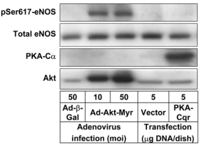
Recently, Michell et al. showed that eNOS-Ser617 was phosphorylated when recombinant eNOS protein was incubated with purified Akt or PKA in kinase assay buffer.14) In their experiments, Akt was shown to phosphorylate eNOS at Ser1179 and Ser617 while PKA phosphorylated Ser1179, Ser635 and Ser617. To determine which of these potential candidates, Akt or PKA, regulates eNOS-Ser617 phosphorylation in cells, BAEC were either infected with a recombinant adenovirus to express a constitutively active Akt mutant (Ad- Akt-Myr)11,15) or transfected with a plasmid construct encoding a constitutively active PKA catalytic subunit mutant (PKA- Cqr).13,16) Control cells were infected with a recombinant adenovirus Ad-β-Gal or transfected with an empty pAdTrackCMV vector. The protein extracts of the cells were subjected to Western blot analysis using phospho-specific antibody for eNOS-Ser617. As shown in Fig. 1, adenovirus-mediated over- expression of Akt-Myr in BAEC markedly enhanced eNOS- Ser617 phosphorylation without significantly affecting total eNOS protein level. This change was not observed in control cells infected with Ad-b-Gal. In contrast, PKA-Cqr expression did not stimulate eNOS-Ser617 phosphorylation at all, similarly to the control cells transfected with an empty vector.
Fig. 1. Akt but not PKA regulates eNOS phosphorylaton at Ser617 in endothelial cells. BAECs were infected with a recombinant adenovirus expressing a constitutively active Akt mutant (Ad-Akt-Myr) or a control adenovirus encoding β-galac- tosidase (Ad-β-Gal), or were transfected with a PKA-Cqr con- struct encoding a constitutively active mutant of mouse PKA catalytic subunit Cα, or an empty pAdTrackCMV vector. The protein extracts from the treated cells were subjected to West- ern blot analysis using antibodies specific for eNOS phosphory- lated at Ser617. The membranes were re-probed with antibodies detecting total eNOS. Expression of Akt-Myr and PKA-Cqr were verified by blotting with Akt and PKA-Cα antibodies, respectively.
These results strongly suggest that Akt, but not PKA, may be the protein kinase responsible for S617 phosphorylationunder physiological conditions. Although it occurs in vitro,14) PKA- dependent phosphorylation of eNOS-Ser617 does not appear physiologically relevant. Robust expression of Akt-Myr and PKA-Cqr was verified by Western blotting with Akt and PKA-Cα antibodies, respectively.
Next, the effects of over-expressed Akt-Myr on phosphorylation of eNOS at sites other than Ser617 were examined. As shown in Fig. 2, Akt-Myr expression stimulated eNOS-Ser1179 phosphorylation, as reported elsewhere.7,11,17) The phosphorylation status of eNOS at The495 and Ser116 appeared to be insensitive to Akt-Myr; however, Ser635 phosphorylation was weakly but clearly stimulated. Because purified Akt protein does not phosphorylate eNOS-Ser635 in vitro,14) it is conceivable that Akt might have stimulated the phosphorylation indirectly, possibly by involving PKA.
eNOS is known to be phosphorylated at Ser1179 and Ser635 by a PKA-dependent mechanism in response to laminar shear stress.11,12) However, many groups, including ours, have noticed a role of Akt in eNOS activation by shear stress.11,18) Therefore I examined if shear stress stimulated the phosphorylation of eNOS-Ser617 which appeared to be a potential Akt-specific site. As shown in Fig. 3, exposure of BAEC to an arterial level of laminar shear stress (15 dyn/cm2) stimulated phosphorylation of eNOS at Ser617 in a time- dependent manner, reaching a maximum after 30 min from the onset of shear stress. Shear stress also stimulated phosphorylation of Akt at Thr308, one of the key regulatory sites 19) as determined by Western blots using phospho-specific
antibodies for that site (Fig. 3). The time course of Akt phosphorylation was similar to that of eNOS-S617 phosphorylation, suggesting that eNOS-Ser617 phosphorylation, probably mediated by Akt, is a physiological response to the mechanical shear stress in endothelial cells. Due to the results from the current study, it became clearer how eNOS is regulated through multiple phosphorylations by multiple kinases in response to shear stress. As summarized in Table 1, Akt and PKA appear to regulate eNOS phosphorylation at Ser617 and Ser635, respectively, in response to shear stress, while both kinases regulate Ser1179 phosphorylation in endothelial cells.
Thus it is suggested that PKA and Akt play critical roles in mediating the effects of laminar fluid shear stress which protect the blood vessel from atherosclerotic lesion formation.
It is not yet perfectly clear how shear stress stimulates Akt and PKA activation which leads to eNOS activation. Acute onset of shear stress may trigger a number of events including Akt activation which is mediated by Src-family and vascular endothelial growth factor receptor 2 (VEGFR2) tyrosine kinases and phosphatidylinositol-3-kinase.4) Supporting this assumption, recent study has identified a mechano-sensory complex that mediates endothelial cell response to fluid shear stress.20) Platelet endothelial cell adhesion molecule-1 (PECAM-1), vascular endothelial cell cadherin and VEGFR2 were proposed to form the complex. Recent studies have demonstrated that cell-cell contact induces the enrichment of eNOS at intercellular junctions.21) At these locations, eNOS interacts with PECAM-1, a protein postulated to act in the mechano-transduction pathway.22, 23) PKA catalytic subunit has been shown to be associated with eNOS at endothelial cell junctions,24) which may help elucidate the yet unclear mechanism by which eNOS is regulated by PKA. However, whether and how PKA is stimulated by shear stress still remains unknown.
The effect of each phosphorylation on the eNOS activity varies; eNOS is activated by phosphorylation at S1179 or Fig. 2. Over-expression of a constitutively active Akt alters
eNOS phosphorylation status. BAECs were infected with a recombinant adenovirus expressing a constitutively active Akt mutant (Ad-Akt-Myr) or a control adenovirus encoding β-galac- tosidase (Ad-β-Gal). The protein extracts from the treated cells were subjected to Western blot analysis using phospho-specific antibodies for each site of eNOS. The membranes were re- probed with antibodies detecting total eNOS. Expression of Akt-Myr was verified by blotting with antibodies detecting total Akt and active Akt phosphorylated at Thr308.
Fig. 3. Shear stress stimulates eNOS phosphorylaton at Ser617 in endothelial cells. Shear stress was imposed to a con- fluent monolayer of BAEC grown in a culture dish at an arte- rial level (15 dyn/cm2) by rotating a Teflon cone (0.5o cone angle) for the specified time. The protein extracts from the treated cells were subjected to Western blot analysis using anti- bodies specific for eNOS phosphorylated at Ser617 or Akt phosphorylated at Thr308. The membranes were re-probed with antibodies detecting total eNOS and total Akt, respectively.
S63512,13,17) while phosphorylation at T497 or S116 provides negative regulation25,26). The biochemical and physiological significances of Ser617 phosphorylation have been studied previously using phospho-mimicking (serine to aspartic acid) and phospho-inhibiting (serine to alanine) mutants. Michell et al. showedthat phosphorylation of Ser617, using phospho- mimicking eNOS mutant protein, increases Ca2+/CaM sensitivity of the enzyme without altering the maximum activity.14) In addition, Bauer et al. showed that phospho- inhibiting eNOS mutant had an attenuated phosphorylation at Ser116 and Ser635. The mutation, however, enhanced its interaction with either Hsp90 or Akt.27) Therefore, eNOS- Ser617 may be important for the modulation of phosphorylation of other sites and protein-protein interactions.
In conclusion, the present study demonstrated that Akt- dependent eNOS phosphorylation at Ser617 is responsive to laminar shear stress, implicating its role in the eNOS activation by shear stress in endothelial cells.
Acknowledgments. This work was supported by BioMedical Research Institute grant, Kyungpook National University Hospital (2005).
References
1. Spain, D. M. (1966) Atherosclerosis. Sci. Am. 215, 48-56.
2. Zarins, C. K., Giddens, D. P., Bharadvaj, B. K., Sottiurai, V.
S., Mabon, R. F. and Glagov, S. (1983) Carotid bifurcation atherosclerosis. Quantitative correlation of plaque localiza- tion with flow velocity profiles and wall shear stress. Circ.
Res. 53, 502-514.
3. Resnick, N., Yahav, H., Shay-Salit, A., Shushy, M., Schu- bert, S., Zilberman, L. C. M. and Wofovitz, E. (2003) Fluid shear stress and the vascular endothelium: for better and for worse. Progress in Biophysics and Molecular Biology 81, 177-199.
4. Cunningham, K. S. and Gotlieb, A. I. (2005) The role of shear stress in the pathogenesis of atherosclerosis. Lab
Invest. 85, 942.
5. Ayajiki, K., Kindermann, M., Hecker, M., Fleming, I. and Busse, R. (1996) Intracellular pH and tyrosine phosphoryla- tion but not calcium determine shear stress-induced nitric oxide production in native endothelial cells. Circ. Res. 78, 750-758.
6. Awolesi, M. A., Widmann, M. D., Sessa, W. C. and Sum- pio, B. E. (1994) Cyclic strain increases endothelial nitric oxide synthase activity. Surgery 116, 439-444.
7. Dimmeler, S., Fleming, I., Fisslthaler, B., Hermann, C., Busse, R. and Zeiher, A. M. (1999) Activation of nitric oxide synthase in endothelial cells by Akt- dependent phos- phorylation. Nature 399, 601-605.
8. Davis, M. E., Cai, H., Drummond, G. R. and Harrison, D.
G. (2001) Shear stress regulates endothelial nitric oxide syn- thase expression through c-Src by divergent signaling path- ways. Circ. Res. 89, 1073-1080.
9. Boo, Y. C. and Jo, H. (2003) Flow-dependent regulation of endothelial nitric oxide synthase: role of protein kinases.
Am. J. Physiol. Cell Physiol. 285, C499-508.
10. Sessa, W. C. (2004) eNOS at a glance. J. Cell Sci. 117, 2427-2429.
11. Boo, Y. C., Sorescu, G., Boyd, N., Shiojima, I., Walsh, K., Du, J. and Jo, H. (2002) Shear stress stimulates phosphory- lation of endothelial nitric-oxide synthase at Ser1179 by Akt-independent mechanisms: role of protein kinase A. J.
Biol. Chem. 277, 3388-3396.
12. Boo, Y. C., Hwang, J., Sykes, M., Michell, B. J., Kemp, B.
E., Lum, H. and Jo, H. (2002) Shear stress stimulates phos- phorylation of eNOS at Ser(635) by a protein kinase A- dependent mechanism. Am. J. Physiol. Heart Circ. Physiol.
283, H1819-1828.
13. Boo, Y. C., Sorescu, G. P., Bauer, P. M., Fulton, D., Kemp, B. E., Harrison, D. G., Sessa, W. C. and Jo, H. (2003) Endothelial NO synthase phosphorylated at SER635 pro- duces NO without requiring intracellular calcium increase.
Free Radic. Biol. Med. 35, 729-741.
14. Michell, B. J., Harris, M. B., Chen, Z. P., Ju, H., Venema,
Table. 1. Regulation of eNOS phosphorylation at various sites by protein kinases and shear stress
Residue Akt PKA Shear stress
In vitro In cells In vitro In cells In cells
Ser116 - (-) - - -
Thr497 - (-) - ↓c -
Ser617 ↑ (↑↑) ↑b (-) (↑)
Ser635 - (↑)a ↑↑ ↑↑ ↑↑
Ser1179 ↑↑ ↑↑ ↑↑ ↑↑ ↑↑
References 14) 11) 14) 13) 12)
Data from the current study are shown in parentheses. Note that Akt stimulates eNOS-Ser635 phosphorylation in cells, probably indirectly, by involving other protein kinases, because purified Akt cannot perform this function in vitro (a). Although purified PKA catalytic subunit phosphorylates eNOS-Ser617 in vitro, this does not occur in cells, indicating it may be an artifact. PKA also stimulates indirect Thr497 dephosphorylation (c). Shear stress appears to stimulate eNOS phosphorylation at Ser1179, Ser635 and Ser617. These findings suggeste that Akt regulates eNOS phosphorylation at Ser1179 and Ser617 while PKA phosphorylates Ser1179 and Ser635 in response to laminar shear stress in endothelial cells. -, no effect; ↑↑, increase; ↑, minor increase; ↓, decrease
V. J., Blackstone, M. A., Huang, W., Venema, R. C. and Kemp, B. E. (2002) Identification of regulatory sites of phosphorylation of the bovine endothelial nitric-oxide syn- thase at serine 617 and serine 635. J. Biol. Chem. 277, 42344-42351.
15. Fujio, Y. and Walsh, K. (1999) Akt mediates cytoprotection of endothelial cells by vascular endothelial growth factor in an anchorage-dependent manner. J. Biol. Chem. 274, 16349- 16354.
16. Orellana, S. A. and McKnight, G. S. (1992) Mutations in the catalytic subunit of cAMP-dependent protein kinase result in unregulated biological activity. Proc. Natl. Acad.
Sci. USA 89, 4726-4730.
17. Fulton, D., Gratton, J. P., McCabe, T. J., Fontana, J., Fujio, Y., Walsh, K., Franke, T. F., Papapetropoulos, A. and Sessa, W. C. (1999) Regulation of endothelium-derived nitric oxide production by the protein kinase Akt. Nature 399, 597-601.
18. Jin, Z.-G., Wong, C., Wu, J. and Berk, B. C. (2005) Flow Shear Stress Stimulates Gab1 Tyrosine Phosphorylation to Mediate Protein Kinase B and Endothelial Nitric-oxide Syn- thase Activation in Endothelial Cells. J. Biol. Chem. 280, 12305-12309.
19. Alessi, D. R. and Cohen, P. (1998) Mechanism of activa- tion and function of protein kinase B. Curr. Opin. Genet.
Dev. 8, 55-62.
20. Tzima, E., Irani-Tehrani, M., Kiosses, W. B., Dejana, E., Schultz, D. A., Engelhardt, B., Cao, G., DeLisser, H. and Schwartz, M. A. (2005) A mechanosensory complex that mediates the endothelial cell response to fluid shear stress.
Nature 437, 426-431.
21. Govers, R., Bevers, L., de Bree, P. and Rabelink, T. J.
(2002) Endothelial nitric oxide synthase activity is linked to its presence at cell-cell contacts. Biochem. J. 361, 193-201.
22. Dusserre, N., L'Heureux, N., Bell, K. S., Stevens, H. Y., Yeh, J., Otte, L. A., Loufrani, L. and Frangos, J. A. (2004) PECAM-1 interacts with nitric oxide synthase in human endothelial cells: implication for flow-induced nitric oxide synthase activation. Arterioscler. Thromb. Vasc. Biol. 24, 1796-1802.
23. Fleming, I., Fisslthaler, B., Dixit, M. and Busse, R. (2005) Role of PECAM-1 in the shear-stress-induced activation of Akt and the endothelial nitric oxide synthase (eNOS) in endothelial cells. J. Cell Sci. 118, 4103-4111.
24. Heijnen, H. F., Waaijenborg, S., Crapo, J. D., Bowler, R. P., Akkerman, J. W. and Slot, J. W. (2004) Colocalization of eNOS and the catalytic subunit of PKA in endothelial cell junctions: a clue for regulated NO production. J. His- tochem. Cytochem. 52, 1277-1285.
25. Fleming, I., Fisslthaler, B., Dimmeler, S., Kemp, B. E. and Busse, R. (2001) Phosphorylation of Thr(495) regulates Ca(2+)/calmodulin-dependent endothelial nitric oxide syn- thase activity. Circ. Res. 88, E68-75.
26. Kou, R., Greif, D. and Michel, T. (2002) Dephosphoryla- tion of endothelial nitric-oxide synthase by vascular endot- helial growth factor. Implications for the vascular responses to cyclosporin A. J. Biol. Chem. 277, 29669-29673.
27. Bauer, P. M., Fulton, D., Boo, Y. C., Sorescu, G. P., Kemp, B. E., Jo, H. and Sessa, W. C. (2003) Compensatory phos- phorylation and protein-protein interactions revealed by loss of function and gain of function mutants of multiple serine phosphorylation sites in endothelial nitric-oxide synthase. J.
Biol. Chem. 278, 14841-14849.