Laminar Shear Stress Up-regulates Peroxiredoxins (PRX) in
Endothelial Cells
PRX 1 AS A MECHANOSENSITIVE ANTIOXIDANT
*Received for publication, September 24, 2007, and in revised form, October 23, 2007 Published, JBC Papers in Press, November 16, 2007, DOI 10.1074/jbc.M707985200
Amy L. Mowbray‡, Dong-Hoon Kang§, Sue Goo Rhee§, Sang Won Kang§¶1, and Hanjoong Jo‡储2
From the‡Wallace H. Coulter Department of Biomedical Engineering, Georgia Institute of Technology and Emory University, Atlanta, Georgia 30322,§Division of Life and Pharmaceutical Sciences and the Center for Cell Signaling and
Drug Discovery Research,¶Department of Life Sciences, Ewha Womans University, Seoul 120-750, Korea, and储Division of Cardiology, Emory University School of Medicine, Atlanta, Georgia 30322
Shear stress plays a significant role in endothelial cell biology and atherosclerosis development. Previous work by our group has shown that fluid flow stimulates important functional changes in cells through protein expression regulation. Perox-iredoxins (PRX) are a family of antioxidant enzymes but have yet to be investigated in response to shear stress. Studies have shown that oscillatory shear stress (OS) increases reactive oxy-gen species (ROS) levels in endothelial cells, whereas laminar shear stress (LS) blocks this response. We hypothesized that PRX are responsible for the anti-oxidative effect of LS. To test this hypothesis, bovine aortic endothelial cells (BAEC) were subjected to LS (15 dyn/cm2), OS (ⴞ5 dyn/cm2, 1 Hz), or static conditions for 24 h. Using Western blot and immunofluores-cence staining, all six isoforms of PRX were identified in BAEC. When compared with OS and static, exposure to chronic LS up-regulated PRX 1 levels intracellularly. LS also increased expression of PRX 5 relative to static controls, but not OS. PRX exhibited broad subcellular localization, with distribution in the cytoplasm, Golgi, mitochondria, and intermediate filaments. In addition, PRX 1 knock down, using specific small interference RNA, attenuated LS-dependent reactive oxygen species reduc-tion in BAEC. However, PRX 5 deplereduc-tion did not. Together, these results suggest that PRX 1 is a novel mechanosensitive antioxidant, playing an important role in shear-dependent reg-ulation of endothelial biology and atherosclerosis.
Shear stress acting on the blood vessel wall plays an impor-tant role in the development of atherosclerosis. Straight regions of the arterial tree are considered “protected” from
atherogen-esis by high levels of unidirectional laminar shear stress (LS)3(1,
2). In contrast, plaque-prone areas in curves and bifurcations of the vasculature correspond to locales exposed to low or unsta-ble shear stress, including oscillatory shear stress (OS) (2– 4). These local mechanical forces have been correlated to the behavior of the exposed endothelium.
Endothelial cells exposed to disturbed flow experience oxi-dative stress, inflammatory molecule expression, and monocyte recruitment as early signatures of atherosclerosis (5–9). In vitro studies have established that OS is a potent stimulator of reac-tive oxygen species (ROS) production in endothelial cells, and quantitative measurements by our group showed a significant increase in both OS-dependent superoxide (O2. ) and hydrogen
peroxide (H2O2) production (9 –12). We found that OS-stimu-lated ROS occurs in an NADPH oxidase-dependent manner and leads to inflammatory responses (ICAM-1) (intercellular adhesion molecule 1) expression and monocyte adhesion (10, 11). Conversely, LS acts to reduce ROS production and subse-quent inflammatory response (10). Nevertheless, the mecha-nism by which LS restricts oxidative stress remains unclear.
Antioxidant defense systems are critical to the protection of cellular macromolecules. They work to maintain a reductive cytosolic environment using both catalytic and non-enzymatic processes (13). In particular, it has been hypothesized that anti-oxidants are likely to regulate intracellular hydrogen peroxide
in a localized manner. Production of H2O2 occurs where
needed for intracellular signaling, while hydrogen peroxide molecules diffusing away from the site of action are destroyed (14). Recently, a new group of ubiquitous antioxidant proteins has been acknowledged in yeast, plant, and animal cells. The peroxiredoxins (PRX) are thiol specific-, non-selenium-con-taining enzymes that use redox-active cysteines to reduce per-oxides and eliminate ONOO2. . They are produced at high levels in the cell, having been reported to comprise 0.1–1% of soluble protein in mammalian cells (15). Based on conserved cysteine residues, six isoforms of peroxiredoxins (PRX 1– 6) have been identified in mammalian systems, and a variety of investiga-tions have described their functional roles in vascular remodel-ing, cancer, and pulmonary and neurodegenerative diseases
*This work was supported by National Institutes of Health Grants HL71014, HL75209, and HL70531 (to H. J.) and 21C Frontier Functional Proteomics Project (FPR05C2-510) from the Korean Ministry of Science and Technol-ogy (to D. H. K. and S. W. K.) as well as an American Heart Association pre-doctoral fellowship (to A. L. M.). The costs of publication of this article were defrayed in part by the payment of page charges. This article must there-fore be hereby marked “advertisement” in accordance with 18 U.S.C. Sec-tion 1734 solely to indicate this fact.
1To whom correspondence may be addressed: Dept. of Life Sciences, 11-1
Daehyun-dong, Seodaemoon-gu, Seoul 120-750, Korea. Tel.: 82-2-3277-3352; Fax: 82-2-3277-3760; E-mail: [email protected].
2To whom correspondence may be addressed: Wallace H. Coulter Dept. of
Biomedical Engineering at Georgia Tech and Emory University, 2005 WMB, Atlanta, GA 30322. Tel.: 404-712-9654; Fax: 404 –727-3330; E-mail: [email protected].
3The abbreviations used are: LS, laminar shear stress; OS, oscillatory shear
stress; ROS, reactive oxygen species; PRX, peroxiredoxin; BAEC, bovine aor-tic endothelial cell; ST, staaor-tic; PBS, phosphate-buffered saline; siRNA, small interference RNA.
at Ewha Medical Library on July 6, 2016
http://www.jbc.org/
(16 –19). However, no study has yet explored the role of shear stress on PRX regulation in endothelial cells.
In this study, we tested the hypothesis that shear stress alters PRX function by regulating protein expression and localization, which in turn affect the redox status of endothelial cells. Our studies show that all six forms of PRX are abundantly expressed in bovine aortic endothelial cells (BAEC) and that atheropro-tective LS increased intracellular PRX 1 levels compared with atherogenic OS. In addition, PRX 1 knockdown experiments implicated PRX 1, but not PRX 5, as an important regulator of shear-dependent cellular redox state.
EXPERIMENTAL PROCEDURES
Endothelial Cell Culture—Bovine aortic endothelial cells were obtained from Cell Applications Inc. Cells were main-tained in a standard humidified incubator (37 °C, 5% CO2) in
Dulbecco’s minimum Eagle’s medium (Invitrogen) supple-mented with 10% fetal bovine serum (Atlanta Biologicals), hep-arin sodium (American Pharmaceutical Partners), endothelial cell growth supplement (isolated by us), and minimum non-essential amino acids (Invitrogen). BAEC from passage 8 –15 were used in the following experimental protocols.
Shear Stress Studies—BAEC were grown to confluent mono-layers in 100-mm tissue culture dishes (Falcon) and were sub-sequently exposed to static (ST) culture conditions or arterial levels of shear stress via cone-and-plate shear apparatus. Atheroprotective LS at 15 dynes/cm2was simulated by rotating
a Teflon cone (0.5ocone angle) unidirectionally in medium as
previously described by us (20). To mimic unstable atherogenic OS, the cone was rotated bidirectionally in medium using a stepping motor (Servo Motor) and computer program (DC
Motor Company). Endothelial cells were exposed to OS at⫾5
dynes/cm2with directional changes of flow at 1 Hz frequency
(7). All shear stress studies were performed in low serum (0.5% fetal bovine serum) growth medium for 24 h.
Preparation of Whole Cell Lysate—Following experimental treatment, cells were washed three times with ice-cold phos-phate-buffered saline and lysed with 600l of lysis buffer (50 mMTris-HCl, pH 7.4, at 4 °C, 1% Nonidet P40, 0.25% sodium
deoxycholate, 150 mMNaCl, 1 mMEDTA, 30 mMNaF, 40 mM
-glycerophosphate, 10 mMNa4P2O7, 2 mM Na3VO4, 1 mM
phenylmethylsulfonyl fluoride, 0.1% SDS). The lysate was fur-ther homogenized by sonication. The protein content of each sample was determined by Bio-Rad DC assay.
Immunoblotting—Aliquots of cell lysate (20 – 40g of pro-tein each) were resolved by size on 12.5 or 15% SDS-polyacryl-amide gels and subsequently transferred to a polyvinylidene difluoride membrane (Millipore). The membrane was incu-bated with primary antibody overnight at 4 °C, followed by incubation with an alkaline phosphatase-conjugated secondary antibody for 1 h at room temperature. Protein expression was detected by a chemiluminescence method, and the intensities of immunoreactive bands were determined via densitometry using the NIH Image program (21). Primary antibodies specific for PRX 1, 2, 3, 4, 5, and 6 (Lab Frontier), phospho-endothelial nitric-oxide synthase (Ser1177) (Cell Signaling Technology), total endothelial nitric-oxide synthase (BD Biosciences), and -actin (Santa Cruz Biotechnology) were used.
Immunocytochemistry—Following shear stress exposure or static culture, BAEC in 100-mm tissue culture dishes were washed three times with phosphate-buffered saline. Cells were fixed with 2% paraformaldehyde and permeabilized in 0.2% Tri-ton X-100. Primary antibody in 3% bovine serum albumin was applied overnight at 4 °C, followed by incubation with second-ary antibody conjugated to Alexa Fluor 488 or 568 (Molecular Probes) for 1 h at room temperature. Nuclei were labeled with Hoechst stain in 3% bovine serum albumin for 15 min at room temperature. To identify mitochondria, cells were incubated
with 100 nMMitotracker Red CMXRos (Molecular Probes) in
growth medium for 15 min at 37 °C prior to fixation. All cells were mounted using the Prolong Antifade kit (Molecular Probes), and fluorescence images were collected via confocal microscope (Zeiss LSM 510). Primary antibodies specific for PRX 1, 2, 3, 4, 5, and 6 (Lab Frontier), the Golgi marker GM 130 (Transduction Laboratories), and the intermediate filament marker vimentin (Sigma) were used.
Small Interfering RNA (siRNA)—Annealed siRNA duplexes and Oligofectamine (Invitrogen) transfection agent were applied to BAEC for 48 –72 h according to the manufacturer’s recommendation. Control non-silencing siRNA (sense,
5⬘-UUCUCCGAACGUGUCACGUtt; antisense,
5⬘-ACG-UGACACGUUCGGAGAAtt) (Qiagen), Alexa Fluor 546-la-beled control non-silencing siRNA (Qiagen), bovine PRX 1
siRNA (sense, 5⬘-GUGCUUCUGUGGAUUCUCAtt;
anti-sense: 5⬘-PhoUGAGAAUCCACAGAAGCACtt) (MWG), and bovine PRX 5 siRNA (sense,
5⬘-GUGGCAUGUCUGACCGU-UAtt; antisense, 5⬘-PhoUAACGGUCAGACAUGCCACtt)
(MWG) were used.
Hydrogen Peroxide Detection—Using a horseradish peroxi-dase-linked Amplex Red fluorescence assay, intracellular hydrogen peroxide production was measured via extracellular
leakage of H2O2 from conditioned BAEC as previously
described (12). Briefly, cells were exposed to either static cul-ture conditions or shear stress in low serum culcul-ture medium. After 24 h, cells were washed twice with Krebs-Ringer
phos-phate buffer and incubated with 5 M Amplex UltraRed
(Molecular Probes) and 0.1 unit/ml horseradish peroxidase type II (Sigma-Aldrich) in Krebs-Ringer phosphate for 40 min. To identify the hydrogen peroxide-specific signal, control sam-ples were coincubated with 500 units/ml catalase. Triplicate readings were taken in a 96-well plate using 100-l samples of medium, and fluorescence was detected via plate reader at exci-tation and emission of 530 and 580 nm, respectively. Hydrogen peroxide levels were calculated in terms of catalase-inhibitable signal and were normalized to cellular protein as measured by the Bio-Rad DC assay. H2O2concentrations were estimated
using a standard curve.
Statistical Analysis—For all quantitative data collected, sta-tistical analysis was assessed by Student’s t test using the Micro-cal Origin statistiMicro-cal package. A significant difference between control and treatment groups was defined as p⬍ 0.05 for three or more independent experiments.
RESULTS
LS Up-regulates PRX 1 Expression in Cell Lysates—Our pre-vious work established that fluid shear stress critically affects
at Ewha Medical Library on July 6, 2016
http://www.jbc.org/
endothelial cell function by regulating protein expression pat-terns. The PRX family is undeniably significant to cellular phys-iology and pathology but remains understudied in the fluid flow field. This led us to investigate whether shear stress regulates the intracellular expression of mammalian PRX in BAEC. To this end, total BAEC lysates were collected after 24 h of static culture, LS (15dynes/cm2), or OS (⫾5dynes/cm2) and analyzed
by Western blot using PRX-specific antibodies. In these studies, ST, cells cultured under no shear stress, were used as a control for the shear system. As previously described, physiologically “normal” arterial endothelial cells are not exposed to chronic static conditions but, rather, experience continuous fluid flow. Therefore, LS is a more relevant control, representing a healthy state, which we will compare with OS, the disease state. With this in mind, all six PRX were detected in BAEC, and
densito-metric analysis indicated the level of PRX 1 was significantly increased by LS compared with OS and static controls. In addi-tion, PRX 5 expression was significantly increased by LS with respect to static conditions but was not statistically different from OS (Fig. 1). Endothelial cell alignment and phosphoryla-ted endothelial nitric-oxide synthase and total endothelial nitric-oxide synthase were included as internal controls. These findings suggest that PRX 1 is mechanosensitive and likely to play an important role in shear-dependent cell biology.
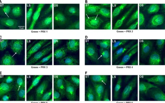
PRX Exist in Various Subcellular Locations throughout BAEC— Via Western blots, we have thus far found that shear stress regulates PRX 1 and 5 expression in BAEC. Based on the large number of PRX family members, we hypothesized that individ-ual PRX likely play important roles in specific subcellular compartments. To investigate the intracellular distribution of PRX, confocal immunofluorescence staining studies were performed. This study revealed data consistent with West-ern blot analysis of shear-induced protein expression in BAEC. Image analysis of staining intensities indicated that PRX 1 increased after 24 h of LS compared with OS and static conditions (Fig. 2A).
In addition to the shear dependence, Fig. 2 also shows that PRX members exhibited assorted staining patterns. This result raised two interesting questions: 1) Are PRX located in specific subcellular locations and 2) does shear stress alter this subcel-lular localization? To clearly characterize the location of each PRX within specific subcellular compartments, colocalization staining was performed in static BAEC using PRX-specific anti-bodies and organelle-specific markers for Golgi (GM 130), endoplasmic reticulum (KDEL receptor, data not shown), lyso-some (cathepsin S, data not shown), intermediate filament (vimentin), and mitochondria (Mitotracker). PRX 1 staining (green) overlapped with the Golgi marker staining (red), shown as yellow in the merged image (Fig. 3A), suggesting that PRX 1 exists in the Golgi apparatus. In addition, PRX 2, 4, 5, and 6 also appeared to be found in the Golgi (Fig. 3, B and E–G). The PRX 3 staining pattern was distinctly dif-ferent from other PRX, showing clear colocalization with the mito-chondria marker (Fig. 3D). Interest-ingly, PRX 2 staining revealed colo-calization with the intermediate filament marker (Fig. 3C). In addi-tion to these subcellular localiza-tions, PRX 1, 2, 4, 5 and 6 were also expressed in the cytosol (Fig. 3, A–C and E–G). Next, we examined whether shear stress stimulated expression of PRX members in other subcellular locations. Sub-cellular location did not appear to change in response to shear stress,
although Golgi were located
upstream of the direction of flow after chronic LS, consistent with previous reports (Fig. 2, A, B, and
FIGURE 1. PRX 1 is up-regulated by LS. Confluent BAEC were exposed to LS, OS, or static conditions for 1 day, and cell lysates were obtained. A, equal aliquots of protein (20 – 40g) were analyzed by Western blot using antibod-ies specific to PRX 1– 6 phospho-endothelial nitric-oxide synthase (peNOS), total endothelial nitric-oxide synthase (teNOS), and-actin blots were used as shear stress controls and internal loading controls, respectively. B, densito-metric analysis was used to quantify the intensity of each band, and the aver-age values (mean⫾ S.E., n ⫽ 12) are shown in bar graphs as % of static control. *, p⬍ 0.05 indicates significance compared with static control. **, p ⬍ 0.05 indicates significance compared with OS.
FIGURE 2. LS stimulated PRX 1 expression in BAEC. Confluent BAEC were exposed to LS, OS, or static condi-tions for 1 day as in Fig. 1. Cells were stained using antibodies specific to PRX 1– 6. Secondary antibodies conjugated to Alexa Fluor 488 (green) were imaged by confocal microscopy. Nuclei were counter-stained with Hoechst dye (blue). Arrows indicate unique subcellular staining patterns of each PRX.
at Ewha Medical Library on July 6, 2016
http://www.jbc.org/
D–F). Taken together, these results clearly indicate that PRX are abundantly expressed throughout the subcellular organelles of endothelial cells.
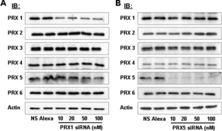
PRX 1 Prevents Oxidative Stress in Endothelial Cells Exposed to LS—PRX 1 is a prominent antioxidant, and our data indicate its expression is highly up-regulated by LS (22). Consequently, we investigated whether PRX 1 was responsible for the decreased ROS levels in endothelial cells exposed to LS. The depletion of individual PRX from cellular systems provides a useful tool to study the functional role of each PRX isoform.
Here, specific siRNAs were used to knock down either PRX 1 or PRX 5 protein levels in order to investigate PRX-dependent ROS accumula-tion. Western blots with isoform-specific PRX antibodies were used to determine the efficacy and speci-ficity of these siRNAs. Compared
with non-silencing siRNA and
Alexa Fluor 546-labeled
non-silenc-ing siRNA (50 nM each), 48 h of
treatment of BAEC with PRX 1 siRNA dramatically reduced (by
75% of non-silencing control)
expression of PRX 1 at a concentra-tion as low as 10 nM(Fig. 4A). As
shown in Fig. 4B, 10 nM PRX 5
siRNA also effectively reduced PRX 5 expression (by 90% of non-silenc-ing control). Via Western blots, the specificity of PRX 1 and 5 siRNAs was assessed by examining the expression of all other PRX family members (Fig. 4, A and B). Using isoform-specific PRX antibodies, we found that these siRNAs had no sig-nificant effect on other PRX, indi-cating that they exclusively targeted PRX 1 or 5, respectively, among all PRX family members (Fig. 4, A and B). Therefore, PRX 1 and 5 siRNAs were confidently used at 10 nMconcentration in
subse-quent functional studies.
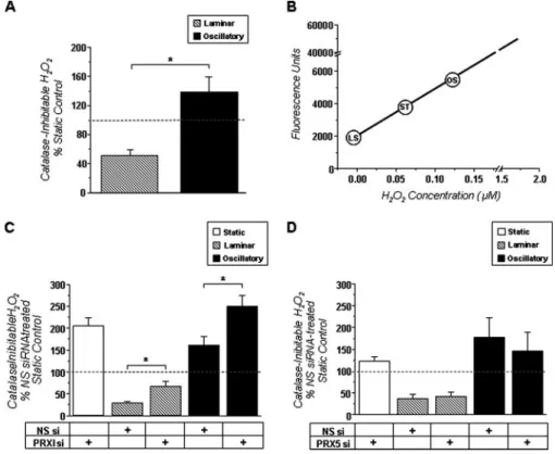
It has been well established that chronic exposure of endo-thelial cells to OS stimulates, while LS inhibits, ROS production (10, 11, 23, 24). Utilizing an Amplex Red assay in the presence or absence of catalase, we verified that hydrogen peroxide levels were 87% less in BAEC exposed to LS compared with those treated with OS (Fig. 5A). Using a standard hydrogen peroxide dose curve, we also found that the relative hydrogen peroxide levels following static culture, LS, and OS were consistent with expected cellular concentrations (Fig. 5B). To determine whether PRX 1 was responsible for the LS-dependent decrease in ROS levels in BAEC, we knocked down PRX 1 using PRX 1 siRNA as indicated above (Fig. 4A). PRX 1 depletion signifi-cantly increased catalase-inhibitable hydrogen peroxide by 2-fold above non-silencing controls in static-, LS-, and OS-treated BAEC (Fig. 5C). To investigate whether this was a global effect of mechanosensitive PRX family members, we also knocked down PRX 5 using PRX 5-specific siRNA. However, when compared with non-silencing controls, PRX 5 depletion had no significant effect on hydrogen peroxide levels in any group. Taken together, these data suggest that PRX 1 is a criti-cally important regulator of ROS levels in both a basal and shear-dependent manner.
DISCUSSION
Through protein expression analysis and subsequent func-tional studies, we have discovered PRX 1 as a mechanosensitive
FIGURE 3. PRX are located in various subcellular organelles in BAEC. Static BAEC were co-stained for PRX 1– 6 and subcellular organelles using PRX-specific antibodies (green), as in Fig. 2, and organelle-specific mark-ers (red). Nuclei were countmark-erstained with Hoechst dye (blue). Center panels: Mitochondria, Golgi, and inter-mediate filaments are stained with Mitotracker CMXRos, GM 130 antibody, and vimentin antibody, respec-tively. GM 130 and vimentin were visualized by secondary antibodies conjugated to Alexa Fluor 568 (red). Merged images are shown in the right panel. Yellow staining indicates colocalization.
FIGURE 4. PRX 1 and PRX 5 siRNAs specifically reduce PRX 1 and PRX 5
protein expression, respectively. BAEC were transfected with either 50 nM non-silencing siRNA (NS), 50 nMAlexa Fluor 546-labeled siRNA (Alexa), or PRX 1 (A) or PRX 5 (B) siRNAs (10, 20, 50, and 100 nM) for 48 h. Cell lysates were analyzed by Western blot with PRX-specific antibodies as indicated.-actin was used as an internal control.
at Ewha Medical Library on July 6, 2016
http://www.jbc.org/
antioxidant. Data to support this concept include: 1) PRX 1 is up-regulated intracellularly by chronic LS compared with OS, 2) PRX exhibit broad staining patterns in BAEC and localize in important cellular structures, 3) ROS production is signifi-cantly reduced in cells exposed to chronic LS, and 4) this effect can be attenuated by PRX 1 depletion, but not PRX 5 depletion. Through this work, we reveal for the first time that PRX are regulated by shear stress in endothelial cells. Previous studies have shown that other antioxidants are also controlled by shear stress. LS has been shown to up-regulate antioxidant genes, including endothelial nitric-oxide synthase, CuZn superoxide dismutase, manganese superoxide dismutase, glutathione per-oxidase, glutathione, and thioredoxin (25–29). In addition, Chen et al. (30) observed that many genes protective against oxidative stress are induced by exposure to prolonged LS. They have also noted that such genes are regulated through a con-served, shear-sensitive antioxidant response element. In sup-port of our finding that PRX 1 expression is LS-dependent, recent work has shown that PRX 1 is a target gene of nuclear factor (erythroid-derived 2)-related factor 2 (Nrf2), a key tran-scription factor that binds to antioxidant response element (31). Collectively, these studies indicate that cells possess an elaborate system of shear-responsive antioxidants and that each may play an independent role to mediate oxidative stress and modulate redox-sensitive signaling pathways.
The ubiquitous nature of the PRX family itself exemplifies this concept. Immunofluorescence microscopy revealed PRX
throughout the cellular milieu of BAEC, colocalizing with the cyto-plasm, Golgi apparatus, mitochon-dria, and intermediate filaments. These observations were consistent with previously reported localiza-tion studies in other cell types, but we are the first to report an apparent PRX 2 colocalization with vimentin (32, 33). This costaining of PRX 2 with vimentin suggests that it may be located in the intermediate fila-ment, but further studies will be necessary to confirm this finding. Although detection of PRX in the Golgi body likely reflects protein processing or packaging, localiza-tion within other organelles indi-cates that PRX may act both globally and in a site-specific manner to reg-ulate ROS in endothelial cells. In the endothelium, ROS, such as O2. and
H2O2, arise from several sources,, including NADPH oxidase, xan-thine oxidase, mitochondrial oxi-dase, cytochrome P450, and uncou-pled nitric-oxide synthase (34, 35). At relatively low concentrations, ROS play critical roles in redox sig-naling and normal cell function. However, higher concentrations of ROS induce oxidative damage of DNA, proteins, carbohy-drates, and lipids (36 –38). This damage has been shown to critically affect cellular function and apoptosis when it occurs in mitochondria, lysosomes, and nuclei (39 – 41). In addition, cytosolic proteins modified by ROS have been shown to affect local cell signaling and, collectively, the redox status of the cell (11, 42, 43). Ubiquitous distribution of PRX in BAEC may reflect diverse sources of ROS throughout the cells and provide protection for important macromolecules and structures against local ROS production. In addition, widespread alloca-tion of PRX may be important for comprehensive management of the overall oxidative state of cells.
Several studies have shown that oxidative stress is regulated by shear stress in endothelial cells (10 –12, 24, 26). We have previously published that both LS and OS stimulate ROS pro-duction acutely but the ROS transiently elevated by LS returns to basal levels within a few hours (10). However, unlike LS, OS continues to increase ROS production, maintaining elevated levels as long as cells are exposed to OS (10, 24, 26). The mech-anism by which ROS levels are lowered in cells exposed to chronic LS is undefined. This study demonstrates that endothe-lial cells exposed to chronic LS express much more PRX 1 com-pared with OS and static conditions. These findings suggest that PRX 1 is up-regulated by LS and that this may be respon-sible for LS-mediated decrease in ROS levels.
As previously determined by electron spin resonance spec-trometry and dichlorofluorescein-diacetate methods,
endothe-FIGURE 5. PRX 1 knock down increases H2O2production in BAEC, whereas PRX 5 knock down does not. Catalase-inhibitable hydrogen peroxide levels were assessed via Amplex Red assay, and average values (mean⫾ S.E., n⫽ 6–12) are shown in bar graphs as % of non-silencing (NS) siRNA-treated static controls. A, confluent BAEC were exposed to ST, LS, or OS for 24 h prior to assay. B, a hydrogen peroxide dose curve was used to estimate relative hydrogen peroxide concentrations in cells conditioned with shear stress. C, BAEC were transfected with either non-silencing or PRX 1 siRNA (10 nM) for 48 h and then exposed to ST, LS, or OS for 24 h prior to assay. D, BAEC were transfected with either non-silencing or PRX 5 siRNA (10 nM) for 48 h and then exposed to ST, LS, or OS for 24 h prior to assay. *, p⬍ 0.05 designates significance between indicated groups.
at Ewha Medical Library on July 6, 2016
http://www.jbc.org/
lial cells exposed to OS produce significantly more superoxide and hydrogen peroxide than those in static culture (10 –12). In contrast, endothelial cells treated with chronic, unidirectional high shear generate considerably less O2. . Here, we used
Amplex Red assay as an independent method to measure ROS levels in BAEC. Consistent with our previous reports, OS increased and LS decreased ROS production (Fig. 5A). The ROS measured by this assay was inhibitable by catalase, indicating that H2O2is the primary ROS component.
PRX 1 is the most abundant and ubiquitously distributed member of mammalian PRX (22). Our current study demon-strated that PRX 1 is dramatically up-regulated by chronic LS compared with OS and is located in the cytoplasm and Golgi. Knock down of PRX 1 using siRNA resulted in significantly higher ROS levels in BAEC exposed to LS, OS, and static con-ditions, whereas PRX 5 depletion did not. Although PRX 1 knock down did not fully abolish the antioxidative outcome of LS, its significant effect was somewhat surprising considering the presence of other PRX family members and additional mechanosensitive antioxidant pathways. In addition, PRX 5 depletion studies provide further evidence that PRX 1 is crucial to shear-dependent ROS regulation. Altogether, these results indicate that chronic exposure to LS up-regulates PRX 1 expression in order to keep ROS levels low in endothelial cells. In summary, we have shown that shear stress regulates expression of the PRX family and that PRX 1 plays a critical role in regulating ROS levels in endothelial cells. Furthermore, this discovery of PRX 1 as a mechanosensitive antioxidant may con-tribute important insights into endothelial cell biology and vas-cular diseases.
Acknowledgments—We thank Kyung Hwa Chang, Hannah Song, Mamta Patel, Chih Wen Ni, and Sarah Tressel at Georgia Institute of Technology and Emory University for helpful comments during these studies.
REFERENCES
1. Davies, P. F., Shi, C., Depaola, N., Helmke, B. P., and Polacek, D. C. (2001)
Ann. N. Y. Acad. Sci. 947,7–17
2. Zarins, C. K., Giddens, D. P., Bharadvaj, B. K., Sottiurai, V. S., Mabon, R. F., and Glagov, S. (1983) Circ. Res. 53, 502–514
3. Caro, C. G., Fitz-Gerald, J. M., and Schroter, R. C. (1969) Nature 223, 1159 –1160
4. Ku, D. N., Giddens, D. P., Zarins, C. K., and Glagov, S. (1985)
Arterioscle-rosis 5,293–302
5. Hajra, L., Evans, A. I., Chen, M., Hyduk, S. J., Collins, T., and Cybulsky, M. I. (2000) Proc. Natl. Acad. Sci. U. S. A. 97, 9052–9057
6. Nerem, R. M., Alexander, R. W., Chappell, D. C., Medford, R. M., Varner, S. E., and Taylor, W. R. (1998) Am. J. Med. Sci. 316, 169 –175
7. Sorescu, G. P., Sykes, M., Weiss, D., Platt, M. O., Saha, A., Hwang, J., Boyd, N., Boo, Y. C., Vega, J. D., Taylor, W. R., and Jo, H. (2003) J. Biol. Chem.
278,31128 –31135
8. Traub, O., and Berk, B. C. (1998) Arterioscler. Thromb. Vasc. Biol. 18, 677– 685
9. Harrison, D., Griendling, K. K., Landmesser, U., Hornig, B., and Drexler, H. (2003) Am. J. Cardiol. 91, 3A, 7A–11A
10. Hwang, J., Saha, A., Boo, Y. C., Sorescu, G. P., McNally, J. S., Holland, S. M., Dikalov, S., Giddens, D. P., Griendling, K. K., Harrison, D. G., and Jo, H.
(2003) J. Biol. Chem. 278, 47291– 47298
11. Sorescu, G. P., Song, H., Tressel, S. L., Hwang, J., Dikalov, S., Smith, D. A., Boyd, N. L., Platt, M. O., Lassegue, B., Griendling, K. K., and Jo, H. (2004)
Circ. Res. 95,773–779
12. McNally, J. S., Davis, M. E., Giddens, D. P., Saha, A., Hwang, J., Dikalov, S., Jo, H., and Harrison, D. G. (2003) Am. J. Physiol. 285, H2290 –H2297 13. Mates, J. M., Perez-Gomez, C., and De Castro, I. N. (1999) Clin. Biochem.
32,595– 603
14. Rhee, S. G., Chae, H. Z., and Kim, K. (2005) Free Radic. Biol. Med. 38, 1543–1552
15. Wood, Z. A., Schroder, E., Robin Harris, J., and Poole, L. B. (2003) Trends
Biochem. Sci. 28,32– 40
16. Kinnula, V. L., Lehtonen, S., Sormunen, R., Kaarteenaho-Wiik, R., Kang, S. W., Rhee, S. G., and Soini, Y. (2002) J. Pathol. 196, 316 –323
17. Kinnula, V. L., Lehtonen, S., Kaarteenaho-Wiik, R., Lakari, E., Paakko, P., Kang, S. W., Rhee, S. G., and Soini, Y. (2002) Thorax 57, 157–164 18. Krapfenbauer, K., Engidawork, E., Cairns, N., Fountoulakis, M., and
Lu-bec, G. (2003) Brain Res. 967, 152–160
19. Choi, M. H., Lee, I. K., Kim, G. W., Kim, B. U., Han, Y. H., Yu, D. Y., Park, H. S., Kim, K. Y., Lee, J. S., Choi, C., Bae, Y. S., Lee, B. I., Rhee, S. G., and Kang, S. W. (2005) Nature 435, 347–353
20. Go, Y. M., Boo, Y. C., Park, H., Maland, M. C., Patel, R., Pritchard, K. A., Jr., Fujio, Y., Walsh, K., Darley-Usmar, V., and Jo, H. (2001) J. Appl. Physiol.
91,1574 –1581
21. Boo, Y. C., Sorescu, G., Boyd, N., Shiojima, I., Walsh, K., Du, J., and Jo, H. (2002) J. Biol. Chem. 277, 3388 –3396
22. Immenschuh, S., and Baumgart-Vogt, E. (2005) Antioxid. Redox. Signal 7, 768 –777
23. Chappell, D. C., Varner, S. E., Nerem, R. M., Medford, R. M., and Alex-ander, R. W. (1998) Circ. Res. 82, 532–539
24. Hwang, J., Ing, M. H., Salazar, A., Lassegue, B., Griendling, K., Navab, M., Sevanian, A., and Hsiai, T. K. (2003) Circ. Res. 93, 1225–1232
25. Topper, J. N., Cai, J., Falb, D., and Gimbrone, M. A., Jr. (1996) Proc. Natl.
Acad. Sci. U. S. A. 93,10417–10422
26. De Keulenaer, G. W., Chappell, D. C., Ishizaka, N., Nerem, R. M., Alex-ander, R. W., and Griendling, K. K. (1998) Circ. Res. 82, 1094 –1101 27. Takeshita, S., Inoue, N., Ueyama, T., Kawashima, S., and Yokoyama, M.
(2000) Biochem. Biophys. Res. Commun. 273, 66 –71
28. Mueller, C. F., Widder, J. D., McNally, J. S., McCann, L., Jones, D. P., and Harrison, D. G. (2005) Circ. Res. 97, 637– 644
29. Yamawaki, H., Pan, S., Lee, R. T., and Berk, B. C. (2005) J. Clin. Investig.
115,733–738
30. Chen, X. L., Varner, S. E., Rao, A. S., Grey, J. Y., Thomas, S., Cook, C. K., Wasserman, M. A., Medford, R. M., Jaiswal, A. K., and Kunsch, C. (2003)
J. Biol. Chem. 278,703–711
31. Kim, Y.-J., Ahn, J.-Y., Liang, P., Ip, C., Zhang, Y., and Park, Y.-M. (2007)
Cancer Res. 67,546 –554
32. Hofmann, B., Hecht, H. J., and Flohe, L. (2002) Biol. Chem. 383, 347–364 33. Kang, S. W., Chae, H. Z., Seo, M. S., Kim, K., Baines, I. C., and Rhee, S. G.
(1998) J. Biol. Chem. 273, 6297– 6302
34. Suzuki, Y., Wang, W., Vu, T. H., and Raffin, T. A. (1992) Biochem. Biophys.
Res. Commun. 184,1339 –1343
35. Weber, C., Erl, W., Pietsch, A., Strobel, M., Ziegler-Heitbrock, H. W., and Weber, P. C. (1994) Arterioscler. Thromb. 14, 1665–1673
36. Beckman, K. B., and Ames, B. N. (1997) J. Biol. Chem. 272, 19633–19636 37. Berlett, B. S., and Stadtman, E. R. (1997) J. Biol. Chem. 272, 20313–20316 38. Gutteridge, J. M. (1995) Clin. Chem. 41, (12B), 1819 –1828
39. Terman, A., Gustafson, B., and Brunk, U. T. (2007) J. Pathol. 211, 134 –143 40. Walford, G. A., Moussignac, R.-L., Scribner, A. W., Loscalzo, J., and
Leopold, J. A. (2004) J. Biol. Chem. 279, 4425– 4432
41. Martinet, W., Knaapen, M. W., De Meyer, G. R., Herman, A. G., and Kockx, M. M. (2002) Circulation 106, 927–932
42. Finkel, T. (1999) J. Leukocyte Biol. 65, 337–340
43. Sykes, M. C., Mowbray, A. L., and Jo, H. (2007) Circ. Res., 100, 152–154
at Ewha Medical Library on July 6, 2016
http://www.jbc.org/
Amy L. Mowbray, Dong-Hoon Kang, Sue Goo Rhee, Sang Won Kang and Hanjoong Jo
doi: 10.1074/jbc.M707985200 originally published online November 16, 2007 2008, 283:1622-1627.
J. Biol. Chem.
10.1074/jbc.M707985200
Access the most updated version of this article at doi: Alerts:
When a correction for this article is posted
•
When this article is cited
•
to choose from all of JBC's e-mail alerts
Click here
http://www.jbc.org/content/283/3/1622.full.html#ref-list-1
This article cites 43 references, 26 of which can be accessed free at
at Ewha Medical Library on July 6, 2016
http://www.jbc.org/