ABSTRACT
The gut is an important organ with digestive and immune regulatory function which consistently harbors microbiome ecosystem. The gut microbiome cooperates with the host to regulate the development and function of the immune, metabolic, and nervous systems. It can influence disease processes in the gut as well as extra-intestinal organs, including the brain. The gut closely connects with the central nervous system through dynamic bidirectional communication along the gut-brain axis. The connection between gut environment and brain may affect host mood and behaviors. Disruptions in microbial communities have been implicated in several neurological disorders. A link between the gut microbiota and the brain has long been described, but recent studies have started to reveal the underlying mechanism of the impact of the gut microbiota and gut barrier integrity on the brain and behavior. Here, we summarized the gut barrier environment and the 4 main gut-brain axis pathways. We focused on the important function of gut barrier on neurological diseases such as stress responses and ischemic stroke. Finally, we described the impact of representative environmental sensors generated by gut bacteria on acute neurological disease via the gut-brain axis.
Keywords: Intestine; Brain; Microbiome; Stroke; Short-chain fatty acid;
Aryl hydrocarbon receptor
GUT BARRIERS
The gut mucosa has multiple layers to provide an efficient barrier against noxious luminal agents. The intestinal epithelium is continuously renewed from pluripotent intestinal stem cells (ISCs) at the base of the crypt. The secretory epithelial cells produce a mucous physicochemical barrier containing antimicrobial peptides to control microbial invasion.
The gut epithelial cells are tightly interconnected with various junctions. Under gut epithelium, an immunological surveillance system finally combats external invaders. Here we summarized a well-organized network of gut mucosa to sustain sterile conditions in most organs within the body.
Review Article
Received: Jun 3, 2021 Revised: Jun 9, 2021 Accepted: Jun 12, 2021
*Correspondence to Sun-Young Chang
Laboratory of Microbiology, College of Pharmacy, Ajou University, 206 Worldcup-ro, Yeongtong-gu, Suwon 16499, Korea.
E-mail: sychang@ajou.ac.kr
Copyright © 2021. The Korean Association of Immunologists
This is an Open Access article distributed under the terms of the Creative Commons Attribution Non-Commercial License (https://
creativecommons.org/licenses/by-nc/4.0/) which permits unrestricted non-commercial use, distribution, and reproduction in any medium, provided the original work is properly cited.
ORCID iDs Min-Gyu Gwak
https://orcid.org/0000-0001-5700-0499 Sun-Young Chang
https://orcid.org/0000-0001-7336-9245 Conflict of Interest
The authors declare no potential conflicts of interest.
Abbreviations
ACTH, adrenocorticotropic hormone; AhR, aryl hydrocarbon receptor; AIS, acute ischemic stroke; ANS, autonomic nervous system; BBB, blood-brain barrier; CCK, cholecystokinin;
CNS, central nerve system; CORT, corticosterone; CRH, corticotropin-releasing
Min-Gyu Gwak , Sun-Young Chang *
Laboratory of Microbiology, College of Pharmacy, and Research Institute of Pharmaceutical Science and Technology (RIPST), Ajou University, Suwon 16499, Korea
Gut-Brain Connection: Microbiome, Gut
Barrier, and Environmental Sensors
hormone; DC, dendritic cell; ENS, enteric nervous system; GABA, γ-aminobutyric acid;
GI, gastrointestinal; GIP, glucose-dependent insulinotropic polypeptide; GLP-1, glucagon- like peptide-1; GPCR, G-protein coupled receptor; HDAC, histone deacetylase; HPA, hypothalamic-pituitary-adrenal; IEC, intestinal epithelial cell; IEL, intraepithelial lymphocyte;
ISC, intestinal stem cell; ITF, intestinal trefoil factor; Lgr5, leucine-rich repeat-containing G-protein coupled receptor 5; Muc2, mucin 2; NMDA, N-methyl-D-aspartic acid; PD, Parkinson's disease; PYY, peptide YY; RELM-β, resistin-like molecule-β; SCFA, short-chain fatty acid; VN, vagus nerve; ZO, zonula occludens.
Author Contributions
Conceptualization: Chang SY; Visualization:
Chang SY; Writing - original draft: Gwak MK;
Writing - review & editing: Chang SY.
INTESTINAL EPITHELIUM
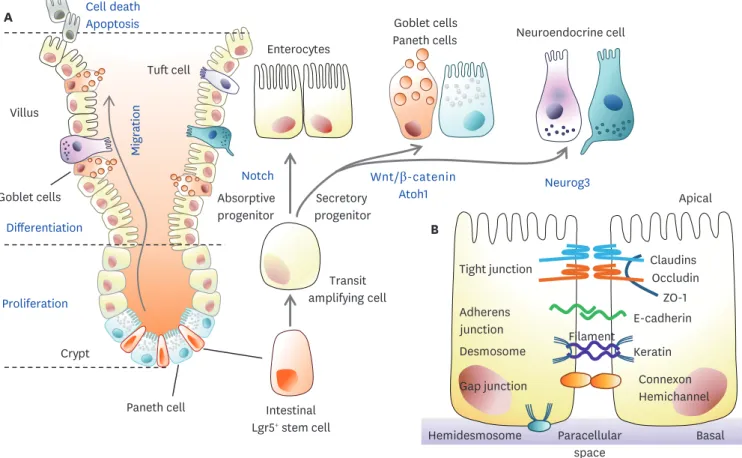
The intestine is covered by an epithelial monolayer consisting of crypts and villi. Although there are differences between the small intestine and colon, the intestinal epithelial cells (IECs) include various cell types (Fig. 1). Besides enterocytes, there are secretory IECs including enteroendocrine cells, goblet cells, Paneth cells, and other types of cells.
Enteroendocrine cells, which are divided into several subtypes according to hormone secretion, regulate physiological function (Table 1). Enterochromaffin cells secrete serotonin, D cells secrete somatostatin, and G cells secrete gastrin (1). Goblet cells and Paneth cells play critical roles in forming physical and biochemical barriers. Goblet cells secrete glycosylated mucins into the intestinal lumen and then form a mucus layer through the disulfide bonds of glycosylated mucins. On the epithelial surface, mucin 2 (Muc2) plays an important role in organizing mucus (2). It maintains intestinal barrier homeostasis and induces anti- inflammatory signals by binding to glycan receptors in lamina propria dendritic cells (DCs). Muc2 secretion is regulated by gut microbes and their metabolites such as short- chain fatty acids (SCFAs). In addition, goblet cell-derived products such as intestinal trefoil factor (ITF) and resistin-like molecule-β (RELM-β), contribute to the regulation of physical barriers (3). ITF is involved in regulating tight junctions, cell apoptosis, and promoting
Wnt/β-catenin Atoh1
Intestinal Lgr5+ stem cell Notch
Crypt Villus
Differentiation
Proliferation
Cell death Apoptosis
Migration
Transit amplifying cell A
Goblet cells
Tuft cell
Paneth cell
Basal Tight junction
Adherens junction Desmosome
Apical
Paracellular space
Claudins Occludin
ZO-1
Connexon Hemichannel E-cadherin Keratin Filament
B
Gap junction
Hemidesmosome
Neurog3 Enterocytes
Goblet cells
Paneth cells Neuroendocrine cell
Absorptive progenitor
Secretory progenitor
Figure 1. Dynamic homeostasis of the gut epithelium and gut barrier integrity. (A) Gross features of the small intestine. Proliferation and differentiation of Lgr5+ ISCs into TA cells occur at the intestinal crypt. TA cells are differentiated into secretory and absorptive epithelial lineages during migration from the crypt to the villi. Secretory lineage cells are more differentiated into mucin/antimicrobial peptide-secreting goblet cells and Paneth cells or neuroendocrine cells producing hormones. When these fully differentiated cells reach the villi tips, apoptotic cell death occurs via cell signaling. (B) The intestinal epithelial barrier is constructed of cellular junctions in the paracellular spaces between adjacent cells. Tight junctions are composed of claudin, occludin, and ZO-1, and adherens junctions are composed of E-cadherin. There are also desmosomes, gap junctions, and hemidesmosomes in the paracellular space, which are involved in transporting nutrients and forming the physical intestinal barrier.
TA, transit-amplifying.
epithelial repair. RELM-β modulates Th2-mediated responses. Microfold cells are found in the follicle-associated epithelia and are involved in the immune response through the absorption of luminal Ags and delivery to Ag-presenting cells. Cup cells account for 6% of the ileum epithelial cells, but their function remains unknown. Tuft cells (taste chemosensory epithelial cells) are involved in the immune response by secreting cytokines (3).
INTERCELLULAR BARRIERS
Tight junctions form a proteinaceous film that regulates the diffusion of ions and solutes between cells in mammals (Fig. 1A). Tight junctions include claudins, occludins, and zonula occludens (ZO) (3). The claudin and occludin proteins are arranged in chains and linked by sealing between adjacent cells. In humans, claudins are comprised of families, which include claudin 1, 2, 3, 4, 5, 7, 8, 12, and 15 (4). Occludin can regulate large molecules, but its specific role remains to be investigated. ZO-1 belonging to the ZO proteins (ZO-1, ZO-2, and ZO-3) acts as a scaffold protein that anchors to the actin cell skeleton through cross- linking (5). The adherens junction, a basal protein complex serves as a pathway for cell-cell adhesion, actin cytoskeleton regulation, cell signaling, and gene transcription regulation.
Cadherin, one of the adherens junction protein complexes, is a major type of transmembrane protein and connects with cadherin in adjacent cells in a calcium-dependent manner. It is indirectly bound to α-catenin through p120 and β-catenin, and α-cadherin connects the actin cytoskeleton. P120-catenin complex linked to the cadherin ternary complex regulates the life cycle of cadherin in the plasma membrane (6).
REGENERATION OF GUT EPITHELIUM
An essential factor in maintaining barrier homeostasis is the controlled supply of new cells.
IECs, one of the most proliferative in the body, constantly regenerate themselves to maintain intestinal homeostasis and barrier integrity against the invasion of many pathogens. The differentiation of these cells is a process that is precisely controlled by several signaling pathways and molecular markers (Fig. 1A). The differentiation of epithelial cells originates from pluripotent leucine-rich repeat-containing G-protein coupled receptor 5 (Lgr5)+ ISCs (7). Lgr5, one of the G-protein coupled receptor (GPCR) class proteins, is critical for the process of self-renewal and differentiation and is a major component of Wnt signaling Table 1. Subtypes of intestinal enteroendocrine cells
Subtypes Functions Marker Function
Enterochromaffin cells Aid in intestinal motility reflexes and secretion 5-HT Appetite, motility
Enterochromaffin-like cells Stimulate gastric acid secretion Histamine Acidity
D cells Inhibit gastrin and somatostatin release SST Acidity and insulin
G cells Stimulate gastric acid secretion Gastrin Acidity
I cells Stimulate pancreatic enzyme secretion CCK Appetite, motility, bile acid
K cells Stimulate insulin release GIP Insulin
L cells Aid in carbohydrate uptake, mucosal enterocyte proliferation, and insulin release GLP-1, GLP-2, PYY Appetite, insulin, motility
Mo cells Initiate myoelectric complex migration Motilin Motility
N cells Inhibit intestinal contractions NTS Motility
S cells Reduce acidity in the upper small intestine SCT Acidity
A cells Secrete ghrelin and nesfatin-1 Ghrelin Appetite, growth hormone release
P cells Secrete leptin Leptin Appetite
5-HT, 5-hydroxytryptamine; SST, somatostatin; NTS, neurotensin; SCT, secretin.
and plays an essential role in intestinal recovery. Crypt base columnar cells multiply at the bottom of the crypt and the daughter cells move up to the villous tip. The rapidly dividing cells differentiate into transit-amplifying cells, which are regulated by 3 main signaling pathways, Wnt, Notch, and BMP (8). Notch-HES1 signaling promotes the absorptive lineage, whereas Math1-Atoh1 signaling promotes the secretory lineage. The process by which each cell is rapidly and precisely differentiated requires much additional research, but it is certainly important in maintaining intestinal homeostasis. At the top of villi, damaged cells are replaced with newly differentiated cells and also inhibit excessive proliferation, thereby inhibiting intestinal tumor formation. Microbial factors influence stem cell differentiation.
The importance of SCFAs, dietary metabolites fermented by microbes, is currently being studied. For example, butyrate as a histone deacetylase (HDAC) inhibitor (9), inhibits crypt proliferation, whereas lactate accelerates stem cell proliferation dependent upon GPR81 (10).
GUT-BRAIN COMMUNICATION
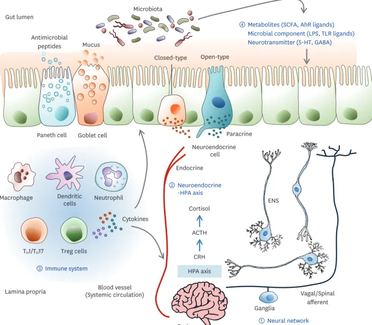
The brain-gut axis has been the subject of research over the past several years. Cross- communication between the brain and gut occurs through multiple biological networks involving the neural network, neuroendocrine system, immune system, and metabolic pathways, which enable bidirectional communication (Fig. 2). In addition, microbial changes in the gut can influence brain physiology and behavioral cognitive functions (11). Here, the complexity and mechanistic modalities for these connections are highlighted in the pathogenesis of human diseases.
Neural network
The intestine physically links the brain through 2 neuroanatomical pathways (12). The first is the direct interaction with the autonomic nervous system (ANS) and vagus nerve (VN). The VN innervates the mucosal and muscle layers of the gut, senses stimuli, and transmits these signals to the brain. These relationships can mediate mechanoreceptors or chemoreceptors, which sense the luminal volume or chemical stimuli such as hormones, neurotransmitters, and metabolites. The efferent VN transfers information from the central nerve system (CNS) to the viscera and plays a critical role in metabolism and immunity. These functions suggest that the VN is an important mediator of bidirectional communication. The second is the bidirectional exchange via the enteric nervous system (ENS) in the intestine, which is connected to the ANS and VN in the spinal cord. This neural network has 4 steps, starting from the ENS and followed by the ganglia that modulate the peripheral visceral reflex response. The next step is the ANS and VN dorsal motor nucleus of the spinal cord and the brain stem nucleus tractus solitarius. The VN dorsal motor nucleus affects the gastrointestinal (GI) tract and, in particular, regulates vagal sensitivity. The final is the higher brain center. Brainstem nuclei control intestinal function according to information from the cortex and the subcortical center. The gut microbiota directly modulates sympathetic neurons (13). Bacteria stimulate the afferent neuron of the ENS, and vagal signals can induce an anti-inflammatory response. Intestinal microbes play an important role in the development and maturation of the immune, endocrine, and nervous systems. Aryl hydrocarbon receptor (AhR) ligands, microbial products, and SCFAs influence ENS activity and regulate gut motility. These findings suggest that gut microbiota mediate neuronal pathways of the gut-brain axis through their metabolites.
Neuroendocrine-hypothalamic-pituitary-adrenal (HPA) axis pathway The gut microbiota aids in the maturation of the neuroendocrine system. The quality and quantity of microbiota and the expression of TLRs may affect neuroendocrine secretion.
The gut microbiota is essential for the postnatal development of the HPA axis in the stress response. Mice lacking a stress response had decreased expression of brain-derived neurotrophic factor and 2A subtype of N-methyl-D-aspartic acid (NMDA) receptors. NMDA Goblet cell
④ Metabolites (SCFA, AhR ligands) Microbial component (LPS, TLR ligands) Neurotransmitter (5-HT, GABA)
Neuroendocrine cell Paneth cell
Open-type Closed-type
Blood vessel (Systemic circulation)
Endocrine
Paracrine Gut lumen
Neutrophil Macrophage Dendritic
cells
③ Immune system TH1/TH17 Treg cells
Cytokines Microbiota
Lamina propria
Brain HPA axis
CRH ACTH Cortisol
① Neural network
② Neuroendocrine -HPA axis
ENS
Vagal/Spinal afferent Ganglia
Mucus Antimicrobial
peptides
Figure 2. The dynamic communication of the gut microbiota-brain axis. Bidirectional communication between the gut and brain can be mediated by several direct and indirect pathways. The communication routes involve ① the nervous system including the ENS and the VN, ② the neuroendocrine and HPA axis, ③ the immune system, and ④ microbiota-derived neuroactive compounds. The gut microbiota can produce neurotransmitters such as GABA, dopamine, and serotonin; amino acids such as tyramine and tryptophan; and microbial metabolites such as SCFAs and AhR ligands. The gut epithelium consists of various kinds of hormone-secreting specialized neuroendocrine cells. These hormone and neuroactive compounds influence the local gut physiology and can travel through the blood circulation to interact with the host immune system and metabolism that directly signals the brain. The gut microbiota can influence epithelial barrier integrity, controlling the transit of signaling molecules from the gut lumen to the lamina propria, which contains various immune cells and neurons, or the blood circulation. Some neuropsychiatric conditions can disrupt gut barrier integrity. Stress can activate an HPA axis response that involves CRH, ACTH, and cortisol sequentially. Cortisol regulates neuro-immune signaling and affects intestinal barrier integrity. Therefore, stress hormones, immune mediators, and CNS neurotransmitters can change the gut environment and alter microbiota composition.
5-HT, 5-hydroxytryptamine.
receptors affect the release and expression of corticotrophin-releasing hormone from the hypothalamus and induce changes in HPA axis function. In turn, the stress-HPA axis also affects the gut microbiota composition. Cortisol is a substance that plays a central role in the stress response. Corticotropin-releasing hormone (CRH) in the hypothalamus influences cortisol through the regulation of adrenocorticotropic hormone (ACTH). Therefore, the HPA axis, a key regulator of the stress response, can influence the regulation of the brain- gut-axis (12). The gut epithelium consists of various kinds of hormone-secreting specialized neuroendocrine cells (Table 1). K-cells regulate insulin through glucose-dependent
insulinotropic polypeptide (GIP) release and promote triglyceride storage (14). GIP stimulates the release of insulin hormone and is involved in the metabolism of dietary fat. In addition, glucagon-like peptide-1 (GLP-1) is responsible for insulin release after glucose intake, which is called the incretin effect. It stimulates fat deposition of adipose tissues, which is related to over-nutrition and obesity. L-cells secrete GLP-1 and peptide YY (PYY) in response to food intake (15). The release of GLP-1 inhibits insulin secretion in pancreatic β-cells and increases satiety. PYY inhibits GI motility, slows food intake, decreases appetite, and increases energy consumption. I-cells release anti-orexigenic and the principal satiety peptide hormone cholecystokinin (CCK) (16). CCK reacts to fatty acids, amino acids, and luminal nutrients and is released by I cells, inhibiting gastric discharge and stimulating gallbladder contraction and the secretion of enzymes in the pancreas to promote digestion. G cells are neuroendocrine cells responsible for the synthesis and secretion of gastrin, which is stimulated by gastrin- releasing peptide neurons or vagal efferent neurons (16). They are mainly located in the pyloric antrum and are also found in the duodenum and pancreas. Enterochromaffin-like cells are found in the gastric pits of the fundus and cardia of the stomach and secrete histamine to bind to H2 receptors in the adjacent parietal cells (1). Gastric acid secretion is further stimulated by parietal cells. S cells found in the jejunum and duodenum release secretin (1). When the internal pH of the small intestine falls below 4, S cells release secretin, which increases the secretion of HCO3− into the lumen. D cells, also known as delta cells, are involved in the production of the hormone somatostatin (1). Somatostatin is involved in inhibiting the production of gastric acid, intestinal motility, and the release of digestive enzymes from the pancreas.
Immune system
The gut microbiota is necessary for the normal development of the peripheral immune system in the gut as well as the brain. For example, the gut microbiota influences microglia, innate immune cells of the brain (17). Germ-free mice have increased numbers of immature microglia while treatment with complex microbiota or SCFAs can rescue mature microglia deficiency (18). Microglia affected by the gut microbiota can influence processes such as stress and human behavioral and neurological disorders. Cytokines produced in the intestine can travel to the brain through the bloodstream and affect the systemic immune system. Altered systemic immunity changes immune signaling and peripheral inflammation in the brain, which is associated with many neuropsychiatric diseases including anxiety, depression, and autism spectrum disorder. The possibility of passing through the blood- brain barrier (BBB) is low, but signals can be transmitted to the brain through the BBB. The production of LPS and cytokines induces a change in the barrier function, and further affects brain development due to the generation of inflammatory factors that can change peripheral vascular permeability. The gut microbiota influences the permeability of the BBB. Increased permeability of the BBB in germ-free mice resulted from the reduced expression of occludin and claudin 5 (19). Injury, infection, and autoimmunity affect BBB permeability and increase the access of microbial products in the circulatory system to the brain (20). Immunological
sensitization in BBB integrity is a characteristic feature of neuropathological symptom, suggesting a connection between the gut environment and the brain. IL-1 and IL-6 in the hypothalamus induce the release of cortisol through activation of the HPA axis (21).
Neurotransmitters, neuropeptides, microbial-derived products
Neuroactive substances such as serotonin (5-hydroxytryptamine), γ-aminobutyric acid (GABA), and SCFAs can affect brain function. Some substances like ammonia and D-lactic acid can produce neurotoxic effects. Bifidobacterium infantis has been shown to affect serotonin by raising tryptophan levels in plasma (22). Gut microbes influence the synthesis and release of neurotransmitters such as noradrenaline, dopamine, and acetylcholine (23). These neurotransmitters originating in the gut are allowed to pass through the intestinal vein but does not directly affect brain function because of BBB. Thus, it can be postulated that the effect of the gut microbiota on brain function is indirect. SCFA is a fermented product of the intestinal microbial metabolism of dietary fiber. SCFAs affect the intestinal epithelium and immunity through GPCRs. These GPCRs are rare but are present in the brain. SCFAs can also affect psychological function through HDACs. Thus, SCFAs can affect the brain in direct and indirect ways such as humoral effects, hormones, immune pathways, and nerve pathways (24). SCFAs modulate microglial immune programming by inducing microglial maturation and morphologic changes in the murine brain (18).
Serotonin is one of the neurotransmitters derived from tryptophan. Tryptophan is converted to 5-hydroxytryptophan by tryptophan hydroxylase and then further converted to serotonin by aromatic amino acid decarboxylase. Serotonin is mainly produced by enterochromaffin cells of the intestine, although it can be produced in both the brain and intestines (25). Therefore, serotonin can participate in communication between them. In the brain, serotonin is involved in feeling a wide range of emotions, including happiness.
Serotonin plays an important role in gut homeostasis, sensory motor function, immune cell function, and angiogenesis following the onset of inflammation (26). Serotonin signaling through its receptor is involved in the regulation of mucus secretion and fluid and intestinal electrolyte transport (27). Serotonin induces gut motility via the extrinsic as well as the intrinsic nervous system. The binding of serotonin to its receptor increases the secretion of acetylcholine and calcitonin gene-related peptide from intrinsic primary afferent neurons.
In addition, intestinal contraction and relaxation (substance p and vasoactive intestinal peptide) are induced by the interaction of the intermediate nerve cells of the stimulation site and motor nerve cells (28). Thus, serotonin is a key component of irritable bowel syndrome (25). Serotonin receptors are expressed in almost all immune cells (29). At the site of inflammation, innate immune cells such as immature DCs, monocytes, mast cells, and eosinophils are activated by serotonin during acute inflammation. It also promotes the proliferation of natural killer cells and helper T cells. Cells stimulated by serotonin increase the secretion of IL-1β, IL-6, and IL-8. Serotonin regulates innate immune responses through reactive oxygen species produced by NADPH oxidase 2 (30). Changes in enterochromaffin cells are related to intestinal inflammation such as ulcerative colitis and irritable bowel syndrome (31). In Crohn's patients, an increase in enterochromaffin cells was observed, and elevated serotonin levels were observed in patients with collagenous colitis (32). Serotonin affects blood vessel synthesis to induce angiogenesis via matrix metalloproteinase and vascular endothelial growth factor induction through activation of the PI3K/AKT/mTOR pathway, suggesting the potential of alternative therapeutic interventions targeting the vascular and lymphatic endothelium in inflammatory bowel disease (33).
IMPACT OF THE GUT BARRIER AND MICROBIOME IN BRAIN DISEASE
Symbiosis of gut bacteria with their hosts impact various neurological function through dynamic communication between gut and brain. These processes may affect human health and behaviors in several neurological disorders. Here we discuss emerging evidences and possible contributions by the gut microbiota to neurological disorders such as host mood and stroke.
Stress and psychiatric disorders
From animal experiments and clinical data, the microbiota has been shown to affect host mood and behavior by producing metabolites and neurotransmitters. Lactobacillus reuteri increased social behavior in the murine model of autism spectrum disorder by upregulating oxytocin in the plasma and brain (34). Lactobacillus rhamnosus generated GABA and regulated its receptors in the CNS to attenuate depression and anxiety-like behavior in the murine model (34,35). Bifidobacterium longum ameliorated mood alterations in irritable bowel syndrome patients via VN signals (36). Bacteroides fragilis improved anxiety-like behavior and communication in a murine model of autism spectrum disorder by reducing 4-ethylphenyl sulfate (37). The gut microbiota affected Parkinson's disease (PD)-like motor symptoms and neuroinflammation in a murine PD model (38). Oral treatment with Proteus mirabilis reproduced PD-like symptoms and increased microglial activation via LPS. Gut bacteria are involved in fear extinction learning via microbial metabolites in mice (39). Depression was associated with impaired control of the HPA axis (40). The comparative analysis of specific pathogen-free mice versus germ-free mice showed that postnatal microbial colonization programmed the HPA system for a stress response via corticosterone (CORT) and ACTH (41). Neonatal mice separated from their mothers underwent a constitutive change in the gut microbiota and impaired growth due to neonatal stress (42). Early life stress alters immunity, which has implications for irritable bowel syndrome and psychiatric illnesses (43).
Stress-related psychiatric disorders induce the breakdown of the gut barrier and increased intestinal permeability (44). Once the gut barrier is dysregulated, luminal bacteria can pass through the intestinal mucosa and directly access immune cells and ENS neurons (45).
The administration of probiotics such as B. infantis or Lactobacillus sp. ameliorated colonic dysfunction via normalizing the levels of norepinephrine and CORT (46). Probiotic treatment such as Lactobacillus farciminis prevents gut leakage by attenuating HPA responses (47). Besides HPA axis regulation, microbes can directly affect CNS through the neuronal network.
Infection with Citrobacter rodentium or Campylobacter jejuni increased anxiety-like behavior through the vagus neuronal system (48,49). The induction of cFOS, a neuronal activation marker, was evident in vagal sensory neurons during the acute phase of C. jejuni infection.
Stroke
Stroke is a disease caused by ischemic or hemorrhagic bleeding followed by brain malfunction due to brain cell death. Acute ischemic stroke (AIS) accounts for 90% of strokes and occurs when blood clots block blood flow through the brain arteries (50). The gut microbiome affects post-stroke outcome and recovery. Microbiome deficiency in germ-free animals had a negative impact on stroke outcomes compared to specific pathogen-free mice (51).
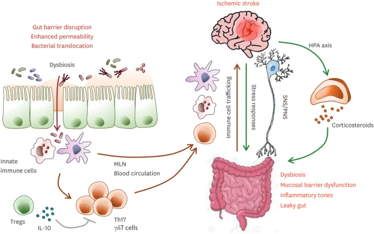
During the acute stage of stroke, extensive infarction results in the impaired regulation of the sympathetic nervous system and parasympathetic nervous system and stress hormone secretion from the HPA axis (52). Thereafter, stroke altered the gut microbiota composition leading to dysbiosis (53), which induced barrier dysfunction and leaky gut followed by enhancing inflammatory tones (Fig. 3). The microbiota-gut-brain axis has a critical role in
immune system regulation after AIS. Especially, T cells are critical players in secondary brain damage following brain injury. Th1, Th17, and IL-17-producing γδ T cells are associated with post-stroke neurotoxicity although Treg cells are closely linked with neuroprotective function via inhibiting pathogenic T cells. Post-stoke effector T cells traffic from the gut to the brain and localize in the leptomeninges and enhance neuroinflammation by secreting IL-17 and the subsequent infiltration of cytotoxic neutrophils (54). Microbiota and their metabolites maintain immune homeostasis by balancing the generation of immune-regulatory Treg cells versus pro-inflammatory T cell subsets in the gut. Stroke-induced gut barrier dysfunction increased translocation from the gut lumen into the lamina propria and the dissemination of commensal bacteria into peripheral tissue, leading to post-stroke infections such as pneumonia and urinary tract infections (55). Taken together, post-stroke dysbiosis enhanced the generation of intestinal pro-inflammatory T cells and the migration of intestinal immune cells into the meninges followed by the exacerbation of brain damage and worse stroke outcomes.
MANIPULATION OF BRAIN HEALTH BY THE GUT ENVIRONMENT
The gut microbiota is emerging as one of important factors in neurological health and disease. Understanding the nature of the microbiome-brain crosstalk, we discuss the current
Immune cell trafficking Stress responses
Dysbiosis
Mucosal barrier dysfunction Inflammatory tones Leaky gut
Ischemic stroke
Gut barrier disruption Enhanced permeability Bacterial translocation
Dysbiosis
Innate
immune cells MLN
Blood circulation
Tregs Th17
γδT cells
HPA axis
IL-10
Corticosteroids
SNS/PNS
Figure 3. Gut-brain system crosstalk via immunity and HPA axis after stroke. Stroke induces dysbiosis, mucosal barrier dysfunction, an increase in gut permeability, and bacteria translocation, leading to post-stroke infection. In the gut, dysbiosis and bacteria translocation induce a pro-inflammatory T cell response via innate immune cells. Immune cells, especially IL-17+ γδ T cells, macrophages, and DCs migrate from the gut to the meninges and the brain after stroke, which leads to post-stroke inflammation and exacerbated brain tissue damage.
SNS, sympathetic nervous system; PNS, parasympathetic nervous system; MLN, mesenteric lymph node.
microbiome-based therapeutic approaches that can improve patient recovery after an ischemic insult in stroke.
Manipulation of microbiome composition
Stroke alters the microbiota composition in the gut, and this dysbiosis has a substantial impact on post-stroke recovery by modulating the immune response. AIS rapidly triggers gut microbiome dysbiosis with Enterobacteriaceae overgrowth, followed by the exacerbation of brain infarction (56). Acute brain ischemia-induced dysbiosis leads to increased gut permeability and motility. This microbiota impact on immunity and stroke outcome was transmissible by microbiota transplantation. Secondary brain damage induced by dysbiosis is treated with antibiotics to reduce the symptoms of brain ischemic disease. Treatment with ampicillin or vancomycin in ischemic brain damage exerted neuroprotection, but treatment with neomycin showed no beneficial effect in mice (53). The antibiotic-dependent microbial shift can be associated with the metabolites from beneficial microbiota. Fecal transplantation from post-stroke mice into germ-free mice induced stroke symptoms by enhancing inflammatory Th1 and Th17 cells (51). Fecal transfer from a healthy donor reduced the brain damage area consistent with neuroprotection by microbial eubiosis (57). Microbial priming of lamina propria DCs led to the expansion of Treg cells in the small intestine and suppression of effector IL-17+ γδ T cell function (54). Representative metabolites for cross-talk between the gut and brain are xenobiotic/aromatic compound derivatives and fermentative SCFAs from dietary fibers. Membrane-derived molecules originating from the gut microbiota have an important impact on host immunity and neurological diseases. Representatively, the consistent introduction of LPS endotoxin into sterile tissues induces chronic inflammatory diseases. Enhanced gut barrier permeability facilitates LPS dissemination to induce a strong inflammatory response, which can disrupt the BBB and activate microglia.
Brain and the AhR
Intestinal cells constantly interact with the luminal milieu originating from host metabolism as well as environmental substances, diet, and commensal flora via multiple environmental sensors. One of these sensors is the AhR, a ligand-activated transcription factor (58). The AhR pathway participates in a broad variety of physiological and pathological processes.
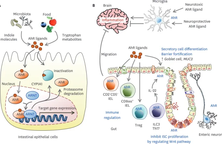
AhR ligands can be broad-spectrum, including endogenous metabolic products from essential aromatic amino acids and tryptophan, and exogenous molecules from artificial xenobiotic or bacterial metabolites (Fig. 4). Upon ligand binding in the cytosol, AhR and some components of the chaperone complex translocate to the nucleus, where the AhR complex binds DNA-responsive elements to control target gene expression. In gut epithelial cells, AhR activation suppresses uncontrolled ISC growth and malignant tumor progression, supporting barrier integrity (59). AhR inhibits pro-inflammatory pathways via the IL-10 receptor and regenerating islet-derived protein IIIγ expression and maintains gut epithelial barrier integrity (60,61). AhR activation has multiple effects on immune cells. In DCs, AhR dampens the induction of pro-inflammatory cytokines and MHC class II expression. AhR induces indoleamine 2,3-dioxygenase 1, leading to kynurenine production, which induces TGF-β-driven Treg cells. In T cells, AhR induces Th17 differentiation and stabilizes Treg and Tr1 cells (62). In intraepithelial lymphocytes (IELs), AhR downregulates T helper-inducing POZ/Krüppel-like factor, leading to differentiation into immunoregulatory TCRαβ+CD8αα+ IELs (63). AhR activation in CD2+CD5+ IELs specific to myelin induces migration into the CNS and limits inflammation through lymphocyte activation gene 3 protein (LAG3) and TGF-β (64). AhR is involved in the homeostasis of TCRγδ+ CD8αα+ IELs (65). AhR mediates the homeostasis of and IL-22 secretion from ILC3s (66). Therefore, AhR deficiency reduces
IL-22 producing ILC3s, leading to impaired protection against C. rodentium infection (67,68).
Taken together, intestinal AhR may control inflammation at distant CNS organs as well as the gut. However, the relevance of AhR signaling in astrocytes and microglial cells of the CNS is still unclear. In astrocytes and microglia of the CNS, AhR activation decreased the NF-κB-driven proinflammatory program and AhR deficiency worsened multiple sclerosis and experimental autoimmune encephalitis (69,70). However, other high-affinity AhR agonists induced Th17 differentiation and caused accelerated onset and increased pathology (71).
Neural cell-specific AhR activation following AIS increased proinflammatory astrogliosis and suppressed restorative neurogenesis, leading to exacerbated symptoms in an experimental mice model (72). Therefore, AhR-dependent neuroprotection seems to be ligand-dependent.
In addition, the kynurenine pathway involved in endogenous tryptophan metabolism is enhanced following ischemic stroke, leading to increased inflammation and worse outcome (73). Decreased inflammation by the inhibition of AhR indirectly favors neurogenesis after stroke (74). The AhR-mediated gut-brain axis controls the inflammatory activity of CNS- resident cells by microbial products in health and disease. Therefore, AhR modulation can be investigated as a therapeutic target for the management of the pro-inflammatory activities of CNS-resident cells. Pharmacological and genetic blockade of AhRs can improve post-stroke Intestinal epithelial cells
Nucleus
ARNT
Proteasome degradation AhR
AhR ARNT AhR ligands Microbiota
Target gene expression CYP1A1 AhR
Inactivation Food
AhR Indole
molecules Tryptophan
metabolites
A B
Secretory cell differentiation Barrier fortification
↑ Goblet cell, MUC2
Inhibit ISC proliferation by regulating Wnt pathway AhR ligands
AhR
Enteric neuron
Treg ILC3
Th17 CD8αα+
IEL Brain
IL-22 AhR
AhR AhR
AhR AhR AhR
CD2+CD5+ IEL
Immune regulation Migration
Microglia
Neurotoxic AhR ligand
Inflammation Neuroprotective
AhR ligand AhR
Gut
Figure 4. The role of AhR signals in the gut and brain. (A) Upon agonist binding, AhR and some components of the chaperone complex translocate to the nucleus, where AhR binds DNA-responsive elements to control target gene expression such as the CYP1A1. CYP1A1 inactivates AhR ligands in the cytosol.
(B) In AhR activation in T cells, AhR induces Th17 differentiation and stabilizes Treg and Tr1 cells. In IELs, AhR leads to differentiation into immunoregulatory TCRαβ+CD8αα+ IELs. AhR activation in CD2+CD5+ IELs specific to myelin induces migration into the CNS and limits inflammation. AhR functions in the homeostasis of TCRγδ+CD8 αα+ IELs. AhR mediates the homeostasis of and IL-22 secretion from ILC3s. AhR controls IEC regeneration, preventing malignant outgrowth.
AhR agonists derived from the diet, gut flora, and host metabolism may affect brain tissues crossing the BBB. In astrocytes and microglia, AhR suppresses or enhances inflammatory tones in the CNS in a ligand-specific manner.
CYP1A1, cytochrome P450 family 1 subfamily A member 1; ARNT, aryl hydrocarbon receptor nuclear translocator.
recovery (72). Laquinimod, an AhR agonist decreases CNS inflammation via reduced myelin loss and increased protective natriuretic factor (75,76). AhR ligand-fermenting probiotics such as L. reuteri or the administration of synthetic AhR ligand represent possible anti- inflammatory therapeutic approaches (77).
Brain and SCFAs
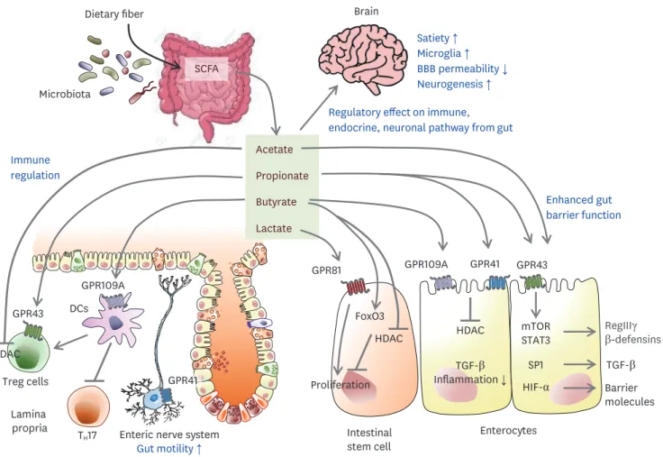
SCFAs are microbiota-derived metabolites fermented from dietary fiber by anaerobic flora and include acetate, propionate, butyrate, and valerate (Fig. 5). SCFAs are agonists for GPR 41, GPR43, and GPR109A on IECs or immune cells (78). In the gut epithelium, SCFAs enhance antimicrobial peptides such as REGIIIγ and β-defensin via the mTOR/
STAT3 pathway (79,80). In the colonic crypts, butyrate suppresses the proliferation of ISCs by inhibiting HDACs and promoting the negative cell-cycle regulator FoxO3, whereas fully differentiated epithelial cells in the villous region reduce butyrate levels through metabolism by acyl-CoA dehydrogenase (9). Butyrate can promote TGF-β expression through HDAC inhibition and SP1 from human enterocytes (81). In contrast, lactate accelerates the proliferation of stem cells dependent on GPR81 (10). In intestinal immune cells, SCFAs
Enteric nerve system Gut motility ↑ Lamina
propria DCs
TH17 Treg cells
GPR109A
GPR41 GPR43
HDAC
Dietary fiber
SCFA Microbiota
Intestinal stem cell
Enterocytes GPR109A
GPR81 GPR41 GPR43
Butyrate Lactate Acetate Propionate
FoxO3
Proliferation HDAC
TGF-β Inflammation ↓
Enhanced gut barrier function
HDAC RegIIIγ
β-defensins
HIF-α Barrier
molecules mTORSTAT3
SP1 TGF-β
Immune regulation
Brain
Satiety ↑ Microglia ↑ BBB permeability ↓ Neurogenesis ↑ Regulatory effect on immune, endocrine, neuronal pathway from gut
Figure 5. Potential gut-brain pathways through SCFAs modulating brain function. Dietary fiber-derived SCFAs fermented by gut microbiota influence gut-brain communication. SCFAs locally interact with IECs and immune cells by their receptors, GPRs, or by inhibiting histone deacetylases, which influence intestinal mucosal immunity and barrier function. In IEC, SCFAs can enhance barrier integrity by upregulating the expression of tight-junction proteins and RegIIIγ.
Butyrate suppresses the proliferation of IEC, but lactate enhances it. SCFAs induce Treg expansion but inhibit Th17 generation via lamina propria DCs. SCFA promotes the secretion of gut hormones from the enteroendocrine cells and indirectly affects the brain via the systemic circulation or vagal pathways. In the brain, SCFAs also increased satiety, microglia, and neurogenesis, and decreased BBB permeability. After a stroke, SCFAs have a critical role in balancing γδT- IL-17+ cells and Tregs and decrease in microglial activation, associated with better recovery after a stroke.
mediate various immune responses in immune cells such as T cells, DCs, and macrophages.
Butyrate enhances IL-10 (79), producing Tregs (82), and reduces the pro-inflammatory cytokine TNF-α from macrophages in the gut (83). After a stroke, reduced levels of plasma SCFAs are associated with worse symptoms (84). Oral supplementation with SCFAs improved post-stroke recovery via immunological modifications. In an independent study, combined supplementation with fermentative bacteria and dietary fiber improved depressive-like behavior following stroke with reduced IL-17+ γδ T cells in the ischemic region of aged mice (85). These data suggest that SCFAs have a beneficial role in post-stroke recovery via immunomodulatory effects. For CNS-resident cells, SCFAs can modulate microglial activity while the molecular mechanism is unclear since the receptor for SCFAs is not expressed in the CNS (18). SCFA supplementation improved motor function and control in post-stroke recovery via regulatory effects on CNS resident microglial activation, which induced the recruitment of circulating T cells to the infarcted brain (84).
CONCLUSION
Here, we summarized the current findings on how the CNS system communicates with the gut environment. The obstacles to understanding the mechanisms of action in this field are connected to the complexities of human neurological disorders and the limitations of animal systems that attempt to model human disease. Although many essential questions regarding the gut-brain axis remain unanswered, the bilateral system of the gut barrier/
microbiome on CNS organ have been established as neuronal network, hormone secretion, neurotransmitters, and immune system. Therefore, brain disease can closely affect the gut environment, and dysregulated gut homeostasis can induce the exacerbation of CNS diseases. Specifically, the outcome of AIS is closely linked to the gut microbiome, barrier integrity, and gut immune system. Besides the neuroprotective effects of probiotic bacterial supplements, the gut environmental sensors need further investigation to understand the molecular mechanism of the gut-brain axis. Studies on SCFAs or AhR ligands may suggest novel strategies to target the gut microbiota to provide new, safe, and effective therapeutic options for neuropsychiatric and neurodegenerative diseases.
ACKNOWLEDGEMENTS
This work was supported by the National Research Foundation of Korea (NRF), funded by the Ministry of Science, ICT, and Future Planning (grant numbers NRF-2020R1A2B5B01001690).
REFERENCES
1. Gribble FM, Reimann F. Enteroendocrine cells: chemosensors in the intestinal epithelium. Annu Rev Physiol 2016;78:277-299.
PUBMED | CROSSREF
2. Kim SH, Jang YS. Recent insights into cellular crosstalk in respiratory and gastrointestinal mucosal immune systems. Immune Netw 2020;20:e44.
PUBMED | CROSSREF
3. Seo K, Seo J, Yeun J, Choi H, Kim YI, Chang SY. The role of mucosal barriers in human gut health. Arch Pharm Res 2021;44:325-341.
PUBMED | CROSSREF
4. Chelakkot C, Ghim J, Ryu SH. Mechanisms regulating intestinal barrier integrity and its pathological implications. Exp Mol Med 2018;50:1-9.
PUBMED | CROSSREF
5. Szakál DN, Gyorffy H, Arató A, Cseh A, Molnár K, Papp M, Dezsofi A, Veres G. Mucosal expression of claudins 2, 3 and 4 in proximal and distal part of duodenum in children with coeliac disease. Virchows Arch 2010;456:245-250.
PUBMED | CROSSREF
6. Davis MA, Ireton RC, Reynolds AB. A core function for p120-catenin in cadherin turnover. J Cell Biol 2003;163:525-534.
PUBMED | CROSSREF
7. Sato T, Vries RG, Snippert HJ, van de Wetering M, Barker N, Stange DE, van Es JH, Abo A, Kujala P, Peters PJ, et al. Single Lgr5 stem cells build crypt-villus structures in vitro without a mesenchymal niche. Nature 2009;459:262-265.
PUBMED | CROSSREF
8. Sato T, Clevers H. Growing self-organizing mini-guts from a single intestinal stem cell: mechanism and applications. Science 2013;340:1190-1194.
PUBMED | CROSSREF
9. Kaiko GE, Ryu SH, Koues OI, Collins PL, Solnica-Krezel L, Pearce EJ, Pearce EL, Oltz EM, Stappenbeck TS. The colonic crypt protects stem cells from microbiota-derived metabolites. Cell 2016;165:1708-1720.
PUBMED | CROSSREF
10. Lee YS, Kim TY, Kim Y, Lee SH, Kim S, Kang SW, Yang JY, Baek IJ, Sung YH, Park YY, et al. Microbiota- derived lactate accelerates intestinal stem-cell-mediated epithelial development. Cell Host Microbe 2018;24:833-846.e6.
PUBMED | CROSSREF
11. Morais LH, Schreiber HL 4th, Mazmanian SK. The gut microbiota-brain axis in behaviour and brain disorders. Nat Rev Microbiol 2021;19:241-255.
PUBMED | CROSSREF
12. Carabotti M, Scirocco A, Maselli MA, Severi C. The gut-brain axis: interactions between enteric microbiota, central and enteric nervous systems. Ann Gastroenterol 2015;28:203-209.
PUBMED
13. Ma Q, Xing C, Long W, Wang HY, Liu Q, Wang RF. Impact of microbiota on central nervous system and neurological diseases: the gut-brain axis. J Neuroinflammation 2019;16:53.
PUBMED | CROSSREF
14. Gutierrez-Aguilar R, Woods SC. Nutrition and l and k-enteroendocrine cells. Curr Opin Endocrinol Diabetes Obes 2011;18:35-41.
PUBMED | CROSSREF
15. Spreckley E, Murphy KG. The l-cell in nutritional sensing and the regulation of appetite. Front Nutr 2015;2:23.
PUBMED | CROSSREF
16. Worthington JJ, Reimann F, Gribble FM. Enteroendocrine cells-sensory sentinels of the intestinal environment and orchestrators of mucosal immunity. Mucosal Immunol 2018;11:3-20.
PUBMED | CROSSREF
17. Abdel-Haq R, Schlachetzki JC, Glass CK, Mazmanian SK. Microbiome-microglia connections via the gut- brain axis. J Exp Med 2019;216:41-59.
PUBMED | CROSSREF
18. Erny D, Hrabě de Angelis AL, Jaitin D, Wieghofer P, Staszewski O, David E, Keren-Shaul H, Mahlakoiv T, Jakobshagen K, Buch T, et al. Host microbiota constantly control maturation and function of microglia in the CNS. Nat Neurosci 2015;18:965-977.
PUBMED | CROSSREF
19. Braniste V, Al-Asmakh M, Kowal C, Anuar F, Abbaspour A, Tóth M, Korecka A, Bakocevic N, Ng LG, Kundu P, et al. The gut microbiota influences blood-brain barrier permeability in mice. Sci Transl Med 2014;6:263ra158.
PUBMED | CROSSREF
20. Amoo M, O'Halloran PJ, Henry J, Husien MB, Brennan P, Campbell M, Caird J, Curley GF. Permeability of the blood-brain barrier after traumatic brain injury; radiological considerations. J Neurotrauma 2021. doi:
10.1089/neu.2020.7545.
PUBMED | CROSSREF
21. El Aidy S, Dinan TG, Cryan JF. Immune modulation of the brain-gut-microbe axis. Front Microbiol 2014;5:146.
PUBMED | CROSSREF
22. Desbonnet L, Garrett L, Clarke G, Kiely B, Cryan JF, Dinan TG. Effects of the probiotic Bifidobacterium infantis in the maternal separation model of depression. Neuroscience 2010;170:1179-1188.
PUBMED | CROSSREF
23. Lyte M. Microbial endocrinology and the microbiota-gut-brain axis. Adv Exp Med Biol 2014;817:3-24.
PUBMED | CROSSREF
24. Silva YP, Bernardi A, Frozza RL. The role of short-chain fatty acids from gut microbiota in gut-brain communication. Front Endocrinol (Lausanne) 2020;11:25.
PUBMED | CROSSREF
25. Mawe GM, Hoffman JM. Serotonin signalling in the gut--functions, dysfunctions and therapeutic targets.
Nat Rev Gastroenterol Hepatol 2013;10:473-486.
PUBMED | CROSSREF
26. Banskota S, Ghia JE, Khan WI. Serotonin in the gut: blessing or a curse. Biochimie 2019;161:56-64.
PUBMED | CROSSREF
27. Fidalgo S, Ivanov DK, Wood SH. Serotonin: from top to bottom. Biogerontology 2013;14:21-45.
PUBMED | CROSSREF
28. Nozawa K, Kawabata-Shoda E, Doihara H, Kojima R, Okada H, Mochizuki S, Sano Y, Inamura K, Matsushime H, Koizumi T, et al. TRPA1 regulates gastrointestinal motility through serotonin release from enterochromaffin cells. Proc Natl Acad Sci U S A 2009;106:3408-3413.
PUBMED | CROSSREF
29. Ahern GP. 5-HT and the immune system. Curr Opin Pharmacol 2011;11:29-33.
PUBMED | CROSSREF
30. Regmi SC, Park SY, Ku SK, Kim JA. Serotonin regulates innate immune responses of colon epithelial cells through Nox2-derived reactive oxygen species. Free Radic Biol Med 2014;69:377-389.
PUBMED | CROSSREF
31. Coates MD, Mahoney CR, Linden DR, Sampson JE, Chen J, Blaszyk H, Crowell MD, Sharkey KA, Gershon MD, Mawe GM. Molecular defects in mucosal serotonin content and decreased serotonin reuptake transporter in ulcerative colitis and irritable bowel syndrome. Gastroenterology 2004;126:1657-1664.
PUBMED | CROSSREF
32. Docherty M. Elevated serotonin associated with collagenous colitis. Am J Gastroenterol 2010;105:1449.
PUBMED | CROSSREF
33. D'Alessio S, Tacconi C, Fiocchi C, Danese S. Advances in therapeutic interventions targeting the vascular and lymphatic endothelium in inflammatory bowel disease. Curr Opin Gastroenterol 2013;29:608-613.
PUBMED | CROSSREF
34. Sgritta M, Dooling SW, Buffington SA, Momin EN, Francis MB, Britton RA, Costa-Mattioli M.
Mechanisms underlying microbial-mediated changes in social behavior in mouse models of autism spectrum disorder. Neuron 2019;101:246-259.e6.
PUBMED | CROSSREF
35. Bravo-Ferrer I, Cuartero MI, Medina V, Ahedo-Quero D, Peña-Martínez C, Pérez-Ruíz A, Fernández-Valle ME, Hernández-Sánchez C, Fernández-Salguero PM, Lizasoain I, et al. Lack of the aryl hydrocarbon receptor accelerates aging in mice. FASEB J 2019;33:12644-12654.
PUBMED | CROSSREF
36. Pinto-Sanchez MI, Hall GB, Ghajar K, Nardelli A, Bolino C, Lau JT, Martin FP, Cominetti O, Welsh C, Rieder A, et al. Probiotic Bifidobacterium longum NCC3001 reduces depression scores and alters brain activity: a pilot study in patients with irritable bowel syndrome. Gastroenterology 2017;153:448-459.e8.
PUBMED | CROSSREF
37. Hsiao EY, McBride SW, Hsien S, Sharon G, Hyde ER, McCue T, Codelli JA, Chow J, Reisman SE, Petrosino JF, et al. Microbiota modulate behavioral and physiological abnormalities associated with neurodevelopmental disorders. Cell 2013;155:1451-1463.
PUBMED | CROSSREF
38. Sampson TR, Debelius JW, Thron T, Janssen S, Shastri GG, Ilhan ZE, Challis C, Schretter CE, Rocha S, Gradinaru V, et al. Gut microbiota regulate motor deficits and neuroinflammation in a model of Parkinson's disease. Cell 2016;167:1469-1480.e12.
PUBMED | CROSSREF
39. Chu C, Murdock MH, Jing D, Won TH, Chung H, Kressel AM, Tsaava T, Addorisio ME, Putzel GG, Zhou L, et al. The microbiota regulate neuronal function and fear extinction learning. Nature 2019;574:543-548.
PUBMED | CROSSREF
40. Barden N. Implication of the hypothalamic-pituitary-adrenal axis in the physiopathology of depression. J Psychiatry Neurosci 2004;29:185-193.
PUBMED
41. Sudo N, Chida Y, Aiba Y, Sonoda J, Oyama N, Yu XN, Kubo C, Koga Y. Postnatal microbial colonization programs the hypothalamic-pituitary-adrenal system for stress response in mice. J Physiol 2004;558:263-275.
PUBMED | CROSSREF
42. García-Ródenas CL, Bergonzelli GE, Nutten S, Schumann A, Cherbut C, Turini M, Ornstein K, Rochat F, Corthésy-Theulaz I. Nutritional approach to restore impaired intestinal barrier function and growth after neonatal stress in rats. J Pediatr Gastroenterol Nutr 2006;43:16-24.
PUBMED | CROSSREF
43. O'Mahony SM, Marchesi JR, Scully P, Codling C, Ceolho AM, Quigley EM, Cryan JF, Dinan TG. Early life stress alters behavior, immunity, and microbiota in rats: implications for irritable bowel syndrome and psychiatric illnesses. Biol Psychiatry 2009;65:263-267.
PUBMED | CROSSREF
44. Kelly JR, Kennedy PJ, Cryan JF, Dinan TG, Clarke G, Hyland NP. Breaking down the barriers: the gut microbiome, intestinal permeability and stress-related psychiatric disorders. Front Cell Neurosci 2015;9:392.
PUBMED | CROSSREF
45. Gareau MG, Silva MA, Perdue MH. Pathophysiological mechanisms of stress-induced intestinal damage.
Curr Mol Med 2008;8:274-281.
PUBMED | CROSSREF
46. Gareau MG, Jury J, MacQueen G, Sherman PM, Perdue MH. Probiotic treatment of rat pups normalises corticosterone release and ameliorates colonic dysfunction induced by maternal separation. Gut 2007;56:1522-1528.
PUBMED | CROSSREF
47. Ait-Belgnaoui A, Durand H, Cartier C, Chaumaz G, Eutamene H, Ferrier L, Houdeau E, Fioramonti J, Bueno L, Theodorou V. Prevention of gut leakiness by a probiotic treatment leads to attenuated HPA response to an acute psychological stress in rats. Psychoneuroendocrinology 2012;37:1885-1895.
PUBMED | CROSSREF
48. Goehler LE, Park SM, Opitz N, Lyte M, Gaykema RP. Campylobacter jejuni infection increases anxiety-like behavior in the holeboard: possible anatomical substrates for viscerosensory modulation of exploratory behavior. Brain Behav Immun 2008;22:354-366.
PUBMED | CROSSREF
49. Lyte M, Li W, Opitz N, Gaykema RP, Goehler LE. Induction of anxiety-like behavior in mice during the initial stages of infection with the agent of murine colonic hyperplasia Citrobacter rodentium. Physiol Behav 2006;89:350-357.
PUBMED | CROSSREF
50. GBD 2016 Lifetime Risk of Stroke CollaboratorsFeigin VL, Nguyen G, Cercy K, Johnson CO, Alam T, Parmar PG, Abajobir AA, Abate KH, Abd-Allah F, et al. Global, regional, and country-specific lifetime risks of stroke, 1990 and 2016. N Engl J Med 2018;379:2429-2437.
PUBMED | CROSSREF
51. Singh V, Roth S, Llovera G, Sadler R, Garzetti D, Stecher B, Dichgans M, Liesz A. Microbiota dysbiosis controls the neuroinflammatory response after stroke. J Neurosci 2016;36:7428-7440.
PUBMED | CROSSREF
52. Huang YY, Li X, Li X, Sheng YY, Zhuang PW, Zhang YJ. Neuroimmune crosstalk in central nervous system injury-induced infection and pharmacological intervention. Brain Res Bull 2019;153:232-238.
PUBMED | CROSSREF
53. Benakis C, Poon C, Lane D, Brea D, Sita G, Moore J, Murphy M, Racchumi G, Iadecola C, Anrather J.
Distinct commensal bacterial signature in the gut is associated with acute and long-term protection from ischemic stroke. Stroke 2020;51:1844-1854.
PUBMED | CROSSREF
54. Benakis C, Brea D, Caballero S, Faraco G, Moore J, Murphy M, Sita G, Racchumi G, Ling L, Pamer EG, et al. Commensal microbiota affects ischemic stroke outcome by regulating intestinal γδ T cells. Nat Med 2016;22:516-523.
PUBMED | CROSSREF
55. Stanley D, Mason LJ, Mackin KE, Srikhanta YN, Lyras D, Prakash MD, Nurgali K, Venegas A, Hill MD, Moore RJ, et al. Translocation and dissemination of commensal bacteria in post-stroke infection. Nat Med 2016;22:1277-1284.
PUBMED | CROSSREF
56. Xu K, Gao X, Xia G, Chen M, Zeng N, Wang S, You C, Tian X, Di H, Tang W, et al. Rapid gut dysbiosis induced by stroke exacerbates brain infarction in turn. Gut 2021; gutjnl-2020-323263.
PUBMED | CROSSREF
57. Pedersen NC, Kim Y, Liu H, Galasiti Kankanamalage AC, Eckstrand C, Groutas WC, Bannasch M, Meadows JM, Chang KO. Efficacy of a 3C-like protease inhibitor in treating various forms of acquired feline infectious peritonitis. J Feline Med Surg 2018;20:378-392.
PUBMED | CROSSREF
58. Rothhammer V, Quintana FJ. The aryl hydrocarbon receptor: an environmental sensor integrating immune responses in health and disease. Nat Rev Immunol 2019;19:184-197.
PUBMED | CROSSREF
59. Metidji A, Omenetti S, Crotta S, Li Y, Nye E, Ross E, Li V, Maradana MR, Schiering C, Stockinger B. The environmental sensor AHR protects from inflammatory damage by maintaining intestinal stem cell homeostasis and barrier integrity. Immunity 2018;49:353-362.e5.
PUBMED | CROSSREF
60. Lanis JM, Alexeev EE, Curtis VF, Kitzenberg DA, Kao DJ, Battista KD, Gerich ME, Glover LE, Kominsky DJ, Colgan SP. Tryptophan metabolite activation of the aryl hydrocarbon receptor regulates IL-10 receptor expression on intestinal epithelia. Mucosal Immunol 2017;10:1133-1144.
PUBMED | CROSSREF
61. Yu M, Wang Q, Ma Y, Li L, Yu K, Zhang Z, Chen G, Li X, Xiao W, Xu P, et al. Aryl hydrocarbon receptor activation modulates intestinal epithelial barrier function by maintaining tight junction integrity. Int J Biol Sci 2018;14:69-77.
PUBMED | CROSSREF
62. Benson JM, Shepherd DM. Dietary ligands of the aryl hydrocarbon receptor induce anti-inflammatory and immunoregulatory effects on murine dendritic cells. Toxicol Sci 2011;124:327-338.
PUBMED | CROSSREF
63. Cervantes-Barragan L, Chai JN, Tianero MD, Di Luccia B, Ahern PP, Merriman J, Cortez VS, Caparon MG, Donia MS, Gilfillan S, et al. Lactobacillus reuteri induces gut intraepithelial CD4+CD8αα+ T cells. Science 2017;357:806-810.
PUBMED | CROSSREF
64. Kadowaki A, Miyake S, Saga R, Chiba A, Mochizuki H, Yamamura T. Gut environment-induced intraepithelial autoreactive CD4+ T cells suppress central nervous system autoimmunity via LAG-3. Nat Commun 2016;7:11639.
PUBMED | CROSSREF
65. Brandstätter O, Schanz O, Vorac J, König J, Mori T, Maruyama T, Korkowski M, Haarmann-Stemmann T, von Smolinski D, Schultze JL, et al. Balancing intestinal and systemic inflammation through cell type- specific expression of the aryl hydrocarbon receptor repressor. Sci Rep 2016;6:26091.
PUBMED | CROSSREF
66. Lee JS, Cella M, McDonald KG, Garlanda C, Kennedy GD, Nukaya M, Mantovani A, Kopan R, Bradfield CA, Newberry RD, et al. AHR drives the development of gut ILC22 cells and postnatal lymphoid tissues via pathways dependent on and independent of Notch. Nat Immunol 2011;13:144-151.
PUBMED | CROSSREF
67. Qiu J, Heller JJ, Guo X, Chen ZM, Fish K, Fu YX, Zhou L. The aryl hydrocarbon receptor regulates gut immunity through modulation of innate lymphoid cells. Immunity 2012;36:92-104.
PUBMED | CROSSREF
68. Qiu J, Guo X, Chen ZM, He L, Sonnenberg GF, Artis D, Fu YX, Zhou L. Group 3 innate lymphoid cells inhibit T-cell-mediated intestinal inflammation through aryl hydrocarbon receptor signaling and regulation of microflora. Immunity 2013;39:386-399.
PUBMED | CROSSREF
69. Rothhammer V, Mascanfroni ID, Bunse L, Takenaka MC, Kenison JE, Mayo L, Chao CC, Patel B, Yan R, Blain M, et al. Type I interferons and microbial metabolites of tryptophan modulate astrocyte activity and central nervous system inflammation via the aryl hydrocarbon receptor. Nat Med 2016;22:586-597.
PUBMED | CROSSREF
70. Rothhammer V, Borucki DM, Tjon EC, Takenaka MC, Chao CC, Ardura-Fabregat A, de Lima KA, Gutiérrez-Vázquez C, Hewson P, Staszewski O, et al. Microglial control of astrocytes in response to microbial metabolites. Nature 2018;557:724-728.
PUBMED | CROSSREF
71. Veldhoen M, Hirota K, Westendorf AM, Buer J, Dumoutier L, Renauld JC, Stockinger B. The aryl hydrocarbon receptor links TH17-cell-mediated autoimmunity to environmental toxins. Nature 2008;453:106-109.
PUBMED | CROSSREF
72. Chen WC, Chang LH, Huang SS, Huang YJ, Chih CL, Kuo HC, Lee YH, Lee IH. Aryl hydrocarbon receptor modulates stroke-induced astrogliosis and neurogenesis in the adult mouse brain. J Neuroinflammation 2019;16:187.
PUBMED | CROSSREF
73. Brouns R, Verkerk R, Aerts T, De Surgeloose D, Wauters A, Scharpé S, De Deyn PP. The role of tryptophan catabolism along the kynurenine pathway in acute ischemic stroke. Neurochem Res 2010;35:1315-1322.
PUBMED | CROSSREF
74. Di Giaimo R, Durovic T, Barquin P, Kociaj A, Lepko T, Aschenbroich S, Breunig CT, Irmler M, Cernilogar FM, Schotta G, et al. The aryl hydrocarbon receptor pathway defines the time frame for restorative neurogenesis. Cell Rep 2018;25:3241-3251.e5.
PUBMED | CROSSREF
75. Kaye J, Piryatinsky V, Birnberg T, Hingaly T, Raymond E, Kashi R, Amit-Romach E, Caballero IS, Towfic F, Ator MA, et al. Laquinimod arrests experimental autoimmune encephalomyelitis by activating the aryl hydrocarbon receptor. Proc Natl Acad Sci U S A 2016;113:E6145-E6152.
PUBMED | CROSSREF
76. Berg J, Mahmoudjanlou Y, Duscha A, Massa MG, Thöne J, Esser C, Gold R, Haghikia A. The immunomodulatory effect of laquinimod in CNS autoimmunity is mediated by the aryl hydrocarbon receptor. J Neuroimmunol 2016;298:9-15.
PUBMED | CROSSREF
77. Zelante T, Iannitti RG, Cunha C, De Luca A, Giovannini G, Pieraccini G, Zecchi R, D'Angelo C, Massi- Benedetti C, Fallarino F, et al. Tryptophan catabolites from microbiota engage aryl hydrocarbon receptor and balance mucosal reactivity via interleukin-22. Immunity 2013;39:372-385.
PUBMED | CROSSREF
78. Dalile B, Van Oudenhove L, Vervliet B, Verbeke K. The role of short-chain fatty acids in microbiota-gut- brain communication. Nat Rev Gastroenterol Hepatol 2019;16:461-478.
PUBMED | CROSSREF
79. Chen J, Vitetta L. The role of butyrate in attenuating pathobiont-induced hyperinflammation. Immune Netw 2020;20:e15.
PUBMED | CROSSREF
80. Zhao Y, Chen F, Wu W, Sun M, Bilotta AJ, Yao S, Xiao Y, Huang X, Eaves-Pyles TD, Golovko G, et al.
GPR43 mediates microbiota metabolite SCFA regulation of antimicrobial peptide expression in intestinal epithelial cells via activation of mTOR and STAT3. Mucosal Immunol 2018;11:752-762.
PUBMED | CROSSREF
81. Martin-Gallausiaux C, Béguet-Crespel F, Marinelli L, Jamet A, Ledue F, Blottière HM, Lapaque N. Butyrate produced by gut commensal bacteria activates TGF-beta1 expression through the transcription factor SP1 in human intestinal epithelial cells. Sci Rep 2018;8:9742.
PUBMED | CROSSREF
82. Furusawa Y, Obata Y, Fukuda S, Endo TA, Nakato G, Takahashi D, Nakanishi Y, Uetake C, Kato K, Kato T, et al. Commensal microbe-derived butyrate induces the differentiation of colonic regulatory T cells.
Nature 2013;504:446-450.
PUBMED | CROSSREF
83. Chang PV, Hao L, Offermanns S, Medzhitov R. The microbial metabolite butyrate regulates intestinal macrophage function via histone deacetylase inhibition. Proc Natl Acad Sci U S A 2014;111:2247-2252.
PUBMED | CROSSREF
84. Sadler R, Cramer JV, Heindl S, Kostidis S, Betz D, Zuurbier KR, Northoff BH, Heijink M, Goldberg MP, Plautz EJ, et al. Short-chain fatty acids improve poststroke recovery via immunological mechanisms. J Neurosci 2020;40:1162-1173.
PUBMED | CROSSREF
85. Lee J, d'Aigle J, Atadja L, Quaicoe V, Honarpisheh P, Ganesh BP, Hassan A, Graf J, Petrosino J, Putluri N, et al. Gut microbiota-derived short-chain fatty acids promote poststroke recovery in aged mice. Circ Res 2020;127:453-465.
PUBMED | CROSSREF