INTRODUCTION
Varicocele is defined as abnormal dilatation of the pampiniform and/or cremasteric venous plexus. It is
the most common treatable cause of male infertility, affecting roughly 15% to 20% of the general population and 40% of men presenting for an infertility evalua- tion. Varicocele is also the most common cause of sec-
Received: Apr 18, 2018 Revised: Jun 11, 2018 Accepted: Jun 25, 2018 Published online Aug 14, 2018 Correspondence to: İlter Alkan https://orcid.org/0000-0001-7530-0890
Department of Urology, Okmeydanı Training and Research Hospital, Kaptanpaşa Mahallesi, Darülaceze Caddesi, No: 25, Okmeydanı, Şişli- Istanbul 34384, Turkey.
Tel: +90-212-314-55-55, Fax: +90-212-221-78-00, E-mail: [email protected] Copyright © 2018 Korean Society for Sexual Medicine and Andrology
pISSN: 2287-4208 / eISSN: 2287-4690
World J Mens Health 2018 September 36(3): 255-262 https://doi.org/10.5534/wjmh.180028
Superoxide Anion Production by the Spermatozoa of Men with Varicocele: Relationship with
Varicocele Grade and Semen Parameters
İlter Alkan1 , Meral Yüksel2 , Halil Lütfi Canat1 , Hasan Anıl Atalay1 , Osman Can1 , Hakan Özveri3 , Mehmet Murad Başar4
1Department of Urology, Okmeydanı Training and Research Hospital, University of Health Sciences, 2Department of Medical Laboratory, Vocational School of Health-Related Services, Marmara University, 3Department of Urology & Andrology, School of Medicine, Acibadem Mehmet Ali Aydinlar University, 4Department of Urology & Andrology, Memorial Şişli Hospital, Istanbul, Turkey
Purpose: To investigate the pathophysiological role of superoxide anion and total reactive oxygen species (ROS) production by the spermatozoa of men with varicocele and its relationship with varicocele grade and semen parameters.
Materials and Methods: This prospective study included 34 men with grade II–III varicocele, regardless of their fertility status.
The control group consisted of 13 healthy men. Semen characteristics were examined according to the 2010 World Health Organization criteria. The swim-up method was used for sperm preparation. Total ROS and superoxide anion production was assayed by luminol- and lucigenin-dependent chemiluminescence (CL), respectively.
Results: The men with varicocele had significantly higher total ROS and superoxide anion levels than the healthy control subjects (2.9±0.4 relative light unit (RLU) vs. 2.4±0.1 RLU, p=0.001 for luminol-dependent CL and 2.8±0.4 RLU vs. 2.3±0.2 RLU, p=0.002 for lucigenin-dependent CL). Cases of grade III varicocele had significantly higher superoxide anion and total ROS levels than grade II cases and control subjects (p<0.001). Superoxide anion and total ROS levels were negatively corre- lated with all semen parameters.
Conclusions: The superoxide anion levels produced by spermatozoa were significantly higher in varicocele patients than in control subjects. ROS production was related to increased varicocele grade, impaired semen concentration, and abnormal morphology in men with varicocele. Our findings suggest that superoxide anion overproduction may be an important step in the cascade of ROS-related damage to spermatozoa, resulting in impaired semen parameters in patients with varicocele.
Keywords: Oxidative stress; Reactive oxygen species; Spermatozoa; Superoxides; Varicocele
This is an Open Access article distributed under the terms of the Creative Commons Attribution Non-Commercial License (http://creativecommons.org/licenses/by-nc/4.0) which permits unrestricted non-commercial use, distribution, and reproduction in any medium, provided the original work is properly cited.
ondary infertility, and has been reported in up to 80%
of cases [1,2]. In previous studies, varicocele was found to be associated with ipsilateral progressive testicular atrophy, deterioration of sperm parameters, decreased serum testosterone levels, and male infertility [3].
The exact pathophysiologic mechanisms that cause varicocele-associated infertility have not been com- pletely elucidated. The etiology and pathophysiology of varicocele can be multifactorial, including elevated tes- ticular temperature, testicular hypoxia, oxidative stress (OS), and apoptosis [4,5]. Studies have demonstrated that varicocele was associated with high levels of semi- nal OS, suggesting that impaired sperm function could be related to OS in patients’ semen [6,7].
Reactive oxygen species (ROS) include the superoxide anion (O2•–), hydrogen peroxide (H2O2), and the hydrox- yl radical (•OH). ROS are products of normal cellular metabolism and produced by both seminal leukocytes and abnormal sperm in semen. Low levels of ROS are necessary for sperm capacitation, the acrosome reac- tion, hyperactivation, and fusion with the oocyte [8-10].
However, in certain pathological cellular conditions, supra-physiologic ROS production overwhelms the an- tioxidant defense mechanisms, resulting in OS. Exces- sive ROS increase the peroxidation of the polyunsatu- rated fatty acids of the sperm cell membrane and can damage spermatozoa. ROS can also induce sperm DNA fragmentation and apoptosis in mature spermatozoa [11,12]. These findings may explain the clinical pre- sentation of impaired semen parameters in men with varicocele and elevated OS.
To date, most clinical studies have investigated total ROS production in the semen of patients with varico- cele. To the best of our knowledge, only 1 clinical study in the literature has investigated superoxide anion production in the untreated semen of patients with varicocele [13]. This prompted us to assay superoxide anion production by the spermatozoa of patients with varicocele using the lucigenin-dependent chemilumi- nescence (CL) method.
The objective of this study was to investigate the pathophysiological role of superoxide anion and total ROS production by the spermatozoa of men with vari- cocele and its relationship with varicocele grade and semen parameters.
MATERIALS AND METHODS
1. Ethics statement
The present study protocol was reviewed and ap- proved by the Institutional Review Board of Okmey- danı Training and Research Hospital (Reg. No. 688).
Informed consent was submitted by all subjects when they were enrolled.
2. Study design and patient selection criteria
This multicenter prospective study was conducted be- tween January and December 2017. Patients who were admitted to Okmeydanı Hospital outpatient clinic with the complaint of left scrotal discomfort were evaluated with a detailed history, physical examination, scrotal Doppler ultrasound, and measurement of serum folli- cle-stimulating hormone (1.27–19.26 mIU/mL) and total testosterone (1.75–7.81 ng/mL) levels. Varicoceles were clinically graded as grade II (palpable while standing upright) or grade III (visible through the scrotal skin) [3,14]. The inclusion criteria for the study were as fol- lows: men aged between 18 and 35 years with clinical grade II/III varicocele on physical examination, as also confirmed by Doppler ultrasound (with the finding of 1 or more veins with a maximal diameter >3 mm and retrograde flow seen either at rest or during the Val- salva maneuver) and normal serum hormone levels, who were non-smokers, had no history of varicocele surgery, genitourinary infections, or medical treatment with antioxidants in last 6 months. All participants were evaluated using semen analysis and ROS assays.
Patients with azoospermia and leukocytospermia on semen analysis were excluded from the study. A total of 34 men who met the inclusion criteria comprised the patient group. The control group included 13 age- matched healthy men with a normal semen analysis who had no history of smoking, urinary infection/
prostatitis, or varicocele surgery, and no varicocele on physical examination.
3. Semen analysis
Semen samples were collected from participants through masturbation into sterile containers after 4 to 5 days of sexual abstinence. After liquefaction, semen specimens were evaluated for semen volume, appear- ance, and viscosity. Semen characteristics were exam- ined according to the 2010 World Health Organization criteria [15].
4. White blood cells in semen
The presence of leukocytes in semen specimens was assessed by a Leucoscreen (Fertipro, Beernem, Belgium) test. Leukocytospermia was defined as the presence of at least 1.0×10⁶ white blood cells (WBCs) per milliliter.
5. Swim-up method
After liquefaction, the sample was transferred into 4 sterile 15-mL tubes using a plastic pipette, and the weight was measured. The specimen was mixed with the same amount of human tubal fluid (HTF; Lifeglob- al, Guelph, ON, Canada) +5% protein medium using a Pasteur pipette. The suspended sample was transferred into 4 sterile 15-mL tubes using plastic pipettes and centrifuged at 800 rpm for 10 minutes. The superna- tant was aspirated and discharged. The samples were
mixed with 0.5 mL of fresh HTF medium for each tube using a Pasteur pipette and centrifuged at 800 rpm for 10 minutes. The supernatant was aspirated and discharged, after which 0.25 mL of HTF medium was slowly added without compromising the pellet in each tube. The tubes were then incubated at an angle of 45 for 1 hour in the incubator at 37°C. After the incuba- tion period, the supernatant was transferred to a ster- ile 5-mL tube using a Pasteur pipette.
6. Determination of reactive oxygen species
Total ROS and superoxide anion levels were mea- sured by the CL method using a luminometer (LB9509;
Berthold, Bad Wildbad, Germany) at room tempera- ture. The sperm pellets were re-suspended in the same medium at a concentration of 1.0×10⁶ spermatozoa/mL.
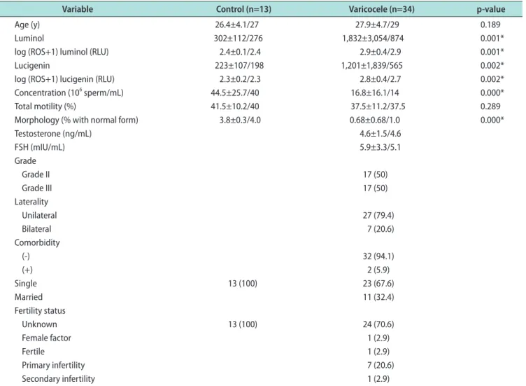
Table 1. General characteristics and statistical data of the study population
Variable Control (n=13) Varicocele (n=34) p-value
Age (y) 26.4±4.1/27 27.9±4.7/29 0.189
Luminol 302±112/276 1,832±3,054/874 0.001*
log (ROS+1) luminol (RLU) 2.4±0.1/2.4 2.9±0.4/2.9 0.001*
Lucigenin 223±107/198 1,201±1,839/565 0.002*
log (ROS+1) lucigenin (RLU) 2.3±0.2/2.3 2.8±0.4/2.7 0.002*
Concentration (106 sperm/mL) 44.5±25.7/40 16.8±16.1/14 0.000*
Total motility (%) 41.5±10.2/40 37.5±11.2/37.5 0.289
Morphology (% with normal form) 3.8±0.3/4.0 0.68±0.68/1.0 0.000*
Testosterone (ng/mL) 4.6±1.5/4.6
FSH (mIU/mL) 5.9±3.3/5.1
Grade
Grade II 17 (50)
Grade III 17 (50)
Laterality
Unilateral 27 (79.4)
Bilateral 7 (20.6)
Comorbidity
(-) 32 (94.1)
(+) 2 (5.9)
Single 13 (100) 23 (67.6)
Married 11 (32.4)
Fertility status
Unknown 13 (100) 24 (70.6)
Female factor 1 (2.9)
Fertile 1 (2.9)
Primary infertility 7 (20.6)
Secondary infertility 1 (2.9)
Values are presented as mean±standard deviation/median only or number (%). ROS levels are given as relative light unit (RLU)/1.0×106 spermato- zoa/mL.
ROS: reactive oxygen species, FSH: follicle-stimulating hormone.
*Asterisks indicate a significant difference (p<0.05).
Luminol (5-amino-2,3-dihydro-1,4-phthalazinedione) or lucigenin (10,10′-dimethyl-9,9′-biacridinium dinitrate) enhancers were added to the test tubes for a final concentration of 0.2 mM. Superoxide anion production was measured by lucigenin-dependent CL. In contrast, luminol was used to detect a broader range of reactive species, (i.e., •OH, H2O2, and HOCl radicals). Counts were made for 5 minutes at 1-minute intervals. After the measurements, the area under the curve was calcu- lated and corrected for the sperm count. Results were given as relative light unit (RLU)/1.0×106 spermatozoa/
mL. In order to normalize values to the same distribu- tion, log transformed values (ROS+1) of the results were used in calculations.
7. Statistical analysis
The statistical results are presented as mean±standard deviation. The distribution of variables was assessed with the Kolmogorov-Smirnov test. The Mann-Whitney U-test was used to compare independent quantitative data. The Kruskal-Wallis test was used for multiple comparisons. Correlations between variables were calculated using the Spearman method. The power of the study was calculated using the G*Power program (University of Dusseldorf, Dusseldorf, Germany) with an effect size convention of 0.8 for the 2-tailed t-test, with an alpha error protection of 0.05. Statistical sig- nificance was assessed using 2-tailed tests, and p-values
<0.05 were considered to indicate statistical signifi- cance. Statistical tests were performed using IBM SPSS ver. 22.0 (IBM Co., Armonk, NY, USA).
RESULTS
The general characteristics and statistical data of the study population are presented in Table 1.
Of the 34 patients, 2 (5.9%) had comorbid patholo- gies. One patient had a history of hypertension and the other had a history of hypothyroidism.
1. Luminol- and lucigenin-dependent reactive oxygen species levels of the study group
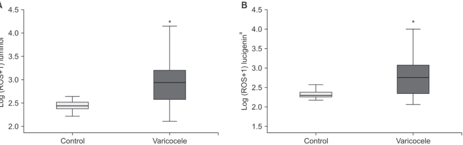
The men with varicocele had significantly higher total ROS levels (2.9±0.4 RLU) than the healthy con- trol subjects (2.4±0.1 RLU) on luminol-dependent CL (p=0.001) (Table 1, Fig. 1A).
The men with varicocele had significantly higher superoxide anion levels (2.8±0.4 RLU) than the healthy control subjects (2.3±0.2 RLU) on lucigenin-dependent CL (p=0.002) (Table 1, Fig. 1B).
2. Luminol- and lucigenin-dependent reactive oxygen species levels and varicocele grade
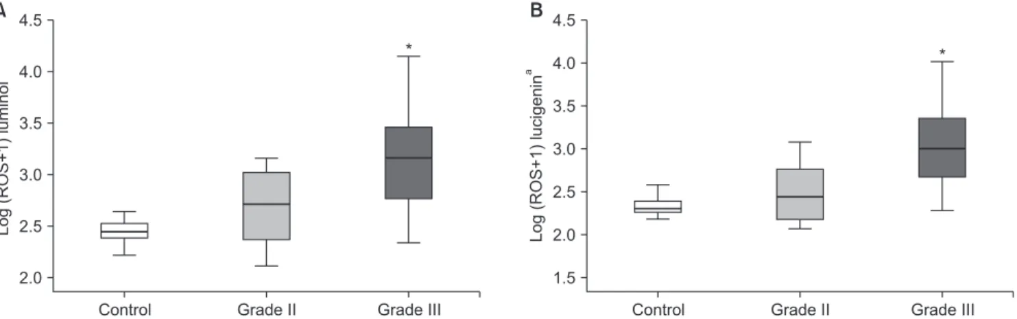
Patients with grade III varicocele had significantly higher ROS levels than patients with grade II vari- cocele and control subjects on luminol-dependent CL (p=0.006 and p<0.001, respectively) (Table 2, Fig. 2A).
Patients with grade III varicocele had significantly higher superoxide anion levels than patients with grade II varicocele and control subjects on lucigenin- dependent CL (p=0.002 and p<0.001, respectively) (Table 2, Fig. 2B).
Log(ROS+1)lucigenina
Control 4.5
4.0 3.5 3.0 2.5
Varicocele 1.5
*
2.0
A B
Log(ROS+1)luminola
Control 4.5
4.0
3.5
3.0
2.5
Varicocele 2.0
*
Fig. 1. Log (ROS+1) levels of patients with varicocele and the control group (mean±standard deviation) assessed using luminol- (A) and luci- genin- (B) dependent chemiluminescence. ROS: reactive oxygen species. *Significant difference with the control group (p=0.001 and p=0.002, respectively). aRelative light unit/1.0×106 spermatozoa/mL.
3. Semen analysis results
The results of the semen analyses are presented in Table 1. Sperm concentrations (10.5±7.9×10⁶/mL vs.
23.0±19.6×10⁶/mL, p=0.013) and normal sperm morphol- ogy (0.35%±0.60% vs. 1.0%±0.61%, p=0.003) were signifi- cantly lower in patients with grade III varicocele than in patients with grade II varicocele. No statistically significant differences were found in sperm motility (34.5%±11.1% vs. 40.4%±10.7%, p=0.262) between patients with grade III and grade II varicocele.
4. Correlations of luminol- and lucigenin- dependent reactive oxygen species levels with semen parameters
There was a positive correlation between luminol- and lucigenin-dependent ROS levels (r=0.84, p<0.001).
Luminol-dependent total ROS levels were negatively correlated with sperm concentration, motility, and nor- mal morphology (r=-0.57, p<0.001; r=-0.33, p=0.048; and r=-0.34, p=0.045, respectively). Lucigenin-dependent su- peroxide anion levels were negatively correlated with sperm concentration, motility, and normal morphology (r=-0.52, p<0.001; r=-0.33, p=0.023; and r=-0.31, p=0.047, respectively).
DISCUSSION
In the present study, we found that lucigenin-depen- dent superoxide anion levels were higher in patients with varicocele than in control subjects. To the best of our knowledge, this is the first study to demonstrate increased superoxide anion production by the sper- matozoa of patients with varicocele assayed using the lucigenin-dependent CL method. A CL assay utilizing luminol is the most commonly described direct method to measure ROS production by the spermatozoa. The amount of oxidation in the spermatozoa cell membrane shows the net oxidative imbalance between ROS pro- duction and the antioxidant levels in semen [16]. The luminol-dependent CL assay measures intracellular and extracellular ROS levels (O2•−, •OH, H2O2) because luminol reacts with a variety of ROS. However, this method does not discriminate these oxidants from one another, meaning that it measures total ROS levels.
Polymorphonuclear leukocyte ROS production accounts for most of the total ROS production in CL assays in untreated semen. Various semen-washing methods can be used to eliminate WBCs from semen in order to as- say spermatozoal ROS production. H2O2 is more reac-
Log(ROS+1)lucigenina
4.5 4.0 3.5 3.0 2.5
1.5 2.0
Control Grade II
*
Grade III
A B
Log(ROS+1)luminola
Control 4.5
4.0
3.5
3.0
2.5
Grade II 2.0
*
Grade III
Fig. 2. Luminol- (A) and lucigenin- (B) dependent log (ROS+1) levels in patients with grade II and grade III varicocele and the control group (mean±standard deviation). ROS: reactive oxygen species. *Significant difference with the other groups (p<0.001). aRelative light unit/1.0×106 spermatozoa/mL.
Table 2. Luminol- and lucigenin-dependent log (ROS+1) levels in patients with grade II and grade III varicocele and the control group
Variable Control (n=13) Grade II (n=17) Grade III (n=17) p-value
Log (ROS+1) luminol (RLU) 2.4±0.1a/2.4 2.7±0.3a/2.7 3.2±0.5/3.2 0.000***
Log (ROS+1) lucigenin (RLU) 2.3±0.2a/2.3 2.5±0.3a/2.4 3.0±0.5/3.0 0.000***
Values are presented as mean±standard deviation/median only. ROS levels are given as RLU/1.0×106 spermatozoa/mL.
ROS: reactive oxygen species, RLU: relative light unit.
aSignificant difference with the grade III varicocele group. ***Asterisks indicate a significant difference (p<0.001).
tive than superoxide anion in the luminol-dependent CL method. Lucigenin-dependent CL is a more specific and validated method for assaying extracellular super- oxide anion levels [17].
Many studies have measured seminal ROS levels in infertile men with varicocele and compared the values with normospermic or fertile donors [6,7]. Those studies, which used a luminol-dependent CL method, showed that ROS levels were significantly higher in varicocele patients than in healthy controls. Our results, show- ing that patients with varicocele had higher total ROS levels than controls, are compatible with the literature.
In fact, most of the studies in the literature about OS in male infertility and varicocele have used luminol- dependent CL or lipid peroxidation products (e.g., malo- ndialdehyde; MDA) and oxidized DNA assays, which are indirect methods of measuring ROS/OS [6,7,18,19].
Indirect methods measure the end products of the per- oxidative process or DNA damage, and mostly provide information about ROS-related damage. Considering those facts, we preferred to use the CL method to as- say ROS production by the spermatozoa of patients with varicocele. For the first time, we used a lucigenin enhancer to assay spermatozoal superoxide anion pro- duction in washed semen samples from varicocele pa- tients, unlike other studies that only utilized a luminol enhancer.
Mazzilli et al [13] studied O2•− overproduction in se- men from 152 subjects, including 18 patients with varicocele. They reported that 88.9% of the varicocele patients had increased semen O2•− production. Super- oxide anion levels were significantly higher in patients with varicocele than in normospermic subjects in their study. They also reported that a close correlation was observed between O2•− levels and WBCs, non-rapid im- motile sperm, and sperm abnormalities. That was the only study in the literature that showed O2•− overpro- duction in the semen of patients with varicocele. They used untreated semen to assay O2•−, meaning that they measured total O2•− production from spermatozoa and infiltrating WBCs in the semen. Since antioxidant enzymes are present in seminal plasma, it can be sup- posed that increased O2•− production overwhelmed the protective antioxidant properties of the seminal plasma of patients with varicocele in their study. Instead, in our study, we washed semen samples using the swim- up procedure to eliminate WBCs, which make a major contribution to ROS production from semen. Therefore,
our aim was to assay spermatozoal O2•− production by reducing the number of WBCs in the semen samples of participants. The other difference was the methodol- ogy used for assaying O2•−. Mazzilli et al [13] used cyto- chrome c reduction in their methodology, whereas we used lucigenin-dependent CL.
Several studies have demonstrated varicocele grade to be correlated with seminal ROS levels. Mostafa et al [18] showed that seminal MDA and H2O2 levels were significantly higher in patients with grade II or III varicocele than in grade I patients. Allamaneni et al [20] demonstrated that luminol-dependent ROS levels were significantly higher in patients with grade II or III varicocele than in men with grade I varicocele. We found that total ROS and superoxide levels of patients with grade III varicocele were significantly higher than the grade II patients and control subjects. Sperm concentrations and the proportion of normal sperm morphology were significantly lower in patients with grade III varicocele than in patients with grade II vari- cocele in our study. Our results suggest that varicocele grade III is associated with higher ROS levels and has a more detrimental effect on sperm concentration and morphology than grade II varicocele.
Animal studies in varicocele models could provide leads for understanding the cellular pathophysiologi- cal mechanisms of varicocele in humans. Cam et al [21]
reported that testicular tissue O2•− levels were higher in a rat left varicocele model group than in the corre- sponding control group. Lucigenin-dependent CL levels were significantly higher in the left testicles of the varicocele group than in the sham group in their study.
They also found a higher apoptotic index in testicular tissue in an experimental varicocele model. Jafari et al [22] showed that mitochondrial superoxide anion pro- duction assayed by the flow cytometric method was the main source of ROS production by sperm cells in a rat left varicocele model. They found higher sperm intra- cellular O2•− production and lower mitochondrial mem- brane potential, sperm count, viability, and motility in rats with experimental left varicocele. They suggested that increased intracellular O2•− production could be an important mechanism in mitochondrial dysfunction in spermatozoa, resulting in functional abnormalities and the death of sperm cells in a varicocele rat model.
Our results are compatible with those experimental studies, as we found that spermatozoal O2•− anion pro- duction levels were higher in patients with varicocele
than in control subjects and that O2•− anion levels were negatively correlated with the semen parameters of patients with varicocele.
The main source of ROS production in somatic cells is electron leakage from the mitochondrial electron transport chain (ETC) during cellular respiration. Su- peroxide anion is the primary ROS generated during cellular respiration, and it is produced through a mon- ovalent reduction of oxygen via the addition of a single electron. Aitken et al [23] reported that the primary ROS produced by human spermatozoa was superoxide anion, which is converted into hydrogen peroxide in a reaction catalyzed by superoxide dismutase. Superoxide can be generated at complex I, II, or III in the ETC in the mitochondria [24]. Studies have suggested that hy- poxia stimulates ROS production by complex I and III in the mitochondrial ETC [25,26]. Agarwal et al [27] em- phasized the importance of hypoxia and hyperthermia in the pathophysiology of varicocele. They proposed that varicocele is essentially associated with a state of energy deprivation, hypoxia, and hyperthermia due to decreased blood supply. They suggested that hypoxia- induced ROS release could be responsible factor for the varicocele pathophysiology because the hypoxia sensor complex III of the ETC was downregulated in their study. Since superoxide generation by the ETC increases during hypoxia, the increased O2•− levels in our patient group support their hypothesis regarding the importance of sperm mitochondrial dysfunction in the pathophysiology of varicocele.
The studies discussed above, in conjunction with our study, suggest that hypoxia-induced mitochondrial dysfunction and increased mitochondrial O2•−/ROS production by the spermatozoa may be responsible for defective sperm function in patients with varicocele.
There are several limitations of our study. First, the study was conducted with a small sample size, which is important drawback despite its prospective design.
Unlike other studies, we did not find a statistically significant difference in sperm motility between the varicocele patients and the control group. Our different findings for the motility parameter could have been because the mean percentage of total sperm motility was at the lower reference limits in the control group, and there was a limited number of individuals in the study cohort. Sperm motility is known to be a major parameter that is negatively affected by ROS overpro- duction, as has been demonstrated in many studies.
In fact, there was a negative correlation between total ROS/superoxide anion levels and motility in our study group, which could be a significant indicator of the harmful effect of ROS on sperm motility. Second, we did not investigate the ameliorative effect of antioxi- dant supplementation on O2•− anion/ROS overproduc- tion or the varicocele-induced detrimental impact on semen parameters in our patient group. Such research, in the form of clinical studies, would help clinicians gain an understanding of the underlying pathophysi- ologic mechanisms of varicocele. The main clinical implication of our study is the possible therapeutic role of antioxidant treatment (monotherapy or adjuvant to varicocelectomy) for the treatment of impairment of semen parameters and resulting infertility in varico- cele patients. Therefore, future studies are needed to clarify the efficacy of antioxidant supplementation to ameliorate ROS-related damage to the spermatozoa in the varicocele patients.
CONCLUSIONS
Superoxide anion and total ROS levels produced by spermatozoa were found to be significantly higher in varicocele patients than in control subjects. ROS production was related to increased varicocele grade, impaired semen concentrations, and abnormal mor- phology in men with varicocele. Our findings suggest that O2•− anion overproduction could be an important step in the cascade of ROS-related damage to the sper- matozoa that results in impaired semen parameters in patients with varicocele.
Disclosure
The authors have no potential conflicts of interest to disclose.
Author Contribution
Conceptualization: Alkan İ, Yüksel M. Data curation: Alkan İ, Can O. Formal analysis: Alkan İ, Başar MM. Funding acquisi- tion: none. Investigation: Alkan İ, Yüksel M. Methodology: Yük- sel M, Başar MM. Project administration: Alkan İ, Başar MM.
Resources: Özveri H, Canat HL. Software: Alkan İ, Atalay HA.
Supervision: Başar MM, Yüksel M. Validation: Atalay HA, Al- kan İ. Visualization: Alkan İ, Özveri H. Writing (original draft):
Alkan İ, Özveri H. Writing (review & editing): Canat HL, Özveri H.
REFERENCES
1. Jarow JP, Coburn M, Sigman M. Incidence of varicoceles in men with primary and secondary infertility. Urology 1996;47:
73-6.
2. Gorelick JI, Goldstein M. Loss of fertility in men with varico- cele. Fertil Steril 1993;59:613-6.
3. World Health Organization. The influence of varicocele on parameters of fertility in a large group of men presenting to infertility clinics. Fertil Steril 1992;57:1289-93.
4. Naughton CK, Nangia AK, Agarwal A. Varicocele and male infertility: part II: pathophysiology of varicoceles in male in- fertility. Hum Reprod Update 2001;7:473-81.
5. Agarwal A, Sharma RK, Desai NR, Prabakaran S, Tavares A, Sabanegh E. Role of oxidative stress in pathogenesis of vari- cocele and infertility. Urology 2009;73:461-9.
6. Hendin BN, Kolettis PN, Sharma RK, Thomas AJ Jr, Agar- wal A. Varicocele is associated with elevated spermatozoal reactive oxygen species production and diminished seminal plasma antioxidant capacity. J Urol 1999;161:1831-4.
7. Pasqualotto FF, Sharma RK, Nelson DR, Thomas AJ, Agarwal A. Relationship between oxidative stress, semen character- istics, and clinical diagnosis in men undergoing infertility investigation. Fertil Steril 2000;73:459-64.
8. de Lamirande E, Jiang H, Zini A, Kodama H, Gagnon C.
Reactive oxygen species and sperm physiology. Rev Reprod 1997;2:48-54.
9. de Lamirande E, Gagnon C. Human sperm hyperactivation and capacitation as parts of an oxidative process. Free Radic Biol Med 1993;14:157-66.
10. Aitken RJ, Irvine DS, Wu FC. Prospective analysis of sperm- oocyte fusion and reactive oxygen species generation as crite- ria for the diagnosis of infertility. Am J Obstet Gynecol 1991;
164:542-51.
11. Barzilai A, Yamamoto K. DNA damage responses to oxidative stress. DNA Repair (Amst) 2004;3:1109-15.
12. Zini A, Dohle G. Are varicoceles associated with increased deoxyribonucleic acid fragmentation? Fertil Steril 2011;96:
1283-7.
13. Mazzilli F, Rossi T, Marchesini M, Ronconi C, Dondero F.
Superoxide anion in human semen related to seminal param- eters and clinical aspects. Fertil Steril 1994;62:862-8.
14. Chiou RK, Anderson JC, Wobig RK, Rosinsky DE, Matam- oros A Jr, Chen WS, et al. Color doppler ultrasound criteria to diagnose varicoceles: correlation of a new scoring system with physical examination. Urology 1997;50:953-6.
15. World Health Organization. WHO laboratory manual for the examination and processing of human semen. 5th ed. Ge- neva: WHO Press; 2010.
16. Cho CL, Esteves SC, Agarwal A. Novel insights into the pathophysiology of varicocele and its association with reac- tive oxygen species and sperm DNA fragmentation. Asian J Androl 2016;18:186-93.
17. Aitken RJ, Buckingham DW, West KM. Reactive oxygen spe- cies and human spermatozoa: analysis of the cellular mecha- nisms involved in luminol- and lucigenin-dependent chemi- luminescence. J Cell Physiol 1992;151:466-77.
18. Mostafa T, Anis T, El Nashar A, Imam H, Osman I. Seminal plasma reactive oxygen species-antioxidants relationship with varicocele grade. Andrologia 2012;44:66-9.
19. Sakamoto Y, Ishikawa T, Kondo Y, Yamaguchi K, Fujisawa M.
The assessment of oxidative stress in infertile patients with varicocele. BJU Int 2008;101:1547-52.
20. Allamaneni SS, Naughton CK, Sharma RK, Thomas AJ Jr, Agarwal A. Increased seminal reactive oxygen species levels in patients with varicoceles correlate with varicocele grade but not with testis size. Fertil Steril 2004;82:1684-6.
21. Cam K, Simsek F, Yuksel M, Turkeri L, Haklar G, Yalcin S, et al. The role of reactive oxygen species and apoptosis in the pathogenesis of varicocele in a rat model and efficiency of vitamin E treatment. Int J Androl 2004;27:228-33.
22. Jafari A, Zahmatkesh M, Sadeghipour HR, Kajbafzadeh A, Sarrafnejd A, Shahrestany T, et al. Flow cytometric evaluation of sperm superoxide anion production in rats with experi- mental varicocele. Urology 2010;75:217-22.
23. Aitken RJ, Clarkson JS, Fishel S. Generation of reactive oxy- gen species, lipid peroxidation, and human sperm function.
Biol Reprod 1989;41:183-97.
24. Smith KA, Waypa GB, Schumacker PT. Redox signaling dur- ing hypoxia in mammalian cells. Redox Biol 2017;13:228-34.
25. Duranteau J, Chandel NS, Kulisz A, Shao Z, Schumacker PT.
Intracellular signaling by reactive oxygen species during hy- poxia in cardiomyocytes. J Biol Chem 1998;273:11619-24.
26. Fernández-Agüera MC, Gao L, González-Rodríguez P, Pin- tado CO, Arias-Mayenco I, García-Flores P, et al. Oxygen sensing by arterial chemoreceptors depends on mitochondrial complex i signaling. Cell Metab 2015;22:825-37.
27. Agarwal A, Sharma R, Samanta L, Durairajanayagam D, Sa- banegh E. Proteomic signatures of infertile men with clinical varicocele and their validation studies reveal mitochondrial dysfunction leading to infertility. Asian J Androl 2016;18:282- 91.