저작자표시-비영리-변경금지 2.0 대한민국 이용자는 아래의 조건을 따르는 경우에 한하여 자유롭게 l 이 저작물을 복제, 배포, 전송, 전시, 공연 및 방송할 수 있습니다. 다음과 같은 조건을 따라야 합니다: l 귀하는, 이 저작물의 재이용이나 배포의 경우, 이 저작물에 적용된 이용허락조건 을 명확하게 나타내어야 합니다. l 저작권자로부터 별도의 허가를 받으면 이러한 조건들은 적용되지 않습니다. 저작권법에 따른 이용자의 권리는 위의 내용에 의하여 영향을 받지 않습니다. 이것은 이용허락규약(Legal Code)을 이해하기 쉽게 요약한 것입니다. Disclaimer 저작자표시. 귀하는 원저작자를 표시하여야 합니다. 비영리. 귀하는 이 저작물을 영리 목적으로 이용할 수 없습니다. 변경금지. 귀하는 이 저작물을 개작, 변형 또는 가공할 수 없습니다.
의학 석사학위논문
Early range of motion exercise in
pediatric patients with olecranon fractures
treated with tension band suture with
double loops and double knots
아주대학교 대학원
의학과 의학전공
Early range of motion exercise in
pediatric patients with olecranon fractures
treated with tension band suture with
double loops and double knots
지도교수 한 경 진
이 논문을 의학 석사학위 논문으로 제출함
2017년 8월
아주대학교 대학원
의학과 의학전공
김 태 훈
김 태 훈의 의학 석사학위논문을 인준함
심사위원장 한경진 ________
심사위원 조재호 ________
심사위원 이두형 ________
아주대학교 대학원
2017년 6월 22일
i Abstract
Background:
Pediatric patients with olecranon fractures are uncommon. The tension band suture technique was introduced to reduce the burden of implant removal and other complications. However, to our knowledge, early range-of-motion exercise has not been introduced in this patient population. Double vicryl loops and knots are used to maintain the benefits of the tension band suture technique and enhance fixation tensile strength. We believe that early range-of-motion exercises could be achieved without nonunion or fixation failure.
Methods
Twelve pediatric patients with olecranon fractures were treated with tension band suture with double loops and knots between 2004 and 2015. Vicryl #1 was used for wiring. Range-of-motion exercises were initiated 1 week postoperatively with a customized functional brace. Early functional outcomes were evaluated using the Mayo Elbow Performance Score 8 and 12 weeks postoperatively before implant removal.
ii Nine boys and 3 girls (average age, 10.6 years; range, 5 years 7 months–16 years 2 months) were included in the study. Initial displacement and angulation of the fractures were 5 mm (2–7) and 12° (4–25), respectively. Two cases had radial neck fractures of the ipsilateral elbow. All patients showed perfect Mayo Elbow Performance Score 8 weeks postoperatively. Pin removals were performed at 13.1 weeks. No complications, including growth arrest, were observed.
Discussion/Conclusion
Tension band suture with double loops and knots, combined with early range-of-motion exercise, may be a complete alternative to tension band wiring.
Level of Evidence: We performed a retrospective case series study (level 4).
Keywords: olecranon, fracture, pediatric, tension band, suture, range-of-motion, double knot
iii Table of contents
Introduction ………... 1
Material and Methods ……… 3
Results ………... 6 Discussion ………. 7 Conclusion ……….. 11 Reference ……… 12 Figures ………. 15 Table ……… 18
1 Introduction
Pediatric olecranon fractures are relatively uncommon and account for approximately 7% of elbow fractures in children.7 In 80% of cases, these fractures are minimally displaced, and patients have a good prognosis without surgical treatment.1 However, displaced olecranon fractures can lead to fixed flexion deformity and significant long-term morbidity in this patient population.2, 7 For these reasons, fractures displaced 2-4 mm are considered
for surgical treatment.
Many surgical options are available in these cases, including closed reduction with pinning, closed reduction with screw fixation, and open reduction with tension band wiring (TBW). TBW with pinning of Arbeitsgemeinschaft für Osteosynthesefragen (AO) is widely used for pediatric and adult patients with olecranon fractures.1, 3 Although it provides stable fixation of the displaced fracture, it also has some pitfalls. For example, wires may irritate the skin during motion and be harmful in children, because they have weaker skin and a higher risk for unexpected early implant removal surgery.12
Evans reported a technique using a degradable suture material for tension band principle in 1999.1 Other surgeons applied that technique and
range-2 of-motion (ROM) is usually initiated after 4 weeks of immobilization in these cases, which is quite longer than usual practice. Passive ROM exercise starts as early as 5 to 7 days postoperatively in adults with olecranon fractures to reduce postoperative flexion contracture.
In this study, we used double vicryl loops and knots to maintain the benefits of the tension band suture technique and enhance fixation tensile strength. Early ROM exercises were achieved without nonunion or fixation failure.
3 Materials and methods
This study was a retrospective case series including 12 pediatric patients with olecranon fractures who were surgically treated with tension band suture with double loops between June 2004 and May 2015. Patients were included in the analysis if they did not have a fused olecranon apophysis and had more than 2-mm displacement of the fracture fragment on the initial radiograph or step off defects. Fractures were classified using the AO Pediatric Comprehensive Classification of Long Bone Fractures (PCCF) and fracture patterns (i.e., transverse, oblique, and longitudinal). Patients with open or severely comminuted fractures requiring external fixation were excluded. Patients with a radial neck fracture around the injured elbow were also treated surgically during surgery if needed.
The procedure followed the TBW technique introduced by AO. In brief, patients were under general anesthesia in a supine position with shoulders forward flexed and elbows flexed. A longitudinal skin incision was made over the tip of the olecranon, exposing the fracture site. The fracture was reduced and compressed with a reduction clamp after removal of any hematoma and debris. Two #3 Kirschner wires (K-wires) were then inserted from the proximal part of the olecranon to the anterior cortex of the proximal ulna. (Fig 1-a) A canal was prepared for the tension band suture, and two holes
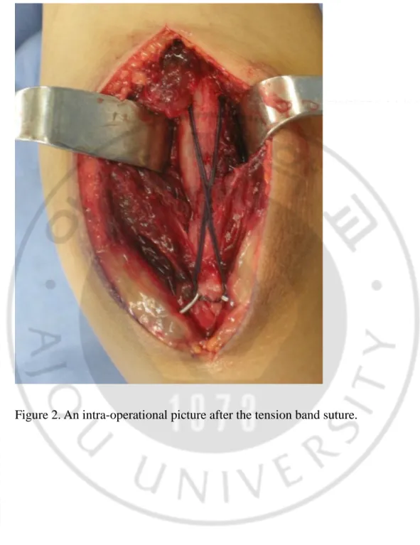
4 were made on the proximal ulna at the same level. The holes were at a sufficient distance from the fracture site and approximately 4 mm anterior of the posterior cortex of the ulna. The holes were made using a 2.5 mm-diameter drill bit medially and laterally. (Fig 1-b) The two arms of a towel clamp were placed and clamped to create a canal through these two holes. Vicryl #1 (Ethicon, Juarez, Mexico) was passed through the canal and wound around the two K-wires creating a figure-of-eight. Hands-tie knots were performed to keep maximal tension, and a knot was placed in the middle of the medial proximal part. (Fig 1-c) Another suture was placed using the same method, but the knot was placed laterally. (Fig 1-d) The K-wires were cut and bent laterally and rotated proximally. (Fig. 2) ROM and stability of the fixation during motion were checked via C-arm fluoroscopy. (Fig. 3) All surgeries were performed by single surgeon Jae ho Cho (JH Cho).
Patients visited the outpatient clinic at 1, 2, 4, 8, and 12 weeks, and then at 6 and 12 months postoperatively. Simple radiographs (elbow anteroposterior, lateral, and both oblique), ROM, degree of pain, and discomfort were evaluated at each visit; the Mayo Elbow Performance Score (MEPS) was evaluated at 8 and 12 weeks. ROM exercises were initiated 1 week postoperatively with a customized functional brace (Fig. 4), and continued for 8 weeks, unless the patient had a combined fracture in the
5
ipsilateral elbow. The brace has hinges with ROM limitation set between-10°
and 110° in 10° increments. It also has a handle at the end to facilitate ROM exercise and limit forearm rotation. Customized braces were used instead of pre-made ones because all of the children were differently sized. Pin removal was performed 12 weeks postoperatively as an outpatient procedure. The second surgery was performed with patients under sedation but without intubation. Incisions measuring less than 5 mm were made on the pin site for the implant removal.
6 Results
Nine boys and 3 girls, with an average age of 10.6 years (5 years 7 months–16 years 2 months) were included in the study. Eight (75%) patients had fractures of the left elbow. The average initial displacement and angulation of the fractures were 5 mm (2–7) and 12° (4–25), respectively. PCCF 21u-M/3 was observed in 11 cases and 21u-M/7.1 in the remaining case. In addition, 8 cases had transverse fractures, while 4 cases had oblique fractures. Two cases had associated radial neck fractures in the ipsilateral elbow, which were managed with closed reduction and percutaneous pinning, and began ROM exercises 2 weeks postoperatively. All patients recovered full ROM of the elbow joint and had perfect MEPS at 8 weeks. In addition, all patients had complete bone union at the 8 weeks’ visit to the outpatient clinic. No complications, including nonunion, loss of reduction, infection, or pin loosening were observed.
The average time of implant removal was 13 weeks. Eight patients had 1-year follow-up after the first surgery, and 4 patients visited the clinic for 6 months. No growth arrest was observed in any of the patients at the last simple radiography (Table 1).
7 Discussion
Olecranon fractures are uncommon among children. In an algorithm for the treatment of pediatric patients with olecranon fractures, Evans suggested that open reduction and internal fixation were needed for fractures with growth plate separation, fractures with more than 4 mm displacement of the articular surface, or step off defects with less than 4 mm of displacement. Displacement of the articular surface of 2-4 mm is considered a grey zone, and the choice for surgical or conservative treatment depends on biomechanical stability of the patient, which is determined clinically by the surgeon. In this algorithm, the use of TBW or tension band suturing in open reduction and internal fixation is at the surgeon’s discretion.1
Traditional K-wire fixation with TBW is considered the gold standard to treat adult and pediatric patients with transverse olecranon fractures.8 However, a high rate of early metal-wire removal has been reported by many authors, and surgeons have avoided using this technique.5, 9 In particular, Jensen reported a 79.2% removal rate.6 In their study, Evans and Graham first introduced tension band suture with degradable material.1 They reported
similar outcomes without complications as TBW in their study.
Surgeons consider various factors when choosing suture materials for tissue repair including the diameter, type of strand, strength, and
bio-8 absorbability of the material appropriate for specific tissues and locations. Vicryl is a commonly used suture material that is made by copolymerization of lactide and glycolide. It has a retention strength of 75% at 2 weeks, 50% at 3 weeks, and 25% at 4 weeks in vivo. Vicryl absorption is essentially completed between 56 and 70 days. It is also a braided material, and a more secure knot can be made with it than with monofilament material. Furthermore, it has the advantage that it is easier to handle than hard wire. Vicryl could replace stainless steel wire if it is of sufficient strength.
Patients with olecranon fractures treated with TBW start ROM exercise approximately 1 week after surgery. Previous studies pertaining to the tension band suture technique started ROM exercise at 4 weeks after the surgery with cast the removed, which is slower than TBW. In particular, one previous study performed in vitro cyclic loading tests of 18-gauge surgical stainless steel wire and #2 Vicryl as tension bands in patients with olecranon fractures.11 After low- and high-cyclic loads, the failure loads for steel (301 ± 61 N and 268 ± 97 N, respectively) were greater than for Vicryl (147 ± 45 N and 117 ± 65 N, respectively). However, there was no significant difference between the groups in fracture displacement during low loads (i.e., 1 to 10 N). This implies that passive ROM exercise could be performed without concern of displacement after surgery. All of our patients started ROM exercise 1 week
9 postoperatively with a motion controlled customized functional brace, and none of them had fixation failure. Patients had full ROM without any flexion contracture with perfect MEPS on their last visit.
Gortzak chose Vicryl #2 for tension band suture in pediatric patients with olecranon fractures with good results.4 In our study, double Vicryl #1 loops and knots were used in pediatric patients with displaced olecranon fractures; no fixation failures were observed. Two bundles of Vicryl #1 may have more strength than a single bundle of Vicryl #2, as the total diameter is larger. Najibi studied material properties of different sizes of Vicryl. In their study, the diameter of Vicryl #1 was 0.51 mm, with incremental differences of 0.09 mm with the diameters of Vicryl 0 and 2-0. In this single loading biomechanical test, the maximal load for failure of Vicryl #1 was 130 ± 9 N; this was within the range of Ethibond and Ticron #2.10 Since the diameter of Vicryl #1 is 0.51 mm, two strands can be passed through a hole made by a 2.5 mm-diameter drill bit, as we used in this study. Three strands may also easily pass through the hole. Double knots were used to enhance the stiffness and to prevent the loosening of one knot.13 Two knots were placed in different
directions to avoid individual and cumulative irritation caused during ROM, which lowers the chance of loosening.
10 As pediatric patients with olecranon fractures are uncommon, it is challenging to accumulate sufficient number of cases for a case controlled study. We are planning to conduct a multicenter study for the prospective case controlled study. Second, this was a retrospective study, and the follow-up duration was 1 year. All patients had complete bone union with good ROM at their last visit. A biomechanical test on the double loops or triple loops with different number of knots of #1 Vicryl would further support this study.
11 Conclusion
Pediatric patients with olecranon fractures displaced more than 2 mm were treated with tension band suture with double loops and knots. This surgical method allowed for early ROM, preserved growth potential, and simple implant removal. It also resulted in good functional outcomes without complications. The double loops and knots tension band suture could be a complete alternative to TBW with secured early ROM exercise.
12 References
1. Evans MC, Graham K. Olecranon fractures in children: part 1: a clinical review. Part 2: a new classification and management algorithm.
J Pediatr Orthop 1999;19:559–569.
https://doi.org/10.1097/01241398-199909000-00001
2. Fabry J, De Smet L, Fabry G. Consequences of a fracture through a minimally ossified apophysis of the olecranon. J Pediatr Orthop B
2000;9:212–214.
https://doi.org/10.1097/01202412-200006000-00013
3. Gaddy BC, Strecker WB, Schoenecker PL. Surgical treatment of displaced olecranon fractures in children. J Pediatr Orthop
1997;17:321–324.
https://doi.org/10.1097/01241398-199705000-00010
4. Gortzak Y, Mercado E, Atar D, Weasel Y. Pediatric olecranon fractures: open reduction and internal fixation with removable Kirschner wires and absorbable sutures. J Pediatr Orthop 2006;26:39–42. http://dx.doi.org/10.1097/01.bpo.0000187988.86892.a2
5. Johnson RP, Roetker A, Schwab JP. Olecranon fractures treated with AO screw and tension bands. Orthopedics 1986;9:66–68. http://dx.doi.org/10.3928/0147-7447-19860101-11
13 6. Jensen CM, Olsen BB. Drawbacks of traction-absorbing wiring (TAW)
in displaced fractures of the olecranon. Injury 1986;17: 174– 5.http://dx.doi.org/10.1016/0020-1383(86)90326-8
7. Landin LA, Danielsson LG. Elbow fractures in children. An epidemiological analysis of 589 cases. Acta Orthop Scand 1986;57(4):309–312. https://doi.org/10.3109/17453678608994398 8. Molloy S, Jasper LE, Elliott DS. Biomechanical evaluation of
intramedullary nail versus tension band fixation for transverse
olecranon fractures. J Orthop Trauma 2004;18:170–174.
https://doi.org/10.1097/00005131-200403000-00008
9. Morrey BF. Current concepts in the treatment of fractures of the radial head, the olecranon, and the coronoid. J Bone Joint Surg Am
1995;77:316–327.
https://doi.org/10.2106/00004623-199502000-00019
10. Najibi S, Banglmeier R, Matta JM. Material properties of common suture materials in orthopaedic surgery. Iowa Orthop J 2010;30;84–88. 11. Parent S, Wedemeyer M, Mahar AT. Displaced olecranon fractures in children: a biomechanical analysis of fixation methods. J Pediatr
Orthop 2008;28:147–151.
14 12. Romero JV, Miran A, Jensen CH. Complications and re-operation rate after tension-band wiring of olecranon fractures. J Orthop Sci
2000;5:318–320. http://dx.doi.org/10.1007/s007760070036
13. Harrell RM, Tong J, Weinhold PS, Dahners LE. Comparison of the mechanical properties of different tension band materials and suture
techniques. J Orthop Trauma 2003; 17(2):119–122.
15 Figures
Figure 1. a) Fracture site was reduced using reduction clamp and 2 K-wires were inserted. b) Two holes for tension band canal was made with 2.5mm drill bit. c, d) Vicryl was passed through canal to make a tension suture in figure of eight technique. One knot was made on lateral side and the other knot was made on medial side.
16
17 Figure 3. Fixation stability and range of motion were confirmed under fluoroscope in operation room.
Figure 4. Customized functrional brace used 1 week after the surgery. Degree of ROM was controlled with hinge at the elbow joint level.
18 Table
TABLE 1. Demographic Data
Num ber Ag e(y rs) S e x S i d e Status of physis AO PCC F Fractur e Pattern Fra ctur e gap( mm ) Asso ciate d fract ure Follow up period (month ) Compl ication s 1 5.7 M R t Before ossificat ion 21u-M/3.1 Oblique 2 No 6 No 2 6.1 M R t Before ossificat ion 21u-M/3.1 Transve rse 4 No 12 No 3 8.2 M L t Open physis 21u-M/3.1 Transve rse 3 Radi us neck 12 No 4 8.8 F L t Open physis 21u-M/3.1 Oblique 5 No 12 No 5 10. 5 F L t Open physis 21u-M/3.1 Transve rse 5 Radi us neck 6 No 6 11. 9 M L t Open physis 21u-M/3.1 Transve rse 4 No 12 No 7 12. 9 M R t Open physis 21u-M/3.1 Oblique 6 No 12 No 8 13. 1 M L t Open physis 21u-M/3.1 Transve rse 6 No 12 No 9 14. 1 M L t Open physis 21u-M/3.1 Transve rse 7 No 12 No 10 14. 2 M L t Near end of fusion 21u-M/7.1 Transve rse 7 No 12 No 11 15. F R Near end of 21u-Oblique 4 No 6 No
19 2 t fusion M/3.1 12 15. 3 M R t Near end of fusion 21u-M/3.1 Transve rse 7 No 6 No
M: male; F: female; R: right; L: left; PCCF: Pediatric Classification of Long Bone Fractures
Table 1