Urological Oncology
Meta-Analysis of Studies Analyzing the Role of Human Papillomavirus in the Development of Bladder Carcinoma
Antonio Jimenez-Pacheco, Manuela Exposito-Ruiz1, Miguel A Arrabal-Polo2, Alfonso J Lopez-Luque
Department of Urology, Santa Ana Hospital, 1Department of Statistic, FIBAO, Virgen de las Nieves University Hospital, 2Department of Urology, San Cecilio University Hospital, Granada, Spain
Purpose: We aimed to ascertain the degree of association between bladder cancer and human papillomavirus (HPV) infection.
Materials and Methods: We performed a meta-analysis of observational studies with cases and controls with publication dates up to January 2011. The PubMed electronic database was searched by using the key words “bladder cancer and virus.” Twenty-one articles were selected that met the required methodological criteria. We implemented an internal quality control system to verify the selected search method. We analyzed the pooled effect of all the studies and also analyzed the techniques used as follows:
1) studies with DNA-based techniques, among which we found studies with polymerase chain reaction (PCR)-based techniques and 2) studies with non-PCR-based techniques, and studies with non-DNA-based techniques.
Results: Taking into account the 21 studies that were included in the meta-analysis, we obtained a heterogeneity chi-squared value of Qexp=26.45 (p=0.383). The pooled odds ratio (OR) was 2.13 (95% confidence interval [CI], 1.54 to 2.95), which points to a sig- nificant effect between HPV and bladder cancer. Twenty studies assessed the presence of DNA. The overall effect showed a significant relationship between virus presence and bladder cancer, with a pooled OR of 2.19 (95% CI, 1.40 to 3.43). Of the other six studies, four examined the virus’s capsid antigen and two detected antibodies in serum by Western blot. The estimated pooled OR in this group was 2.11 (95% CI, 1.27 to 3.51), which confirmed the relationship between the presence of virus and cancer.
Conclusions: The pooled OR value showed a moderate relationship between viral in- fection and bladder tumors.
Key Words: Human papillomavirus; Infection; Meta-analysis; Urinary bladder neoplasms;
Viruses
This is an Open Access article distributed under the terms of the Creative Commons Attribution Non-Commercial License (http://creativecommons.org/licenses/by-nc/3.0) which permits unrestricted non-commercial use, distribution, and reproduction in any medium, provided the original work is properly cited.
Article History:
received 23 November, 2011 accepted 28 December, 2011
Corresponding Author:
Antonio Jimenez-Pacheco Department of Urology, Santa Ana Hospital, Calle Párroco José Rodríguez no 65, Bloque A-1, 3oB, Granada 18014, Spain
TEL: +34-958154355 FAX: +34-958023084 E-mail: [email protected]
INTRODUCTION
Bladder carcinoma is the most common of the malignant neoplasias of the urinary tract and is characterized by wide variability in prognosis. Carcinomas of transitional cells account for over 90% of bladder tumors [1]. The majority of patients tend to relapse, but a group of patients progress toward muscle-invasive disease [2]. To date, the mecha- nisms associated with the initiation and progression of these tumors, along with possible risk factors, are not well
known [3].
Many studies have tried to assess the carcinogenic risk of viruses such as human papillomavirus (HPV), only to find conflicting results. The papillomaviruses are a group of small, double-stranded DNA viruses that infect the stratified epithelium of the skin and mucosa [4]. Over 120 different types of HPV have been described, only some of which implant their DNA into the host cell’s genome. HPVs have been classified as “low risk” (types 6 and 11) and “high risk” (types 16 and 18) on the basis of their implantation
FIG. 1. Meta-analysis of all included studies.
properties and their tendencies to associate with benign processes or invasive carcinomas [1].
It has been shown that infection with HPV is implicated in the pathogenesis of several intraepithelial lesions and cancers, such as anal, cervical, and oral cancer [5].
However, the association of HPV with the development of tumors of the urinary tract continues to be controversial, given that in most cases, the evaluation of the results was difficult because appropriate control groups were lacking [4].
In relation to bladder carcinoma, until now, few studies have found an association between HPV infection and an increased risk for the development of neoplasias. This pos- sible relationship is based on the epithelial tropism of HPV and the anatomical proximity of the urethra (which is con- sidered a reservoir for the virus) and the bladder [1,6].
For this reason, it is open for debate whether the associa- tion between chronic HPV infection and bladder carcinoma is a coincidence or whether the relationship is causal. To address this, our study identified and analyzed interna- tionally published studies (published before January 2011) that analyzed the relationship between bladder car- cinoma and HPV and that described defined materials and methods. We conducted a meta-analysis of all of the results presented in these studies to analyze the currently avail- able data on the relationship between this virus and blad- der cancer.
MATERIALS AND METHODS
We conducted a search of the PubMed database by using the keywords “bladder cancer and virus.” From this search, we identified 763 studies published before January 2011.
We then selected a total of 22 studies [4,7-27] that had well-established groups of cases and controls; that were published in English, French, or Spanish; and that ana- lyzed the relationship between HPV and bladder cancer.
Of these studies, two were eliminated, in which the number of positive cases was zero in both studied groups (which would not allow for the calculation of any effect measure- ment between virus presence and bladder cancer) [26,27].
Additionally, we added results associating HPV and blad- der carcinoma that were found in a doctoral thesis [28].
We conducted an internal quality check system to verify the selected search method, which consisted of tracing the bibliography of each one of the selected studies. Using this method, we were able to confirm that no study was omitted.
Given the diversity of the studies, before the meta-analy- sis, the studies were classified on the basis of the laboratory test used.
The heterogeneity of the studies was compared by using the Cochran chi-squared test (heterogeneity chi-squared- Qexp) and the consistency index (I2). The odds ratio (OR) was calculated for each study with a 95% confidence interval (95% CI). The mean of the pooled OR was calculated by us- ing the random effects model by the DerSimonian-Laird method. We considered the effect significant when the CI for the OR did not contain the value 1.
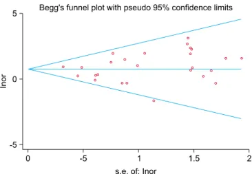
FIG. 2. Funnel Plot of all included studies.
Publication bias was examined by using Begg’s and Egger’s tests.
A forest plot was used to graphically represent the esti- mated ORs of all studies, including the pooled effects. The analyses were conducted by using Metadisc software (Informer Technologies Inc.).
RESULTS
We analyzed the pooled effect of all the studies and also ana- lyzed the techniques used as follows: 1) studies with DNA-based techniques, among which we found studies with polymerase chain reaction (PCR)-based techniques and studies with non-PCR- based techniques, and 2) stud- ies with non-DNA-based techniques.
1. Pooled analysis of all studies
Taking into account the 21 studies included in the pooled meta-analysis, we obtained a heterogeneity chi-squared value of Qexp=26.45, with 25 degrees of freedom (p=0.383), which allowed us to confirm that the studies were suffi- ciently homogeneous compared with each other. Additionally, the inconsistency index I2 for the 21 studies had a value of 5.7%, which confirmed that the percentage of hetero- geneity was very low. The pooled OR was 2.13 (95% CI, 1.54 to 2.95), which demonstrated a significant effect between HPV presence and bladder cancer (Fig. 1).
The result of the Begg’s test was not significant (p=
0.508), allowing us to conclude that there were no indica- tions of publication bias, which was confirmed graphically by the Funnel Plot (Fig. 2).
2. Studies that used DNA-based techniques
Table 1 lists the authors, the total numbers of cases and controls with the numbers of positives found in both groups, and the methods used. In the cases, 79.4% were transitional cell bladder tumors; in the controls, the sam- ples were predominantly healthy bladder tissue (63.7%).
PCR was used in the majority of the cases (80%). The ampli-
fied regions varied and tended to specify the detected viral genotype (the majority were type 16 or 18). Only three sam- ples (15%) used frozen tissue biopsies.
The prevalence of HPV infection in the cases ranged from 3.12 to 100%. In the controls, the prevalence ranged from 0 to 100%.
We included a total of 20 studies that used DNA-based techniques for determination of infection. These studies met the hypothesis of homogeneity, with a Qexp=21.17 with 19 degrees of freedom (p=0.3277) and an I2=10.2%, which allowed us to assume comparability between the studies.
The pooled effect showed a significant relationship be- tween viral presence and bladder cancer, with a pooled OR of 2.19 (95% CI, 1.40 to 3.43). Fig. 3 shows the OR and 95%
CI values for each of the studies.
There was no evidence of publication bias by Begg’s test (p=1).
1) Studies that used PCR-based techniques: The 16 stud- ies that used PCR-based techniques also met the homoge- neity criteria with a Qexp=16.46, with 15 degrees of freedom (p=0.352) and an I2=8.9%. The pooled OR also confirmed a significant relationship between viral presence and blad- der cancer, with a pooled OR of 2.40 (95% CI, 1.51 to 3.81) (Fig. 4).
Begg’s test did not show evidence of publication bias for this subgroup (p=0.558).
2) Studies that used non-PCR-based techniques: The subgroup of studies that used non-PCR-based techniques was significantly smaller than the others, including only four studies. Despite the low number, this group also met the homogeneity hypothesis with a Qexp=2.78, with 3 de- grees of freedom (p=0.426) and an I2=0.0%. In this sub- group, the pooled OR was 0.84 (95% CI, 0.21 to 3.31). Stati- stical significance was not reached, which was probably re- lated to the small number of studies included (Fig. 5).
This subgroup also did not show evidence of publication bias by Begg’s test (p=0.734).
3. Studies that used non-DNA-based techniques
Table 2 lists the authors, the total numbers of cases and controls with the numbers of positives found in both groups, the materials used for the studies (carcinoma biop- sies of transitional cells in 93.4% of the cases and samples of healthy bladder tissue in 42.5% of controls), and the methods used (four studies assessed the viral capsid anti- gen, and two evaluated serum antibodies by use of Western blot).
The prevalence of HPV infection in these cases ranged from 17.3 to 88.8%. In the controls, the prevalence ranged from 0 to 66.6%.
Despite the fact that the subgroup of studies using non-DNA-based techniques was small, these six studies could be considered homogeneous with a value of Qexp=5.39, with 5 degrees of freedom (p=0.370) and a low I2 of 7.3%.
Although the number of studies was not very large, the esti- mated pooled OR was statistically significant at 2.11 (95%
CI, 1.27 to 3.51), which confirms the relationship between
TABLE 1. Characteristics of human papillomavirus DNA-based studies included in the meta-analysis AuthorNo. of positive casesTotal casesNo. of positive controlsTotal controlsTissueAnalysisStudied genotype (genome region) Aglianò et al. [7] (1994)2346 (TCC) 010 (healthy bladder)FixedPCR + Dot blot H16,18 (not indicated) Anwar et al. [8] (1992)3948 (46 TCC, 2 SCC) 721 (healthy bladder)FixedPCR, Dot blot H+SB-H6, 11, 16, 18, 33 (E6) Badawi et al. [9] (2008)1120 (17 TCC, 3 SCC) 34 (cystitis)Not indicatedPCR16, 18 (E6) Barghi et al. [10] (2005)2159 (TCC) 120 (non-tumoral bladder)FixedPCR6, 11,16, 18, 33, 35 (L1) Bryant et al. [11] (1991)1292 (66 TCCp, 10 TCCs, 3 SSC, 4 Adenoc, 7 C. undifferentiated, 2 TCC d/s)
08 (3 dysplasia, 5 benign tumors)FixedH. in situ6b, 11, 16 y 18 (not indicated) Chan et al.[12] (1997)1320 (TCC) 710 (inverted papilloma)FixedPCR+Dot blot H.6, 11, 16, 18, 31 y 33 (E6) Fioriti et al. [13] (2003) 132 (not indicated) 020 (healthy bladder)Not indicatedH. in situ16, 18, 33 (not indicated) Gazzaniga et al. [14] (1998)1635 (TCC) 010 (healthy bladder)FrozenPCR+Dot blot H.16,18 (not indicated) Gould et al. [4] (2010) 25 (TCC) 428 (4 healthy bladder, 6 cystitis, 18 inverted papilloma)FixedMPG+H. mixed6, 11, 16, 18, 31, 33, 35, 39, 42, 43, 44, 45,51, 52, 56, 58, 59, 66, 68, 70, 73, 82 Guo et al. [15] (2006) 19 (CIS) 118 (13 non-tumoral bladder tissue, 5 healthy bladder)Not indicatedH. in situ6, 11, 16, 18, 31, 33, 45, 51 Kerley et al. [16] (1991) 122 (18 TCC, 3 SCC, 1 Adenoc.) 05 (healthy bladder)FixedH. in situ+PCR selective6, 11, 16, 18, 31, 33, 35 (not indicated) LaRue et al. [17] (1995)2871 (not indicated) 08 (healthy bladder)FrozenPCR+H. in situ6, 11, 16, 18, 31, 33, 35 (not indicated) Ludwing et al. [18] (1996) 623 (21 TCC, 1 SCC, 1 Adenoc.) 341 (32 Healthy bladder, 9 chronic cystitis)Not indicatedPCR+RFLP6b, 11, 16 y 18 (L1) Noel et al. [20] (1994) 22 (TCC) 02 (healthy bladder)Not indicatedSelective PCR6b, 11, 16 y 18 (E7) Oft et al. [21] (1993) 11 (TCC) 02 (healthy bladder)FrozenDifferential PCR; H. in situ P32 ARNm of region E7 6 (L1, E6) Shibutani et al. [22] (1992) 421 (20 TCC, 1 SCC) 22 (1 dysplasia, 1 inflamed tissue)Not indicatedDifferential PCR; H. in situ P32 ARNm of region E7
Not indicated Smetana et al. [23] (1995)2059 (TCC) 041 (non-tumoral bladder tissue)FixedSB-H6/11, 16/18 (E1) Tekin et al. [24] (1999) 242 (TCC) 010 (healthy bladder)Not indicatedH. in situ+PCR16 y 18 (L1) Yu et al. [25] (1993)3053 (TCC) 020 (healthy bladder)Not indicatedPCR16, 18 (E6, E7) Pareja [28] (2008)1234 (31 TCC, 3 SCC)1451 (healthy bladder)FixedPCR35 genotypes (L1) TCC, transitional cell carcinoma; PCR, polymerase chain reaction; SCC, squamous cell carcinoma; Adenoc., adenocarcinoma; SB-H, Southern blot H; H, hybridization; RFLP, restriction fragment length polymorphism; MPG, multiplex HPV genotyping; d/s, deep/superficial; CIS, squamous cell carcinoma in situ.
FIG. 4. Meta-analysis of studies that used PCR-based techniques.
FIG. 5. Meta-analysis of studies that used non-PCR-based techniques.
FIG. 3. Meta-analysis of the studies that used DNA-based techniques.
TABLE 2. Characteristics of studies based on viral antigen and antibody detection included in the meta-analysis Author
No. of positive
cases Total cases
No. of positive
controls Total controls Tissue Analysis Studied genotype (genome region) Badawi et al.
[9] (2008) 7 20 (17 TCC, 3
SCC) 6 24 (cystitis) Serum ELISA, AC anti-capsid L1
VHP 16 y 52 16, 52 (not indicated) Gould et al. [4]
(2010)
2 5 (TCC) 15 28 (4 healthy bladder, 6 cystitis, 18 Inverted papilloma)
Fixed Overexpression of P16 + AC anti-P16
AC anti-P16
Guo et al. [15]
(2006)
8 9 (CIS) 12 18 (13 non-tumoral bladder tissue, 5 healthy bladder)
Serum Immunohistochemistry of EGFR + AC anti-EGFR Ludwig et al.
[18] (1996)
5 23 (21 TCC, 1 SCC, 1 Adenoc.)
2 41 (9 chronic cystitis, 32 healthy bladder)
Serum Western blot Ag types 6b (L1, L2), 16 (L2, E4, E7), 18 (L2, E7)
Mantovani et al. [19]
(1994)
27 45 (TCC) 28 121 (75 healthy bladder, 46 non-neoplastic bladder tissue)
Serum ELISA and Western blot Ag genus
Smetana et al.
[23] (1995)
19 110 (TCC) 0 41 (non-neoplastic bladder tissue)
Fixed IPO + AC anti-capsid
TCC, transitional cell carcinoma; SCC, squamous cell carcinoma; Adenoc., adenocarcinoma; IPO, immunoperoxidase; AC, antibody;
Ag, antigen; ELISA, enzyme-linked immunosorbent assay; EGFR, immunohistochemistry for epithelial growth factor receptor; CIS, squamous cell carcinoma in situ.
FIG. 6. Meta-analysis of studies that used non-DNA-based techniques.
viral presence and cancer in this group (Fig. 6).
This subgroup did not show evidence of publication bias by Begg’s test (p=0.707).
DISCUSSION
Bladder transitional cell carcinoma is one of the most com- mon neoplasias in developed countries [10]. It is a clinical entity of which certain aspects are unknown, such as the existence of a genetic factor that determines its predis- position, despite the existence of various involved onco- genes, or the identity of the main risk factor involved in its genesis. These unknowns prevent us from implementing an effective prevention campaign. In this study, we eval- uated HPV, which has been mentioned in the literature as a possible etiological agent of genital tumors, although its exact effect on bladder carcinoma is still vague, and numer- ous hypotheses exist regarding its role in this regard.
The integration of HPV frequently occurs in the vicinity of known proto-oncogenes in human epithelial cells, al- though it is still unknown whether this occurrence favors the progression of pre-cancerous lesions towards cancer.
The viral circular double-stranded DNA must be broken for the oncogenic phenomenon of integration to take place. It has been shown that this breakage is specifically produced in the viral genome region encoding the E2 gene, which then loses its functional capacity. In the most oncogenic HPVs (types 16 and 18), the E2 gene seems to act mainly as a repressor of the transcription of genes E6 and E7, whose fundamental activities are to promote cellular pro- liferation and transformation by producing centrosome amplification and chromosomal delay (i.e., lag) during mi- tosis, which leads to chromosomal instability [10]. This ge- nomic instability is thought to be an essential part of the conversion of a normal cell to a cancerous one.
The HPV E6 and E7 genes, especially in high-risk HPVs
(types 16 and 18), acquire their transforming activities as a result of encoding E6 and E7 oncoproteins, which specifi- cally interact with proteins regulating cell proliferation. It has been shown that oncoprotein E7 interacts with the cel- lular protein p105 Rb, which is encoded by the retino- blastoma gene Rb1. The interaction of the HPV E7 oncopro- tein with the regulator protein p105-Rb presumably in- activates p105-Rb, with similar consequences as those that occur in tumors encoding retinoblastoma gene alterations.
In contrast, the association of E6 with p53 may give rise to an effect similar to what happens after mutation of the p53 gene. It is thought that this constitutes one of the oncogenic pathways of HPV [29].
Some authors have reported that the sensitivity of HPV detection greatly depends on several technical factors, such as tissue fixation, DNA preparation, and amplifica- tion conditions [19].
Over the years, microbiological tests used for the de- tection of infection have changed. PCR and in situ hybrid- ization are techniques with high sensitivities and specific- ities for viral genome detection [1]; these techniques have been used most commonly in studies. This, however, should not detract from the value of other techniques for which specificities and sensitivities have been reported.
Li et al. [30] presented the outcomes of a meta-analysis of articles published from January 1989 until August 2010 in which they showed an association between HPV and bladder cancer. The authors discussed important issues that were not analyzed in previous meta-analyses, such as the geographic variation in risk of bladder cancer with HPV infection; the most common HPV types identified, which are similar to those identified in other tumors, mainly cer- vical tumors; and the sensitivity of detection of infection as a function of the method used.
In the present meta-analysis, in addition to the techni- ques based on DNA detection (PCR-based methods and non-PCR-methods) noted by Li et al. [30], we included the analysis of seven studies that used virus detection techni- ques that were not based on DNA (i.e., the detection of viral capsid antigen and antibodies in serum by Western blot- ting). In this case, the joint point estimate of the prevalence of infection through the detection of antigen or antibody was 32.4%.
We can observe that the association between HPV and bladder cancer varies with geographical location. Elevated percentages of HPV detection have been documented in bladder tumors in Southeast Asia, reaching up to a re- ported 81% in Hong Kong [31]. Lower percentages have been detected in Europe and North America, not exceeding 39% [1]. It is possible that these differences are related to problems in diagnostic techniques, such as not using a wide enough range of specific primers for PCR, or having sam- ples containing DNA from less-common types of HPV, lead- ing to false-negatives [1].
Another issue to consider is that of justifying the associa- tion, given that the virus could be present in a lesion before it appears as an inductor to be acquired during sexual
intercourse. This does not rule out that the tumor may be secondarily colonized as part of the mucosal microbiota [6].
In our study, the Qexp values obtained from the analysis of studies using non-DNA-based and DNA-based techni- ques, viral genome PCR detection methods, and non-PCR detection methods showed homogeneity of data and a small dispersion of results. Of the 21 selected studies, we ob- tained a pooled OR of 2.13, allowing us to conclude that there is a clear and moderate association between virus ex- posure and the presence of bladder cancer.
CONCLUSIONS
As of January 2011, there were only 21 studies with both case and control groups that analyzed the relationship be- tween HPV and bladder cancer. The methods used varied, but PCR use predominated. The studies demonstrated the presence of infection in the majority of cases, although an important dispersion was observed. The pooled OR value showed a moderate relationship between infection by the virus and the tumor. There is a lack in the literature of a sufficiently large number of cases and samples that are compared with controls and that use a combination of mi- crobiological techniques in the same subjects and samples to obtain a definitive answer to this question. For this rea- son, the relationship between the virus and this tumor should continue to be evaluated through pathogenic stud- ies of the disease.
CONFLICTS OF INTEREST
The authors have nothing to disclose.
REFERENCES
1. Ben Selma W, Ziadi S, Ben Gacem R, Amara K, Ksiaa F, Hachana M, et al. Investigation of human papillomavirus in bladder cancer in a series of Tunisian patients. Pathol Res Pract 2010;206:740-3.
2. Moonen PM, Bakkers JM, Kiemeney LA, Schalken JA, Melchers WJ, Witjes JA. Human papilloma virus DNA and p53 mutation analysis on bladder washes in relation to clinical outcome of blad- der cancer. Eur Urol 2007;52:464-8.
3. Erill N, Colomer A, Verdú M, Román R, Condom E, Hannaoui N, et al. Genetic and immunophenotype analyses of TP53 in bladder cancer: TP53 alterations are associated with tumor progression.
Diagn Mol Pathol 2004;13:217-23.
4. Gould VE, Schmitt M, Vinokurova S, Reddy VB, Bitterman P, Alonso A, et al. Human papillomavirus and p16 expression in in- verted papillomas of the urinary bladder. Cancer Lett 2010;
292:171-5.
5. Spano JP, Marcelin AG, Carcelin G. HPV and cancer. Bull Cancer 2005;92:59-64.
6. Gutiérrez J, Jiménez A, de Dios Luna J, Soto MJ, Sorlózano A.
Meta-analysis of studies analyzing the relationship between bladder cancer and infection by human papillomavirus. J Urol 2006;176(6 Pt 1):2474-81.
7. Aglianò AM, Gradilone A, Gazzaniga P, Napolitano M, Vercillo R, Albonici L, et al. High frequency of human papillomavirus de- tection in urinary bladder cancer. Urol Int 1994;53:125-9.
8. Anwar K, Naiki H, Nakakuki K, Inuzuka M. High frequency of
human papillomavirus infection in carcinoma of the urinary bladder. Cancer 1992;70:1967-73.
9. Badawi H, Ahmed H, Ismail A, Diab M, Moubarak M, Badawy A, et al. Role of human papillomavirus types 16, 18, and 52 in re- current cystitis and urinary bladder cancer among Egyptian patients. Medscape J Med 2008;10:232.
10. Barghi MR, Hajimohammadmehdiarbab A, Moghaddam SM, Kazemi B. Correlation between human papillomavirus infection and bladder transitional cell carcinoma. BMC Infect Dis 2005;5:102.
11. Bryant P, Davies P, Wilson D. Detection of human papillomavirus DNA in cancer of the urinary bladder by in situ hybridisation. Br J Urol 1991;68:49-52.
12. Chan KW, Wong KY, Srivastava G. Prevalence of six types of hu- man papillomavirus in inverted papilloma and papillary transi- tional cell carcinoma of the bladder: an evaluation by polymerase chain reaction. J Clin Pathol 1997;50:1018-21.
13. Fioriti D, Pietropaolo V, Dal Forno S, Laurenti C, Chiarini F, Degener AM. Urothelial bladder carcinoma and viral infections:
different association with human polyomaviruses and papill- omaviruses. Int J Immunopathol Pharmacol 2003;16:283-8.
14. Gazzaniga P, Vercillo R, Gradilone A, Silvestri I, Gandini O, Napolitano M, et al. Prevalence of papillomavirus, Epstein-Barr virus, cytomegalovirus, and herpes simplex virus type 2 in uri- nary bladder cancer. J Med Virol 1998;55:262-7.
15. Guo CC, Fine SW, Epstein JI. Noninvasive squamous lesions in the urinary bladder: a clinicopathologic analysis of 29 cases. Am J Surg Pathol 2006;30:883-91.
16. Kerley SW, Persons DL, Fishback JL. Human papillomavirus and carcinoma of the urinary bladder. Mod Pathol 1991;4:316-9.
17. LaRue H, Simoneau M, Fradet Y. Human papillomavirus in tran- sitional cell carcinoma of the urinary bladder. Clin Cancer Res 1995;1:435-40.
18. Ludwig M, Köchel HG, Fischer C, Ringert RH, Weidner W.
Human papillomavirus in tissue of bladder and bladder carcino- ma specimens. A preliminary study. Eur Urol 1996;30:96-102.
19. Mantovani G, Cermelli C, Malagoli M, Provvisionato A, Ferrari P, Castagnetti M, et al. IgG antibodies to papillomavirus ge- nus-antigens in adult men with cancer of the urinary bladder.
New Microbiol 1994;17:1-8.
20. Noel JC, Peny MO, Mat O, Antoine M, Firket C, Detremmerie O,
et al. Human papillomavirus type 16 associated with multifocal transitional cell carcinomas of the bladder in two transplanted patients. Transpl Int 1994;7:340-3.
21. Oft M, Böhm S, Wilczynski SP, Iftner T. Expression of the differ- ent viral mRNAs of human papilloma virus 6 in a squamous-cell carcinoma of the bladder and the cervix. Int J Cancer 1993;
53:924-31.
22. Shibutani YF, Schoenberg MP, Carpiniello VL, Malloy TR.
Human papillomavirus associated with bladder cancer. Urology 1992;40:15-7.
23. Smetana Z, Keller T, Leventon-Kriss S, Huszar M, Lindner A, Mitrani-Rosenbaum S, et al. Presence of human papilloma virus in transitional cell carcinoma in Jewish population in Israel. Cell Mol Biol (Noisy-le-grand) 1995;41:1017-23.
24. Tekin MI, Tuncer S, Aki FT, Bilen CY, Aygün C, Ozen H. Human papillomavirus associated with bladder carcinoma? Analysis by polymerase chain reaction. Int J Urol 1999;6:184-6.
25. Yu ST, Wu MM, Li LM. Prevalence of human papillomaviruses 16 and 18 in transitional cell carcinoma of bladder. Chin Med J (Engl) 1993;106:494-6.
26. Yavuzer D, Karadayi N, Salepci T, Baloglu H, Bilici A, Sakirahmet D. Role of human papillomavirus in the development of urothelial carcinoma. Med Oncol 2011;28:919-23.
27. Knowles MA. Human papillomavirus sequences are not detect- able by Southern blotting or general primer-mediated polymer- ase chain reaction in transitional cell tumours of the bladder. Urol Res 1992;20:297-301.
28. Pareja Vilchez M. Detection of the human papillomavirus in sam- ples of bladder cancer by a DNA hybridization test. Clinical Relations [tesis doctoral]. Granada: Granada Univ.; 2008.
29. Scheffner M, Werness BA, Huibregtse JM, Levine AJ, Howley PM. The E6 oncoprotein encoded by human papillomavirus types 16 and 18 promotes the degradation of p53. Cell 1990;63:1129-36.
30. Li N, Yang L, Zhang Y, Zhao P, Zheng T, Dai M. Human papil- lomavirus infection and bladder cancer risk: a meta-analysis. J Infect Dis 2011;204:217-23.
31. Furihata M, Inoue K, Ohtsuki Y, Hashimoto H, Terao N, Fujita Y. High-risk human papillomavirus infections and over- expression of p53 protein as prognostic indicators in transitional cell carcinoma of the urinary bladder. Cancer Res 1993;53:4823-7.