669
Evaluation of Renal Microvasculature Using Micro-computed Tomography in Rat
Seung Hyun You, Kwon Ha Yoon1, Ill Young Seo
From the Department of Urology, Wonkwang University School of Medicine,
1Department of Radiology, Wonkwang University School of Medicine and the Institute for Radiological Imaging Science, Iksan, Korea
Purpose: Rodent models that mimic human renal diseases are being increasingly recognized as powerful tools in the development of new drugs and for evaluating the efficacy of novel therapeutics in a preclinical setting. However, there are few reports on microvasculature imaging of the urinary system in small animals. An experimental study was per- formed to evaluate the microvasculature in a rat kidney using micro- computed tomography (CT) with three-dimensional images.
Materials and Methods: Five Sprague-Dawley male rats (age: 10-12 weeks, weight: 200-250g) underwent a laparotomy under anesthesia with an intra- muscular injection of 0.5cc xylazine hydrochloride and ketamine mixed solution (1:10). After ligation of the abdominal aorta and inferior vena cava immediately above the renal artery, a 24 gazed catheter was inserted into the abdominal aorta. A physiological solution and heparin (500U) were infused through the catheter to flush the blood from the renal vasculature.
The kidney was enhanced using self-made contrast material. The excised kidney was frozen for the micro-CT scan.
Results: The mean longitudinal diameter and weight of the 10 resected kidneys was 1.95±0.15cm and 2.0±0.28g, respectively. The images were represented by three-dimensional arrays of cubic voxels with opacities in the blood vessels. In the section taken from the arrays, four regions of the kidney could be identified easily by their characteristic vascular features.
Conclusions: Micro-CT is a promising method for evaluating the renal microvascular architecture in a rat kidney. It can for the foundation of an experimental study aimed at providing quantitative information on the urinary system in a rodent model. (Korean J Urol 2008;49:669-674)
Key Words: Kidney; Imaging, three-dimensional; Microcirculation; Vas- culature
대한비뇨기과학회지 제 49 권 제 8 호 2008
원광대학교 의과대학 비뇨기과학교실,
1영상의학교실 및
익산방사선영상과학연구소
유승현ㆍ윤권하1ㆍ서일영
접수일자:2008년 4월 2일 채택일자:2008년 7월 1일
교신저자: 서일영
원광대학병원 비뇨기과 전북 익산시 신용동 344-2
570-711
TEL: 063-859-1333 FAX: 063-842-1455 E-mail: seraph@
wonkwang.ac.kr 이 논문은 정부 (교육과학기술부)의 재원으로 한국과학재단의 지원을 받아 수행된 연구임 (No. M2070403001-07M0403-00110).
서 론
작은 동물을 이용한 다양한 연구들이 진행되고 있으며, 비뇨생식기에 대한 기초 연구에서도 쥐 등의 설치류를 이 용한 연구들이 많다.1,2 그러나 아직은 작은 동물들에서 해 부학적인 영상정보가 부족한 상황이며, 특히 비뇨생식기 장기에 대한 영상 정보는 매우 적다. 최근에 개발된 영상기 기인 마이크로-computed tomography (CT)는 작은 동물의 뼈
구조를 관찰하기 위해 사용되기 시작하여, 최근에는 종양 모델에서 종양세포의 변화를 관찰하거나 병적 모델에서 약 물에 대한 반응을 관찰하는 등의 미세구조와 기능 간의 연 구에 많은 도움이 되고 있다.3-6 그러나 작은 동물에서 신장 의 미세혈관구조 등의 비뇨생식기 장기에 대한 마이크로- CT 영상은 아직은 초보적인 수준이다.
저자들은 자체연구소에서 개발한 마이크로-CT를 이용하 여 흰쥐 신장의 미세혈관구조를 관찰해 보았고, 이를 3차원 적으로 재조합하여 미세혈관구조에 대한 입체적인 정보를
Table 1. Characteristics of micro-computed tomography X-ray tube
Spatial resolution (MTF) Field of view
Reconstruction image Voxel size
Estimated radiation dose Reconstruction algorithm
40-130kVp, 65μA 11.3cycles/mm 56x59mm
1,024x1,024 pixels, 512 slices 50x50x100μm
84mGy
Filtered Feldkamp back projection7 MTF: modulation transfer function
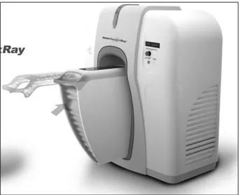
Fig. 1. Photograph of the developed micro-computed tomography system for in vivo small animal imaging.
얻고자 하였다.
대상 및 방법 1. 실험 대상
대한실험동물센터 (대전, 한국)로부터 공급받은 출생 후 10-12주의 Sprague-Dawley 흰쥐 (체중 200-250g)를 대상으로 하였다.
2. 마이크로-CT 시스템
원광대학교 익산방사선영상과학연구소에서 개발한 마이 크로-CT 스캐너 (NFR-Polaris-G90: Nano Focus Ray, 익산, 한 국)를 이용하였다 (Table 1) (Fig. 1). 이 CT는 원추-빔형 (cone- beam type)의 평판형 X-선 영상센서 (flat-panel detector)를 기 반으로 하는 볼륨 CT로써 엑스선 발생장치의 관전압 및 관 전류는 각각 40-130kVp, 그리고 65μA이었다. CT는 한 조사 (projection) 당 1o 씩 회전하여 360o 회전 시 총 360개의 이미
지를 획득하였다. 한 번의 조사에 걸린 시간은 1초로 총 조 사 시간은 6분이 소요되었다. 조사영역 (field of view; FOV) 은 56x59mm이고, 재조합 방법 (reconstruction algorithm)은 Feldkamp back projection 방법을 이용하였다.7 재조합된 영 상은 화상을 구성하는 최소 단위의 화소가 1,024x1,024 pixels이었으며, 512개의 단층 영상을 얻을 수 있었다. 따라 서 입체화상을 구성하는 삼차원영상의 화소인 voxel의 크 기는 약 50x50x100μm로 계산되었다. 재조합된 영상을 삼 차원의 영상으로 보기 위하여 3D-렌더링 소프트웨어 프로 그램 (Lucion: Infinitt Technology, 서울, 한국)을 이용하였다.
3. 혈관조영제
신장의 미세혈관을 조영하기 위해 조영제를 제조하였다.
먼저 4g의 barium sulfate (BaSO4)와 0.5g의 gelatin에 증류수 10ml를 이용하여 40% volume/volume의 BaSO4와 5% weight/
volume의 gelatin의 혼합물을 제조하여 사용하였다. Toyota 등8의 연구에서는 8% weight/volume의 gelatin을 사용하였으 나, gelatin마다 약간의 점도차이가 존재하고 이전의 실험 결과에서 5% 농도의 gelatin에서 가장 좋은 영상을 얻을 수 있어서 저자들의 연구에서는 0.5g의 gelatin을 10ml의 증류 수에 혼합하여 사용하였다.
4. 동물실험방법
Xylazine hydrochloride와 ketamine을 1:10으로 섞은 용액 0.5cc를 허벅지 근육에 주사하여 마취시킨 후 개복하였다.
장을 젖히고 후복막을 박리한 후에 상장간막 동맥과 하장 간막 동맥, 복강동맥을 결찰한 후 장을 절제하여 시야를 확 보하였다. 우측 신동맥 분지부의 직상부 복부대동맥과 하 대정맥을 결찰하고 좌측 신동맥 분지부의 약 1cm 하방의 복부 대동맥에 24게이지의 카테터를 삽관한 후 카테터와 대동맥을 함께 결찰하였다. 또한 주입액이 신장을 거쳐 정 맥으로 흘러나오는 것을 확인하기 위해 대동맥 카테터 삽 관부위에 인접한 하대정맥에 수직으로 약 0.5cm의 절개를 가하였다. 먼저 상온의 생리식염수 10cc에 heparin 500U를 혼합하여 미리 삽관한 카테터를 통해 자동주입펌프를 이용 하여 250mmHg의 압력으로 주입하였다.8 절개한 하대정맥 을 통해 혈액 및 생리식염수가 충분히 빠져나간 것을 확인 하였다. 미리 준비한 조영제 (BaSO4+gelatin+증류수)를 42oC까지 데운 후 삽관된 대동맥 카테터를 통하여 같은 압 력으로 자동주입펌프를 이용하여 주입하였다. 조영제가 신 동맥을 거쳐 신장을 지나 신정맥으로 배출되고 이후 하대 정맥의 절개창으로 용액이 충분히 유출되는 것을 확인한 후 주입을 중지하였다. 이후 신문부 전체를 봉합사 (5-0 silk)로 결찰한 후 신장을 절제하고 약 30초 이내에 40oC로
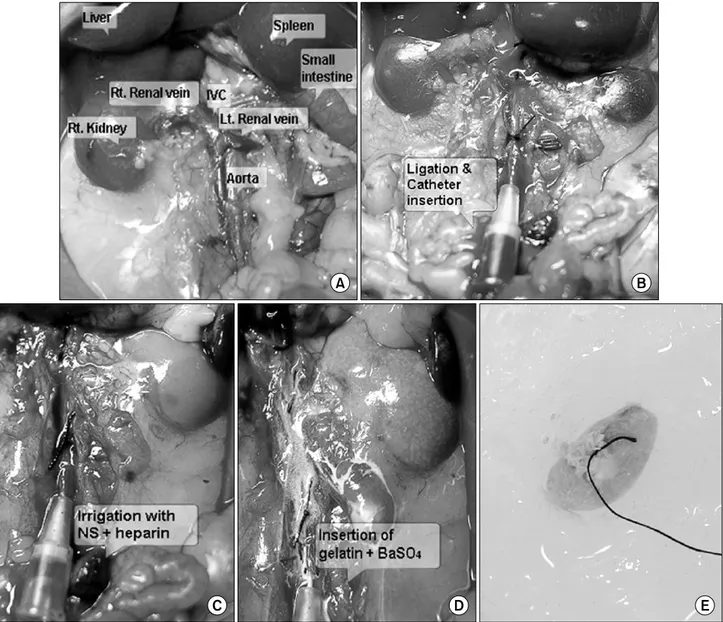
Fig. 2. The preparation steps for micro-computed tomography (CT) scanning. (A) Laparotomy was performed after anesthesia by an intramuscular injection of 0.5cc xylazine hydrochloride and ketamine mixed solution (1:10). (B) After the ligation of the abdominal aorta and inferior vena cava immediately above the renal artery, a 24 gauged catheter was inserted into the abdominal aorta at 1cm below to the renal artery. (C) A physiologic solution (10cc) and heparin (500U) were infused through the catheter to flush the blood from the renal vasculature. (D) The colors of the kidneys were changed because of the fixation with the contrast material, gelatin and barium sulfate (BaSO4). (E) The excised kidney was frozen for the micro-CT scan.
급속 냉동시켰다. 급속 냉동을 함으로써 gelatin을 겔 형태 로 변화시켜 조영제의 입자가 치우침 없이 혈관 내에 고루 분포할 수 있었다. 냉동된 조직을 마이크로-CT를 이용하여 촬영하였다 (Fig. 2).
결 과
실험 과정에서 실패한 경우는 신상부의 대동맥과 대정맥 결찰이 정확하게 이루어지지 않아 충분한 조영이 되지 않
은 경우와 대동맥 안으로 카테터 삽관이 완전히 되지 못한 경우였다. 실험 술기가 완전히 이루어진 총 5마리의 흰쥐에 서 10개의 신장을 절제하여 영상을 얻을 수 있었다. 절제한 흰쥐 신장의 장축의 평균 길이는 1.95±0.15cm, 횡축의 평균 길이는 1.43±0.11cm, 평균 무게는 2.0±0.28g으로 측정되었 다. 신동맥과 정맥은 모든 신장에서 각각 1개씩 관찰되었다.
여러 방향의 단면 영상을 통해 흰쥐의 신동맥에서부터 신분절동맥을 거쳐 엽간동맥, 사구체까지 조영된 미세혈관 구조를 관찰할 수 있었으며, 사구체를 지나 직혈관을 통해
Fig. 4. Renal microvasculature images of the ex-vivo mouse in the micro-computed tomography (CT). (A) projection image, (B) 3 dimensional-CT reconstruction image.
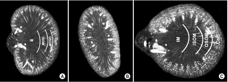
Fig. 3. Coronal (A), sagittal (B), and transverse (C) sections through a volume image of a rat kidney. The cortex (Cort), outer stripe of the outer medulla (OSOM), inner stripe of outer medulla (ISOM), and the inner medulla (IM) were distinguished by their vascular features.
Interlobar vessels (*) are evident in the sections.
조영제가 배출되는 것을 관찰할 수 있었다. 또한, 혈관분포 에 따라 신피질, 바깥수질의 바깥줄무늬층 (outer stripe of outer medulla), 바깥수질의 속줄무늬층 (inner stripe of inner medulla), 속수질층 (inner medulla) 등의 네 구역으로 신단면 을 구분할 수 있었다 (Fig. 3).
이후, 전반적인 신장의 미세혈관구조를 관찰하기 위해 영상을 3차원으로 조합하였고, 그 결과 입체적인 흰쥐 신장 의 미세혈관구조를 관찰할 수 있었다 (Fig. 4).
고 찰
비뇨기과 영역의 기초 실험 과정에서 쥐 등 설치류가 다 양하게 이용되고 있으나 이들에 대한 영상 정보가 부족한
현실이다. 특히, 신장의 미세혈관구조에 대한 정보는 제한 적인 방법에 의해서만 일부 얻을 수 있다.
신장은 기능적인 단위인 100만 개의 신원으로 구성되어 있으며, 적절한 신원의 기능은 미세혈관과 세뇨관의 3차원 적 상호관계에 따라 결정된다.9 기존에는 흰쥐 신장의 미세 혈관구조를 관찰하기 위해서 혈관 내로 india ink나 방사선 비투과성의 조영물질을 주입한 후, 광학 또는 전자 현미경 으로 관찰하는 방법을 사용하거나 미세혈관조영술을 이용
하였다.10-12 그러나 이런 방법으로는 2차원적 관찰만이 가
능하기 때문에, 표층혈관이 심층혈관과 겹쳐져 심층혈관이 가려지거나 서로 연결된 것처럼 관찰되는 등의 여러 단점 이 있었다. 이 단점들을 보완하기 위해서 미세절제술을 사 용하여 신장의 단면을 제작한 후 관찰하였다. 그러나 단면
제작 중 인접 단면이 손상될 수 있고, 다수의 단면을 제작해 야만 하며, 온전한 신장 조직이 단면 제작 과정에서 손상되 어 다른 방향의 연구가 불가능하다는 점 등으로 전반적인 신장의 미세혈관구조를 알기에는 미흡하였다.13
최근 개발된 영상기기인 마이크로-CT는 온전한 신장의 미세혈관구조를 관찰할 수 있고, 이를 재조합하여 3차원의 영상 정보를 얻을 수 있어서 신장의 미세혈관구조 연구에 대한 새로운 이정표가 되고 있다.14 또한, 양성자 가속기 (synchrotron)를 이용하면 고농도로 집적된 X-선을 이용할 수 있어 말초혈관보다 작은 크기의 고해상도 영상도 얻을 수 있고 수입세동맥, 수출세동맥 등의 신원의 구성 요소 하 나하나의 구별까지도 가능하다.15 이 때, 조영물질을 세뇨관 과 혈관에 주사하면, 신장의 기능적 단위인 신원에서 세뇨 관과 주위의 세동맥들과의 복잡한 상호관계들을 가시화할 수 있다.13 따라서 더 많은 연구와 마이크로-CT 개발이 이루 어지면, 작은 동물에서 신기능 이상의 원인, 정도 등과 미세 혈관 구조변화의 관계를 영상학적 방법을 통해 밝힐 수 있 을 것으로 생각한다.
흰쥐의 신장은 기존의 전자현미경 등을 사용한 여러 연 구에서 알려진 바와 같이 미세혈관분포에 따라 네 구역으 로 나눌 수 있다. 일반적으로 신피질은 다수의 사구체로 특 징지워지며, 신수질은 평행하게 주행하는 수많은 직혈관 다 발들에 의해 신피질과 구분된다. 신수질은 직혈관 다발들의 밀도에 따라 바깥수질의 바깥줄무늬층, 바깥수질의 속줄무 늬층, 속수질층의 세 구역으로 다시 구분된다.16 Garcia-Sanz 등14은 흰쥐에서 마이크로-CT를 이용하여 미세혈관 분포에 따라 신장을 네 구역으로 구분한 결과를 보고하였으며, 저 자들의 영상에서도 흰쥐의 신장을 네 구역으로 구분할 수 있었다.
또한 마이크로-CT 기술의 발달에 따라 신장의 각각의 네 구역의 특징을 정량화하여 서로 비교할 수 있다. 기존의 전 자현미경, 미세혈관조영술을 이용한 방법으로는 정량 분석 이 거의 불가능하였으나, 마이크로-CT를 사용하면 전체 신 장의 부피에서 혈관의 부피의 비율, 각 구역 당 혈관 부피 비율, μl 당 사구체의 수 등과 같은 정량화된 정보를 얻을 수 있으며, 신장 조직의 손상없이 온전한 상태로 이들 정보 의 획득이 가능하다.13,14,17 Garcia-Sanz 등14은 신장의 미세혈 관구조를 삼차원적으로 관찰하였고, 이를 혈관분포에 따라 네 구역으로 나누었으며, 또한 이를 정량적으로 분석하여 신피질, 바깥수질의 바깥줄무늬층, 바깥수질의 속줄무늬층, 속수질층에서 혈관 부피의 비율을 각각 27%, 18%, 30%, 18%라 보고하였다.
마이크로-CT를 이용함으로써 설치류 등 작은 동물에 대 한 많은 영상 정보를 얻을 수 있지만 아직까지 해결되지
못한 몇 가지 문제점이 있다. 마이크로-CT를 이용하여 신장 의 미세혈관 구조에 대한 정보를 얻기 위해서는 신장을 절 제해야만 하고, 전 처치가 필요하다. Paulus 등5은 살아있는 생쥐에서 마이크로-CT를 사용하여 관찰해 보았으나 저배 율의 영상만을 획득할 수 있었고, 미세혈관 구조를 관찰하 기에는 한계가 있었다고 보고하였다. Jorgensen 등18은 살아 있는 대상으로 미세혈관 구조를 관찰하기 위해서는 주위 조직에 의한 간섭 현상이 없이 긴 촬영시간과 많은 방사선 노출이 필요하다고 하였다.
마이크로-CT로 흰쥐 신장의 미세혈관들을 조영하기 위 해서는 조영제의 입자가 전반적으로 고르게 분포해야 하 며, 중력에 의해서 조영제의 입자가 치우치지 않아야 한다.
이런 이유로 Fortepiani 등6과 Bentley 등13은 크롬산납 (lead chromate; PbCrO4)을 포함하고 있는 방사선 비투과성 mi- crofil 실리콘 고무 (silicon rubber)를 사용하였고, Toyota 등8 은 증류수에 BaSO4와 gelatin을 혼합하여 사용하였다. Toyota 등8의 연구에서는 8% weight/volume의 gelatin을 사용하였으 나, gelatin마다 약간의 점도차이가 존재하고 이전의 실험 결과에서 5% 농도의 gelatin에서 가장 좋은 영상을 얻을 수 있어서 저자들의 연구에서는 0.5g의 gelatin을 10ml의 증류 수에 혼합하여 사용하였다. 먼저 증류수와 혼합된 BaSO4와 gelatin을 42oC까지 가열하여 졸 (sol) 형태로 혈관 내에 주입 하였고, 충분히 주입한 후에는 40oC로 급속 냉동하여 gelatin을 겔 (gel) 형태로 변화시켜 조영제의 입자가 치우침 없이 혈관 내에 고루 분포하도록 하였고, 이를 통해 성공적 으로 흰쥐 신장의 미세혈관 구조를 관찰할 수 있었다.
결 론
마이크로-CT는 쥐 등 설치류에서 신장의 미세혈관 구조 를 관찰하는 데 유용한 영상진단 기구이다. 이 방법을 사용 하면 신장을 얇은 두께로 절단할 필요가 없으므로 절단면 제작에 의한 조직의 손상을 막을 수 있어 온전한 상태로 신장의 구조를 관찰할 수 있다. 저자들은 자체연구소에서 개발한 마이크로-CT를 이용하여 흰쥐 신장의 미세혈관 구 조를 관찰해 보았고, 이를 3차원적으로 재조합하여 흰쥐 신 장의 미세혈관 구조에 대한 입체적인 정보를 얻을 수 있었다.
마이크로-CT를 이용한 영상검사 방법은, 쥐 등 설치류의 병적 모델에서 신장 내 미세혈관의 변화, 신원 내의 말초혈관 들과 세뇨관 사이의 상호 관계의 변화 등을 연구하는 등 비뇨 기계 기초 실험 분야에 유용하게 사용될 것으로 생각한다.
REFERENCES
1. Seo IY, Jeong CS, Rim JS. The anti-inflammatory effect of cobalt-protoporphyrin for rats with epididymitis induced by Escherichia coli infection. Korean J Urol 2006;47:656-60 2. Nam JK, Lee SD, Kim BO. HIF-1alpha, iNOS and VEGF
expression of contralateral kidney during acute stage in complete ureteral obstruction of rat model. Korean J Urol 2005;46:518-25
3. Ritman EL. Micro-computed tomography-current status and developments. Annu Rev Biomed Eng 2004;6:185-208 4. Feldkamp LA, Goldstein SA, Parfitt AM, Jesion G, Kleerekoper
M. The direct examination of three-dimensional bone ar- chitecture in vitro by computed tomography. J Bone Miner Res 1989;4:3-11
5. Paulus MJ, Gleason SS, Kennel SJ, Hunsicker PR, Johnson DK. High resolution X-ray computed tomography: an em- erging tool for small animal cancer research. Neoplasia 2000;
2:62-70
6. Fortepiani LA, Ruiz MC, Passardi F, Bentley MD, Garcia-Estan J, Ritman EL, et al. Effect of losartan on renal microvas- culature during chronic inhibition of nitric oxide visualized by micro-CT. Am J Physiol Renal Physiol 2003;285:F852-60 7. Feldkamp LA, Davis LC, Kress JW. Practical cone-beam algo-
rithm. J Opt Soc Am A 1984;1:612-9
8. Toyota E, Ogasawara Y, Fujimoto K, Kajita T, Shigeto F, Asano T, et al. Global heterogeneity of glomerular volume distribution in early diabetic nephropathy. Kidney Int 2004;66:
855-61
9. Gossl M, Bentley MD, Lerman LO. Review-3D micro CT
imaging of renal micro-structural changes. Nephron Clin Pract 2006;103:c66-70
10. Ludders JW, Wilson JW, Ribble GA. Microangiography and correlated histology: a research technique for examining renal microcirculation. Am J Vet Res 1985;46:2536-8
11. Evan AP, Dail WG Jr. Efferent arterioles in the cortex of the rat kidney. Anat Rec 1977;187:135-45
12. Sung KT, Yoon JB. Comparative studies of the renal vas- culature of the human and the experimental animals by renal microangiography and corrosion casts. Korean J Urol 1990;
31:471-80
13. Bentley MD, Ortiz MC, Ritman EL, Romero JC. The use of microcomputed tomography to study microvasculature in small rodents. Am J Physiol Regul Integr Comp Physiol 2002;282:
F1267-79
14. Garcia-Sanz A, Rodriguez-Barbero A, Bentley MD, Ritman EL, Romero JC. Three-dimensional microcomputed tomography of renal vasculature in rats. Hypertension 1998;31:440-4 15. Ritman EL, Jorgensen SM, Lund PE, Thomas PJ, Dunsmuir
JH, Romero JC, et al. Synchrotron-based micro-CT of in-situ biological basic functional units and their integration. Proc Soc Photo Opt Instrum Eng 1997;3149:13-24
16. Lemley KV, Kriz W. Structure and function of the renal vasculature. In: Tisher CC, Brenner BM, editors. Renal pathology with clinical and functional correlations. 1st ed. Philadelphia:
Lippincott Williams & Wilkins; 1989;926-64
17. Pfaller W. Structure function correlation on rat kidney. Quan- titative correlation of structure and function in the normal and injured rat kidney. Adv Anat Embryol Cell Biol 1982;70:1-106 18. Jorgensen SM, Demirkaya O, Ritman EL. Three-dimensional imaging of vasculature and parenchyma in intact rodent organs with X-ray micro-CT. Am J Physiol 1998;275:H1103-14