Blood Res2016;51:204-14. bloodresearch.or.kr
210 Letters to the Editor
Thromb Haemost 2005;3:1432-6.
4. Dashe JS, Ramin SM, Cunningham FG. The long-term con- sequences of thrombotic microangiopathy (thrombotic throm- bocytopenic purpura and hemolytic uremic syndrome) in pregnancy. Obstet Gynecol 1998;91:662-8.
5. Miner PE, Nutt RL, Thomas ME. Thrombotic thrombocytopenic purpura occurring in pregnancy. Am J Obstet Gynecol 1955;70:611-7.
6. Weiner CP. Thrombotic microangiopathy in pregnancy and the postpartum period. Semin Hematol 1987;24:119-29.
7. Shepard KV, Bukowski RM. The treatment of thrombotic throm- bocytopenic purpura with exchange transfusions, plasma in- fusions, and plasma exchange. Semin Hematol 1987;24:178-93.
8. Scully M, Hunt BJ, Benjamin S, et al. Guidelines on the diagnosis and management of thrombotic thrombocytopenic purpura and other thrombotic microangiopathies. Br J Haematol 2012;158:
323-35.
9. George JN. The association of pregnancy with thrombotic thrombocytopenic purpura-hemolytic uremic syndrome. Curr Opin Hematol 2003;10:339-44.
10. Scully M, Thomas M, Underwood M, et al. Thrombotic thrombo- cytopenic purpura and pregnancy: presentation, management, and subsequent pregnancy outcomes. Blood 2014;124:211-9.
11. Moatti-Cohen M, Garrec C, Wolf M, et al. Unexpected frequency of Upshaw-Schulman syndrome in pregnancy-onset thrombotic thrombocytopenic purpura. Blood 2012;119:5888-97.
12. Martin JN Jr, Bailey AP, Rehberg JF, Owens MT, Keiser SD, May WL. Thrombotic thrombocytopenic purpura in 166 pregnancies:
1955-2006. Am J Obstet Gynecol 2008;199:98-104.
13. Altuntas F, Aydogdu I, Kabukcu S, et al. Therapeutic plasma ex- change for the treatment of thrombotic thrombocytopenic pur- pura: a retrospective multicenter study. Transfus Apher Sci 2007;36:57-67.
14. Tun NM, Villani GM. Efficacy of rituximab in acute refractory or chronic relapsing non-familial idiopathic thrombotic throm- bocytopenic purpura: a systematic review with pooled data analysis. J Thromb Thrombolysis 2012;34:347-59.
15. Jiang Y, McIntosh JJ, Reese JA, et al. Pregnancy outcomes follow- ing recovery from acquired thrombotic thrombocytopenic purpura. Blood 2014;123:1674-80.
A case of atypical hemolytic uremic syndrome associated with the
c.1273C > T mutation in the complement C3 gene
TO THE EDITOR: Hemolytic uremic syndrome (HUS) is characterized by non-immune hemolytic anemia, thrombo- cytopenia, and renal impairment [1]. The disease typically develops in children under the age of 5 years and is preceded by bloody diarrhea caused by infection associated with Shiga
toxin-producing Escherichia coli or Shiga-like toxin-pro- ducing bacteria (STEC-HUS) [2].
Atypical HUS (aHUS), which comprises 5–10% of HUS cases, is not associated with a prodrome of diarrhea and has a worse prognosis than that caused by STEC-HUS [3].
Uncontrolled complement activation, whether sporadic or familial, plays a major role in the pathogenesis of aHUS.
Genetic abnormalities in the complement system that lead to uncontrolled complement activation have been demon- strated in 60% of aHUS cases [4]. The most common muta- tion in aHUS occurs in complement factor H (CFH), followed by membrane cofactor protein (MCP), complement factor I (CFI), thrombomodulin, and complement component 3 (C3) [3]. So far, only 11 patients with aHUS associated with mutations in CFH, CFI, MCP, and diacylglycerol kinase epsilon have been reported in Korea, owing to the rarity of this syndrome and the lack of a suitable laboratory for its genetic diagnosis [5–7].
C3 mutation occurs in about 4–10% of all aHUS cases [3]. This mutation results in a resistance to C3b inactivation caused either by decreased regulatory binding of MCP, CFH, and CFI to C3, or by increased binding of C3 to complement factor B (CFB) to produce a high amount of C3 convertase [8]. The prognosis of aHUS with a C3 mutation is known to be poor, where about 50% of the cases recur after the initial treatment, and the rate of death or development of end-stage renal disease is about 60% [3].
Although C3 mutations are a well-known etiology of aHUS, the c.1273C>T mutation has never been reported in Caucasians and Koreans. Herein, we report a case of aHUS with C3 mutation in Korea and review of the literature.
CASE
A 66-year-old woman was admitted to the hospital owing to altered mental status. She had previously been diagnosed with systemic lupus erythematosus (SLE) and diabetes melli- tus and was on medications. She was treated with a 1,000 mg intravenous steroid pulse regimen for cerebral infarction and SLE. Five days after steroid treatment, she was dis- charged home with clinical improvement.
A month after the discharge, her mental status became aggravated again and was re-admitted to the department of nephrology under the impression of thrombotic thrombo- cytopenic purpura. Her complete blood count showed a hemoglobin level of 11.2 g/dL, reticulocyte count of 0.2%, platelet count of 60,000 /mm3, and white blood cell count of 13,450 /mm3. The prothrombin time was 12 s, and the activated partial thromboplastin time was 42 s. The periph- eral blood smear revealed many schistocytes. In the blood chemistry, her blood urea nitrogen and creatinine levels were increased to 46.3 mg/dL and 1.25 mg/dL, respectively.
Her total and direct bilirubin levels were 0.58 mm/dL and 0.19 mm/dL, respectively. Her lactated dehydrogenase (LDH) level was elevated to 1,192 U/L. The complement levels (reference ranges in parentheses) were as follows:
bloodresearch.or.kr Blood Res 2016;51:204-14.
Letters to the Editor 211
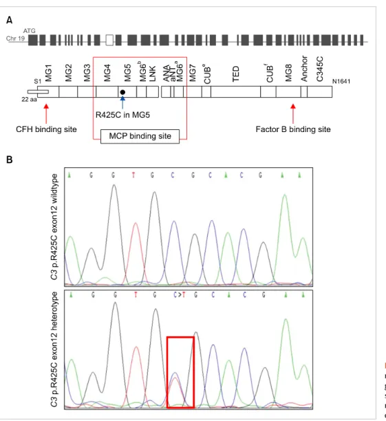
Fig. 2. Localization of the R425C C3 mutation. (A) Position within the C3 gene and the protein primary structure. (B) Representative ge- nome sequencing histogram for our patient (red square).
Fig. 1. Restriction fragment length polymorphism analysis of our patient with heterozygote c.1273C>T mutation in C3 gene showing 250 bp product (solid arrow).
Abbreviations: P, patient; WT, wild type.
C3, 40.1 mg/dL (90–180 mg/dL) and C4, 17.9 mg/dL (16–49 mg/dL). Brain MRI showed a subtle diffusion-restricted le- sion in the right corpus callosum and left cerebellum, sugges- tive of cerebral infarction. The ADAMTS13 (a disintegrin and metalloproteinase with a thrombospondin type 1 motif, member 13) activity was within the normal range. She had no history of bloody diarrhea. The Shiga-toxin assay on stool samples showed negative results. Immediately after admission, the patient received plasma exchange (daily for 2 days), followed by hemodialysis and steroids, and her consciousness improved to the level that she could recognize her family. The hemoglobin, platelet, and creatinine levels recovered to the normal range. Her LDH level also decreased to 456 U/L. The patient completely recovered and was dis- charged 56 days after admission. She is currently doing well without recurrence of disease under outpatient manage- ment on day 340 after the initial diagnosis.
Detection of complement C3 gene mutation
Genomic DNA was extracted from the patient’s peripheral
Blood Res2016;51:204-14. bloodresearch.or.kr
212 Letters to the Editor
blood leukocytes using a G-DEX II Genomic DNA Extraction Kit (iNtRON Biotechnology, Korea). For mutation detection, the coding exons and intron flanking regions of C3 (NM_000064.2) were amplified by the polymerase chain reaction (PCR), using a forward primer (5'-CAA TTC CCA GGT CTC AGG GA-3') and a reverse primer (5'-GAG AGA AAA GGA GAA AGG G-3'). The 743 bp PCR product was digested with 5 U BanII for 16 h at 37oC for restriction fragment length polymorphism analysis. In contrast to the wild type (171 bp only), an additional 273-bp band was detected in our patient (Fig. 1). Direct sequencing of the C3 gene revealed that she had heterozygous mutation 1273 C>T in exon 12, that affects codon 425 resulting in sub- stitution of arginine with cysteine (R425C) in the -chain of C3 (Fig. 2). The family study could not be done due to proband’s refusal.
DISCUSSION
To the best of our knowledge, this is the first case report of a Korean patient with aHUS associated with the c.1273C> T mutation in the complement C3 gene. Although direct evidence to define this polymorphism as a causative muta- tion for aHUS onset is lacking, in silico analyses assumed it as the potentially causative mutation [9].
The mutation was first demonstrated in an 8-month-old Japanese male infant, who had developed aHUS after cardiac surgery [10]. The infant died because of progressive renal and neurologic damage on postoperative day 50. Plasma infusions and hemodialysis were not effective. The response rate to short-term plasma therapy is known to be 40–50%
[3]. In contrast, our patient experienced less severe renal damage than the infant and was responsive to plasma exchange. It suggests that the clinical response to plasma therapy in patients with a C3 mutation is variable, depending on the stage of renal or neurologic damage. The father and aunt of the Japanese patient had the same heterozygous mutation but they had no history of aHUS. Our patient also has no family members who experienced symptomatic aHUS. It suggests that the penetrance of aHUS associated with the c.1273C>T mutation in the complement C3 gene is low. This is consistent with a previous finding of only a 10% penetrance of aHUS in individuals with a mutation in C3 as compared with higher penetrance rates in patients with mutations in the CFH, CFI, MCP, and CFB genes [11]. This means that a heterozygous carrier with the c.1273C>T mutation of C3 itself may not develop aHUS without an additional environmental trigger or genetic modifier as a second hit, as suggested by Fan et al. [9].
The c.1273C>T mutation is located within the C3 gene domain that binds with MCP. Therefore, we suspect that the pathogenesis of aHUS caused by c.1273C>T is likely to be associated with dysregulation of the alternative path- way, caused by prevention of proper binding of the MCP regulator to C3b.
Recently, eculizumab, a humanized monoclonal antibody that blocks complement C5 activation and the formation
of the terminal complement component, has been widely used for the treatment of aHUS in the USA, Europe, and Japan. Eculizumab has been reported to induce long-term remission in patients with aHUS associated with C3 gene mutations [12, 13]. In conclusion, we describe the first Korean case of aHUS associated with the c.1273C>T muta- tion in the complement C3 gene, where the patient was responsive to plasma exchange. Given the poor prognosis of aHUS and its low penetrance of development, identifying carriers in the family of this proband and properly educating them on how to avoid risk factors that trigger aHUS would be required to manage the patient and her carrier family members.
Hye Jeong Cho1, Jung O Kim2, Ji Young Huh3, Yong Park4, Myung-Gyu Kim4, Doyeun Oh1
1Department of Internal Medicine, 2Institute for Clinical Research, 3Laboratory Medicine, School of Medicine, CHA University, Seongnam, 4Department of Internal Medicine, Korea University College of Medicine, Seoul, Korea
Correspondence to: Doyeun Oh Department of Internal Medicine, School of Medicine,
CHA University, 59 Yatap-ro, Bundang-gu, Seongnam 13496, Korea
E-mail: [email protected]
Received on Jul. 28, 2015; Revised on Aug. 21, 2015; Accepted on Sep. 10, 2015 http://dx.doi.org/10.5045/br.2016.51.3.210
Acknowledgments
This work was supported by a grant from the Korean Ministry of Education (2013R1A1-A2060778).
AuthorsÊ Disclosures of Potential Conflicts of Interest No potential conflicts of interest relevant to this article were reported.
REFERENCES
1. Gasser C, Gautier E, Steck A, Siebenmann RE, Oechslin R.
Hemolytic-uremic syndrome: bilateral necrosis of the renal cortex in acute acquired hemolytic anemia. Schweiz Med Wochenschr 1955;85:905-9.
2. Gordjani N, Sutor AH, Zimmerhackl LB, Brandis M. Hemolytic uremic syndromes in childhood. Semin Thromb Hemost 1997;23:281-93.
3. Noris M, Remuzzi G. Atypical hemolytic-uremic syndrome. N Engl J Med 2009;361:1676-87.
4. Kavanagh D, Goodship TH. Atypical hemolytic uremic syn- drome, genetic basis, and clinical manifestations. Hematology Am Soc Hematol Educ Program 2011;2011:15-20.
5. Lee JM, Park YS, Lee JH, et al. Atypical hemolytic uremic syn- drome: Korean pediatric series. Pediatr Int 2015;57:431-8.
6. Jung S, Kang ES, Ki CS, Kim DW, Paik KH, Chang YS. Successful therapeutic plasma exchange in a 3.2-kg body weight neonate with atypical hemolytic uremic syndrome. J Clin Apher
bloodresearch.or.kr Blood Res 2016;51:204-14.
Letters to the Editor 213
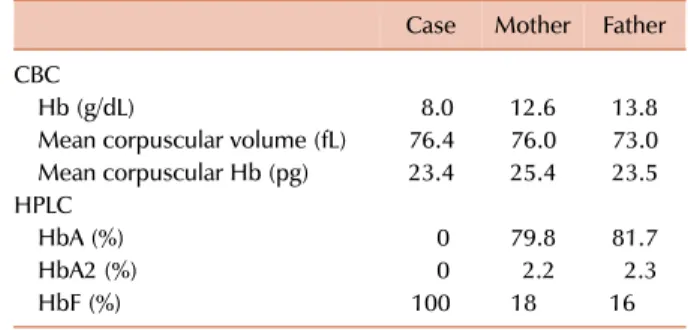
Table 1. Laboratory parameters of the case and parents.
Case Mother Father CBC
Hb (g/dL) 8.0 12.6 13.8
Mean corpuscular volume (fL) 76.4 76.0 73.0 Mean corpuscular Hb (pg) 23.4 25.4 23.5 HPLC
HbA (%) 0 79.8 81.7
HbA2 (%) 0 2.2 2.3
HbF (%) 100 18 16
Abbreviations: CBC, complete blood count; Hb, hemoglobin;
HPLC, high-performance liquid chromatography.
2011;26:162-5.
7. Cho HY, Lee BS, Moon KC, Ha IS, Cheong HI, Choi Y. Complete factor H deficiency-associated atypical hemolytic uremic syn- drome in a neonate. Pediatr Nephrol 2007;22:874-80.
8. Frémeaux-Bacchi V, Miller EC, Liszewski MK, et al. Mutations in complement C3 predispose to development of atypical hemo- lytic uremic syndrome. Blood 2008;112:4948-52.
9. Fan X, Yoshida Y, Honda S, et al. Analysis of genetic and predis- posing factors in Japanese patients with atypical hemolytic ure- mic syndrome. Mol Immunol 2013;54:238-46.
10. Matsukuma E, Imamura A, Iwata Y, et al. Postoperative atypical hemolytic uremic syndrome associated with complement c3 mutation. Case Rep Nephrol 2014;2014:784943.
11. Lhotta K, Janecke AR, Scheiring J, et al. A large family with a gain-of-function mutation of complement C3 predisposing to atypical hemolytic uremic syndrome, microhematuria, hyper- tension and chronic renal failure. Clin J Am Soc Nephrol 2009;4:1356-62.
12. Köse O, Zimmerhackl LB, Jungraithmayr T, Mache C, Nürnberger J. New treatment options for atypical hemolytic uremic syn- drome with the complement inhibitor eculizumab. Semin Thromb Hemost 2010;36:669-72.
13. Al-Akash SI, Almond PS, Savell VH Jr, Gharaybeh SI, Hogue C.
Eculizumab induces long-term remission in recurrent post-transplant HUS associated with C3 gene mutation. Pediatr Nephrol 2011;26:613-9.
Delta beta thalassemia:
a rare hemoglobin variant
TO THE EDITOR: Delta beta ()-thalassemia results from a deletion in both the delta and beta genes on chromosome 11. The gamma genes on the affected chromosome increase their production of gamma globin, thereby increasing the amount of hemoglobin F (HbF). -Thalassemia hetero- zygotes clinically show characteristics of thalassemia minor.
However, homozygous -thalassemia may give a clinical picture of thalassemia intermedia with a mild anemia.
A 12-month-old boy presented to the hematology out- patient department for evaluation of pallor and jaundice that had been for the past 2 months. He had no history of a blood transfusion. His family history was insignificant for congenital anemia. His parents had a consanguineous marriage. Physical examination revealed pallor and palpable spleen 2 cm below the left costal margin. Other examination findings were unremarkable. A complete blood count (CBC) revealed an Hb level of 8.0 g/dL, WBC count of 8.9×10⁹/L, and platelet count of 341×109/L (Table 1). A peripheral blood smear revealed anisopoikilocytosis with hypochromic mi- crocytic red cells, target cells, and basophilic stippling (Fig.
1A). The corrected reticulocyte count was 1.6%. Liver func- tion tests showed raised levels of serum total bilirubin (3.5 mg/dL) and indirect bilirubin (3.0 mg/dL). High-perform-
ance liquid chromatography (HPLC) showed 100% HbF and an absence of HbA and HbA2 (Fig. 1B). The Kleihauer- Bekte test revealed a pancellular pattern (Fig. 1C). Conse- quently, a CBC followed by HPLC was performed for both parents who were apparently healthy and had no history of blood transfusions (Table 1). Kleihauer-Betke tests of both parents showed a heterocellular distribution of HbF.
Hence, the patient was diagnosed with homozygous
-thalassemia, whereas the parents with heterozygous
-thalassemia. Unfortunately, mutational analysis could not be performed because the patient was lost to follow-up.
-Thalassemia results from the deletion of both and
genes. Homozygotes for -thalassemia have 100% HbF and, because of the increased synthesis of HbF, may have thalassemia intermedia rather than thalassemia major [1].
However, the phenotype of heterozygotes resembles that of the -thalassemia trait, but the HbA2 percentage is not increased and is often normal. HbF in such individuals is consistently elevated, varying from 5% to 20%. Peripheral blood film findings are similar to those for the -thalassemia trait, and the distribution of HbF is heterocellular, which is best observed via flow cytometry. It is necessary to dis- tinguish it from hereditary persistence of fetal hemoglobin (HPFH). The two groups of disorders are distinguished by the phenotype of heterozygous individuals. Heterozygotes of -thalassemia mutations have 5% to 20% HbF, which is heterocellularly distributed in red cells, whereas hetero- zygotes of HPFH mutations have 17% to 30% HbF, with a pancellular distribution. In addition, homozygotes of HPFH are asymptomatic, whereas -thalassemic homo- zygotes have thalassemia intermedia-like features [2].
At least nine mutations can result in -thalassemia. This type of thalassemia is observed in many ethnic groups, in- cluding some Mediterranean populations (Italians, Greeks, and Turks). Although the exact diagnosis of -thalassemia requires genetic analysis for mutations, Hb electrophoresis or HPLC findings of markedly elevated HbF may be suggestive. An extensive PubMed search was done to de- termine the incidence of -thalassemia in different parts of the world, but owing to the rarity of this Hb variant,