225
책임저자: 정상영, 광주시 동구 학1동 8번지
501-746, 전남대학교 의과대학 외과학교실 Tel: 062-220-6456, Fax: 062-227-1635 E-mail: [email protected]
접수일:2009년 3월 31일, 게재승인일:2009년 7월 21일
FK506에 의한 Jurkat 세포자멸사 유전자 발현분석
한일병원 외과, 1식품의약품안전청, 2전남대학교 의과대학 외과학교실
장태영ㆍ이재숙
1ㆍ우고운ㆍ김현철ㆍ이호균
2ㆍ정상영
2Apoptosis Gene Expression Pattern Analysis of Jurkat Cells Treated with FK506
Tae Young Jang, M.D., Jae Sook Lee, M.D.1, Go Woon Woo, M.D., Hyun Chul Kim, M.D., Ho Kyun Lee, M.D.2, Sang Young Chung, M.D.2
Department of Surgery, Hanil Hospital, 1Korea Food & Drug Administration, Seoul,
2Department of Surgery, Chonnam National University Medical School, Gwangju, Korea
Purpose: FK506 (tacrolimus) is a widely used immunosuppressive agent in the treatment of various medical conditions, including autoimmune disease, bone marrow and organ transplantations. Previously FK506 was known to cause apoptotic death of human Jurkat T cells.
Methods: The current study was designed to analyze the gene expression pattern of Jurkat T cells after FK506 application by using cDNA microarray. Treatment of Jurkat T cells with FK506 resulted in a decrease of cell viability in a time- and dose-dependent manner. Next, total RNA of Jurkat T cells was extracted by using TRIzol reagent and used to carry out a confirmation test for the purity and integrity of total RNA.
Results: Gene expression levels related to apoptosis and cell cycle process were mainly focused to analyze in FK506-treated Jurkat T cells. According to the inhibition of calcineurin activity, MARCKS in PKC substrates and Sp3 transcription factor was markedly increased in FK506-treated cells. Also, cell cycle control gene Id1 and Id3 were induced in expression from FK506-treated Jurkat T cells. However, FK506 decreased the expression of Src homology 2, G protein, and MEK 2 genes in bioactive peptide induced signaling pathway. It also reduced the expression level of the insulin receptor, DRPLA and Bai1-associated protein 2 genes, which are involved in the regulation of cell motility and morphology control.
Conclusion: The author will continue to pursue the exact functional roles of genes that are markedly changed in expression by FK506 in human Jurkat T cells in vitro and in vivo experimental models. (J Korean Surg Soc 2009;77:225-237)
Key Words: Tacrolimus, Gene expression, Apoptosis, Jurkat cell 중심 단어: 타크로리무스, 유전자 발현, 세포자멸사, Jurkat 세포
서 론
FK506 (tacrolimus)은 토양 진균류인 Streptomyces tsuku-
baensis에서 분리된 macrolide 계통의 항생제와 유사한 구조 를 가지고 있으며 1982년 일본에서 처음 개발되어 1990년 간이식에 처음으로 사용된 후 현재는 신장, 췌장, 폐 및 골 수 이식 등 전반적인 기관 이식 수술 후에 급성 거부 반응 을 억제하는데 매우 효과적인 면역 억제제로 널리 사용되 고 있다.(1) FK506의 효과는 세포내 calcineurin과 calcium에 의한 신호전달 과정을 저해하여 일차적으로 T세포를 억제 하는데 관여한다. 이는 FK506이 세포질 내에 존재하는
FK506 수용체인 FK506-binding proteins (FKBPs)과 결합하 여 heterodimeric 복합체를 형성하여 calcineurin의 활성을 억 제하고 calcineurin에 의해 촉진되는 일련의 과정을 억제함 으로서 이루어진다.(2,3) FK506은 칼슘 및 칼모듈린 의존성 serine/threonine 단백인산화 효소인 calcineurin의 활성화를 억제하여 nuclear factor of activated T cells (NFATs)의 탈인 산화를 억제함으로서 NFATs에 의한 각종 염증 유발성 사 이토카인의 전사를 억제하고 T 림프구 활성화를 억제하여 면역 기능을 저해하는 것으로 알려져 있다.(4) 그러나 FK506의 장기적인 사용은 심혈관 질환, 신장 기능의 저하 와 신경독성과 같은 부작용을 일으키며 특히 FK506에 의한 당뇨병의 발병이 흔하게 발생하는 것으로 알려졌다.(5,6) 본 연구에서는 사람 Jurkat T 세포를 대상으로 FK506에 의한 세포독성과 관련하여 cDNA microarray 분석을 통하여 세포자멸사와 관련된 유전자의 발현 양상 및 생물학적 경 로(biological pathway) 등을 분석함으로써 면역 억제제인 FK506의 분자 생물학적 기반을 제공하고자 하였다. 그 결 과 FK506은 사람 Jurkat T세포에서 세포자멸사, 세포주기 조절에 관련된 유전자들의 발현이 증가 또는 감소하였고 thrombin, bradykinin 및 angiotensin II와 같은 bioactive pep- tide에 의한 MAP kinase 관련 유전자, insulin 및 dentatoru- bral-pallidoluysian atrophy 신호전달 관련 유전자의 발현 감 소 등 유의한 결과를 얻었기에 보고하는 바이다.
방 법
1) 재료
(1) 세포주: 사람 T 림프구 세포주인 Jurkat 세포는 한국세 포주 은행(KNCC, 서울대학교)으로부터 구입하여 계대 배 양하면서 실험을 실시하였다.
(2) 시약 및 기기: 실험에 필요한 RPMI 1640, 항생제 및 우태아 혈청(Fetal Bovine Serum, FBS)은 GIBCO BRL사 (Grand Island, NY, USA) 제품을, 배양용기(24-well plate와 10 cm dish)는 Falcon사(Becto Dickinson, San Jose, CA, USA) 에서 구입하여 사용하였다. 3-[4,5-dimethylthiazol-2-yl]-2,5- diphenyl tetrazolium bromide (MTT)는 Sigma사(St, Louis, MO, USA)에서 구입하였고 TRIzol은 Invitrogen사(Calsbed, CA, USA)에서 구입하였다.
2) 방법
(1) 세포주 배양 및 시약 처리: 세포는 CO2 세포 배양기(3
7℃, 5% CO2)에서 10% 우태아 혈청이 포함된 RPMI 1640 배지로 배양하였으며, 24시간 간격으로 배양액을 교체하여 지수기(Log Phase)에 있는 세포에 FK506을 처리한 후 세포 독성 현상과 Microarray를 위한 실험을 수행하였다. FK506 은 10 mM 농도로 DMSO에 녹여 -20oC에 보관하였고, RPMI 1640 배지에 희석하여 사용하였다.
(2) 세포생존율 측정: 세포의 생존율은 MTT 분석법으로 측정하였다. 세포 배양판(24-well plate)에 세포(1×10⁵/ml)를 1 ml씩 분주하여 3시간 이상 CO2 세포배양기 안에서 안정 화시킨 후 FK506을 1.25∼40μM/ml의 다양한 농도로 처리 하였다. MTT 용액(5 mg/ml; phosphate buffered saline, PBS;
pH7.4)은 각각의 배양세포에 배양액의 1/10을 첨가하여 4시 간 반응 후 well-plate를 1,500 rpm으로 15분 원심 분리하여 상층액을 제거하였다. 바닥에 부착된 불용성의 보라색 for- mazan을 완전히 건조한 후 1 ml의 DMSO (Dimethyl sulf- oxide)용액으로 충분히 용해한 다음, 분광광도계(ELISA reader, Molecular Devices Co., Sunnyvale, CA, USA)를 이용 하여 545 nm 파장에서 흡광도를 측정하여 정상 대조군의 값과 비교하여 백분율(%)로 표시하였다.
(3) 세포내 총 RNA 추출: 세포를 10 cm dish에 2×10⁶/10 ml의 농도로 분주하여 3시간 안정화하고 10 및 20μM/ml 농도의 FK506을 각각 48시간 처리하여 15 ml conical 튜브 에 옮겨서 1,200 rpm으로 5분간 원심 분리하여 상등액을 제 거하고 1 ml의 TRIzol로 세포를 용해하였고 제조사의 기술 된 내용에 의거하여 total RNA을 추출하였다. 얻어진 상등 액과 동일양의 isopropanol을 첨가하여 혼합하고 −20oC에 서 12시간 반응시켜 14,000 rpm으로 20분간 원심 분리하여 상등액을 제거한 후 침전물을 70% 에탄올에 세척하여 자연 건조하였다. 여기에 TE 완충용액(10 mM Tris-HCl, pH8.0, 1 mM EDTA, pH8.0)을 가하여 RNA pellet을 용해한 후 260 nm 와 280 nm의 Spectriohotometer (Beckman사)로 optical density (OD)값 측정하여 RNA를 정량하였다. 추출된 RNA 표본의 순도는 EtBr이 포함된 1.5%의 formamide-agrose gel을 이용하 여 전기영동하고 UV 시스템을 이용하여 확인하였다.
(4) Microarray용 RNA 순도 측정: 얻어진 총 RNA의 cDNA microarray 적합성 여부를 확인하기 위하여 국내의 Geno- micTree사에 분석 의뢰하였다. GenomicTree사의 분석 기준 은 migration, peak pattern, 28s/18s ribosomal RNA의 비율 및 OD260/230비율을 고려하여 평가하여 RIN (RNA Integrity Number) 값을 결정한다. RNA의 quality는 통계적인 수치로 1∼10의 범위를 가지며 고순도의 시료를 요구하는 DNA
Fig. 1. FK506 decreased the viability of Jurkat cells in dose- and time-dependent manners. Cells were treated with various concentration of FK506 for 96 hours, then cell viability was measured by MTT assay after FK506 treatment. Data represent the mean±standard deviation (SD) of triplicates.
Table 1. RNA furification for microarray
Sample μg/μl OD260/230 Total (μg) Ratio (28s/18s) RIN*
Jurkat Con-48h 2.5062 1.94 120.2976 2.0 9.9
Jurkat F10-48h 2.2160 2.30 106.3680 2.0 9.9
Jurkat F20-48h 1.7765 2.04 85.2720 1.9 10.0
*RIN = RNA integrity number.
microarray analysis를 위한 권장 값은 7 이상이 요구된다. 본 실험에서 얻어진 RNA의 순도는 10에 근접하여 다음 분석 에 적합함을 확인하였다(Table 1).
(5) cDNA microarray method: Agilent’s Human whole ge- nome 4×44K glass slide cDNA microarrays를 이용하여 분석 하였다. FK506 10μM과 20μM 농도의 실험군은 Cy5로 표 지하여 각각 Cy3로 표지된 대조군과 비교하여 다음의 과정 을 거쳐 분석하였다.
① 증폭 및 라벨링: Agilent’s Low RNA Input Linear Amplification kit PLUS를 이용하여 증폭 및 라벨링 과정을 수행하였다.
② Microarray hybridization: Agilent’s Gene Expression Hybridization Kit를 이용하여 수행하였다.
③ Microarray wash: Agilent’s Gene Expression Wash Buffer Kit를 이용하였다.
④ Scan and image analysis: Agilent’s DNA microarray scan- ner 및 Feature Extraction Software를 이용하였다.
(6) Data Analysis: Agilent's GeneSpring Software를 이용하 여 normalization, clustering 및 filtering하여 2배 이상의 증감 을 보이는 유의한 유전자를 선별하였고 이들 유전자의 기 능을 웹사이트의 DAVID's program을 이용하여 biological function을 분석하였다.
결 과
1) FK506이 Jurkat 세포의 생존율에 미치는 영향 Jurkat 세포에 대한 면역억제제 FK506의 독성을 조사하 기 위하여 FK506을 다양한 농도로 96시간 처리한 후 세포 생존율을 MTT 방법으로 분석하였다. Jurkat 세포에 10μM 농도의 FK506을 72시간 처리시 62.5%, 20μM 농도에서는 45.7%, 30μM 농도에서는 33.5% 및 40μM 농도에서는 18.8%의 세포생존율을 나타내었다. 또한 Jurkat 세포에 1.25 μM 농도의 FK506을 96시간 처리 시 세포생존율은 91.7%, 2.5μM 농도에서는 84.3%, 5μM 농도에서는 72.9%, 10μM
농도에서는 46.2%, 20μM 농도에서는 22.4%, 30μM 농도에 서는 12.9% 및 40μM 농도에서는 6.6%의 세포생존율을 보였 다. 이상의 결과 Jurkat 세포에서 FK506에 의한 세포 독성은 농도 및 시간 의존적으로 증가하는 것으로 나타났다(Fig. 1).
2) FK506 처리 후 Jurkat 세포에서 total RNA 추출 및 Scatter와 MA plot 분석
총 RNA의 integrity는 EtBr이 함유된 1.5% formamide- agarose gel로 확인하였으며, Agilent's 2100 Bioanalyzer System을 이용하여 total RNA의 quality 및 integrity를 재확 인하였다.
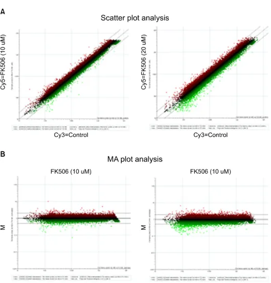
분산그래프에서는 chip 상의 모든 spots들을 표시한 것으 로 Cy3-channel (가로축)과 Cy5-channel (세로축)에서 각 sig- nal intensity값을 나타낸다. 가운데 사선은 각 channel에서 같은 signal intensity 값을 가진 probe들이 분포하고 있는 것 을 나타낸다. 그래프 상에서 점선 밖의 spot은 2배 이상 발 현양의 증가 혹은 감소를, 점선 안쪽은 2배 이하 발현양의
Fig. 2. Modulation of gene expre- ssion following FK506- treated Jurkat cells. Total RNA was prepared from Jurkat using TRIzol as de- scribed by manufacturer. (A) Scatter plot analysis. The x and y axes represent Cy3 (Control) and Cy5 (FK506 10μM or 20μM) signal in- tensity values, respectively.
These represent up-regu- lated (upper lines; red spots) or down-regulated (under lines; green spots) genes. (B) MA plot analysis. The x and y axes represent A (R+G/2) and M (R/G) signal in- tensity values. The R and G repesent Cy5-background and Cy3-background signal in- tensity, respectively. These represent up-regulated (upper lines; red spots) or down- regulated (under lines; green spots) genes.
증가 또는 감소를 나타낸 것으로 microarray chip data에서 취하지 않고 버린다(Fig. 2A). 또한 MA plot은 대조군에 대 한 각 실험군 유전자 발현의 변이에 대한 각 channel의 sig- nal intensity의 평균값과 ratio (R/G: R=Cy5 signal−back- ground/G=Cy3 signal−background) 값 사이의 상관관계를 보 여주는 그림으로 가로축은 A=(R+G/2), 세로축은 M=(R/G) 값을 나타낸다. 가운데 선(M=1)은 R=G 선을 표시한 것이 며, M=2 선은 G 값 보다 R 값이 2배 높은 값을 가진 probe들 이 분포하고 있는 것을 나타낸 것이고, 그 이상의 spots는 2배 이상 높은 signal intensity 값을 가진 probe들의 분포를 나타내고 있으며, 아래 M=0.5 선은 반대의 경우를 나타낸 다(Fig. 2B).
3) FK506에 의한 Jurkat 세포 사멸에서 유전자 발현양에 따른 데이터 분석
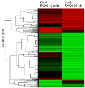
데이터 분석을 위하여 chip의 데이터들은 global normal- ization한 후 각 유전자들의 발현양을 분석하였다. 대조군 유전자 발현양을 1로 하여 실험군의 상대적 발현양을 나타 내어 2배 이상 증감한 유전자들만 데이터로 나타내었다. 전 체적으로 각 cDNA microarray에서 증가된 background의 sig- nal을 제거한 결과 29,163의 real gene을 얻었고, chip분석 결 과 값으로 이용할 수 없는 bad spots를 제거하고 대조군과 비교하여 10μM 농도의 FK506 처리군은 28,881개, 20μM 농도의 FK506 처리군은 27,756개의 유전자가 chip분석 결 과 값으로 유의하게 나타났다. 이 중에서 대조군과 비교하 여 2배 이상 증가 또는 감소된 유전자를 선별하여 hier- archical clustering하여 TreeView 프로그램으로 dendrogram 을 그렸다. 발현양 증가는 적색, 발현양 감소는 녹색으로 표
Fig. 3. Cluster image showing the different classes of total gene expression profiles.
Table 2. Two-fold increase of biological processes
Term Gene
number % Term Gene
number %
Positive regulation of programmed cell death 7 2.79 DNA repair 7 2.79
Amine biosynthetic process 4 1.59 Establishment of tissue polarity 2 0.80
Cellular macromolecule metabolic process 51 20.32 Translational initiation 4 1.59
Cellular localization 17 6.77 Regulation of locomotion 4 1.59
Positive regulation of microtubule polymerization 2 0.80 Chromatin assembly 5 1.99
Cell morphogenesis 12 4.78 Somatic muscle development 2 0.80
Cellular biosynthetic process 21 8.37 Regulation of biological quality 18 7.17
Cellular developmental process 32 12.75 Biological regulation 74 29.48
Basic amino acid transport 2 0.80 Multicellular organismal development 38 15.14
Chromatin assembly or disassembly 5 1.99 Protein-DNA complex assembly 8 3.19 Establishment and/or maintenance of 10 3.98 Macromolecular complex assembly 18 7.17
chromatin architecture Vasculature development 6 2.39
Primary metabolic process 119 47.41 mRNA metabolic process 9 3.59
Cellular metabolic process 113 45.02 DNA metabolic process 22 8.76
Nucleic acid metabolic process 58 23.11 Regulation of cellular component 4 1.59 Monocyte differentiation 2 0.80 organization and biogenesis
Interphase of mitotic cell cycle 4 1.59 B cell activation 4 1.59
Regulation of biosynthetic process 6 2.39 Coenzyme metabolic process 6 2.39
Cofactor metabolic process 7 2.79 Cell differentiation 32 12.75
Vasculogenesis 3 1.20 Organelle organization and biogenesis 24 9.56
Cellular structure morphogenesis 12 4.78 Regulation of biological process 66 26.29
Cellular component assembly 18 7.17 Regulation of cell size 6 2.39
Macromolecule metabolic process 106 42.23 Anatomical structure morphogenesis 24 9.56
Developmental process 52 20.72 Chromosome organization and biogenesis 11 4.38
Blood vessel development 6 2.39 Cell growth 6 2.39
Interphase 5 1.99 Nitrogen compound biosynthetic process 5 1.99
Metabolic process 126 50.20 Cellular component organization and biogenesis 54 21.51
Multicellular organismal process 53 21.12 Establishment of cellular localization 17 6.77
Blood vessel morphogenesis 6 2.39 Cellular protein metabolic process 50 19.92
Regulated secretory pathway 4 1.59 G1/S transition of mitotic cell cycle 3 1.20 시하였으며 chip에 사용된 total gene들과 각 기능에 관련된 각각의 cluster를 모아서 분류하였다(Fig. 3).
4) FK506에 의한 Jurkat 세포 사멸에서 발현 증가된 유 전자의 biological processing 분류
대조군과 비교하여 10μM 농도의 FK506 처리한 실험군 에서 2배 이상 증가한 유전자는 308개중 223개였으며 20μM 농도의 FK506을 처리한 실험군과 비교시 2배 이상 증가한 유전자는 289개중 202개였다. 이들 유전자는 각각 GenBank 를 통해 확인하였으며, FK506 처리에 의해 2배 이상 증가한 총 348개중 251개의 유전자는 DAVID's Bioinformatics Resource Analysis를 통해 분석하였으며, Functional gene on- cology에 따라 182개의 유전자를 총 58종류의 다양한 bio- logical processing과 관련하여 표로 분류하였다(Table 2).
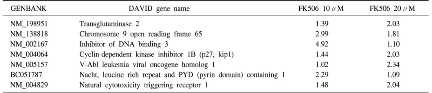
Table 3. Two-fold positive regulation of programmed cell death (7 Genes)
GENBANK DAVID gene name FK506 10μM FK506 20μM
NM_198951 Transglutaminase 2 1.39 2.03
NM_138818 Chromosome 9 open reading frame 65 2.99 1.81
NM_002167 Inhibitor of DNA binding 3 4.92 1.10
NM_004064 Cyclin-dependent kinase inhibitor 1B (p27, kip1) 1.44 2.03
NM_005157 V-Abl leukemia viral oncogene homolog 1 1.02 2.34
BC051787 Nacht, leucine rich repeat and PYD (pyrin domain) containing 1 2.29 1.09
NM_004829 Natural cytotoxicity triggering receptor 1 1.48 2.04
Table 4. Two-fold G1/S transition of mitotic cell cycle (3 Genes)
GENBANK DAVID gene name FK506 10μM FK506 20μM
NM_000944 Protein phosphatase 3 (formerly 2B), catalytic subunit, 1.48 2.16 alpha isoform (Calcineurin A alpha)
NM_005504 Branched chain aminotransferase 1, cytosolic 1.23 2.03
NM_004064 Cyclin-dependent kinase inhibitor 1B (p27, kip1) 1.44 2.03
5) FK506에 의한 Jurkat 세포 사멸에서 세포자멸사 촉진 유전자의 발현 변화
cDNA microarray chip에 사용된 total gene들과 각 기능에 관련된 각각의 cluster를 모아서 분류하였다. 세포고사의 양 성 제어(positive regulation of programmed cell death) 유전자 중 FK506처리에 의해 Jurkat 세포에서 2배 이상 증가한 유 전자들을 Table 3에 나타내었다. FK506에 의해 증가한 유전 자는 7개였으며 이 중 Transglutaminase 2와 Natural cytotox- icity triggering receptor 1은 FK506 처리 농도에 의존적으로 발현이 증가하였다(Table 3).
6) FK506에 의한 Jurkat 세포 사멸에서 G1/S transition of mitotic cell cycle 관련 유전자의 발현 변화 cDNA microarray chip에 사용된 total gene들과 각 기능에 관련된 각각의 cluster를 모아서 분류하였다. 세포의 주기와 관련된 G1/S transition of mitotic cell cycle 유전자중 FK506 처리에 의해 Jurkat 세포에서 2배 이상 증가한 유전자들을 Table 4에 나타내었다. FK506에 의해 증가한 유전자는 3개 였으며 이들 유전자는 FK506 처리 농도에 의존적으로 발현 이 증가하였다(Table 4).
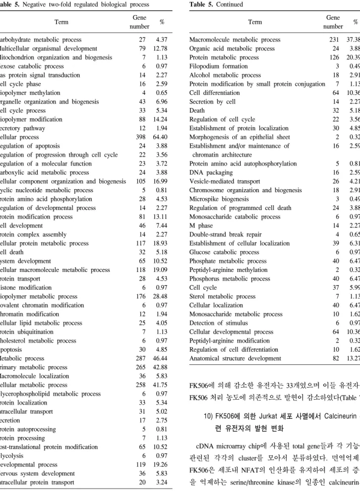
7) FK506에 의한 Jurkat 세포 사멸에서 발현 감소된 유 전자의 biological processing 분류
대조군과 비교하여 10μM 농도의 FK506 처리한 실험군
에서 2배 이상 감소한 유전자는 713개 중 598개였으며 20μM 농도의 FK506을 처리한 실험군과 비교 시 2배 이상 증가한 유전자는 397개 중 337개였다. 이들 유전자는 각각 GenBank 를 통해 확인하였으며, FK506 처리에 의해 2배 이상 감소한 총 744개의 유전자중 618개의 유전자는 DAVID's Bioinfor- matics Resource Analysis를 통해 분석하였으며, Functional gene oncology에 따라 443개의 유전자를 다양한 biological processing과 관련하여 표로 분류하였다(Table 5).
8) FK506에 의한 Jurkat 세포 사멸에서 cell cycle proc- ess 관련 유전자의 발현 변화
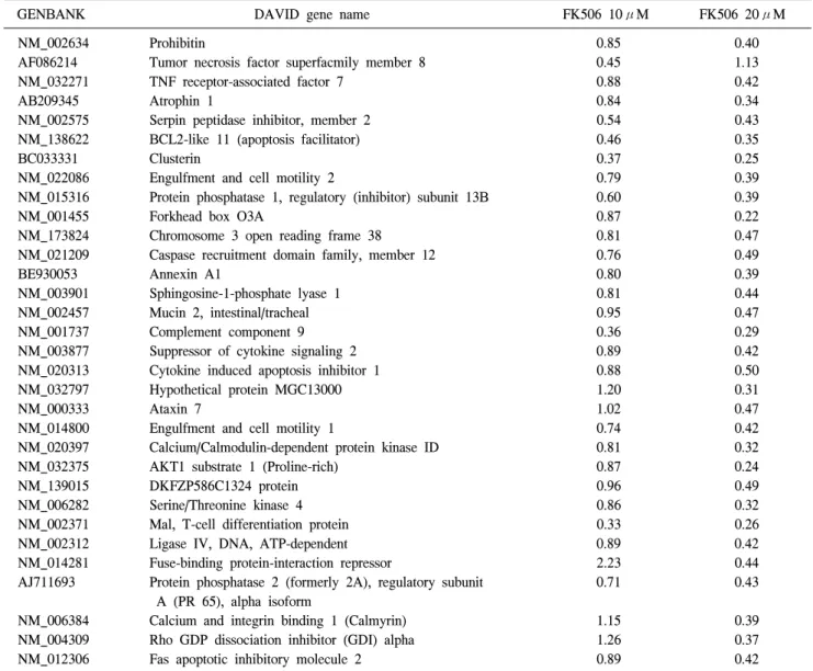
cDNA microarray chip에 사용된 total gene들과 각 기능에 관련된 각각의 cluster를 모아서 분류하였다. 세포의 주기와 관련된 cell cycle process 유전자중 FK506 처리에 의해 Jurkat 세포에서 2배 이상 감소한 유전자들을 Table 6에 나 타내었다. FK506에 의해 감소한 유전자는 32개였으며 이들 유전자는 FK506 처리 농도에 의존적으로 발현이 감소하였 다(Table 6).
9) FK506에 의한 Jurkat 세포 사멸에서 cell death 관련 유전자의 발현 변화
cDNA microarray chip에 사용된 total gene들과 각 기능에 관련된 각각의 cluster를 모아서 분류하였다. 세포의 사멸과 관련된 cell death 유전자중 FK506 처리에 의해 Jurkat 세포 에서 2배 이상 감소한 유전자들을 Table 7에 나타내었다.
Table 5. Continued
Term Gene
number %
Macromolecule metabolic process 231 37.38 Organic acid metabolic process 24 3.88
Protein metabolic process 126 20.39
Filopodium formation 3 0.49
Alcohol metabolic process 18 2.91
Protein modification by small protein conjugation 7 1.13
Cell differentiation 64 10.36
Secretion by cell 14 2.27
Death 32 5.18
Regulation of cell cycle 22 3.56
Establishment of protein localization 30 4.85 Morphogenesis of an epithelial sheet 2 0.32 Establishment and/or maintenance of 16 2.59 chromatin architecture
Protein amino acid autophosphorylation 5 0.81
DNA packaging 16 2.59
Vesicle-mediated transport 26 4.21
Chromosome organization and biogenesis 18 2.91
Microspike biogenesis 3 0.49
Regulation of programmed cell death 24 3.88 Monosaccharide catabolic process 6 0.97
M phase 14 2.27
Double-strand break repair 4 0.65
Establishment of cellular localization 39 6.31
Glucose catabolic process 6 0.97
Phosphate metabolic process 40 6.47
Peptidyl-arginine methylation 2 0.32
Phosphorus metabolic process 40 6.47
Cell cycle 37 5.99
Sterol metabolic process 7 1.13
Cellular localization 40 6.47
Monosaccharide metabolic process 10 1.62
Detection of stimulus 6 0.97
Cellular developmental process 64 10.36 Peptidyl-arginine modification 2 0.32 Regulation of cell differentiation 10 1.62 Anatomical structure development 82 13.27 Table 5. Negative two-fold regulated biological process
Term Gene
number %
Carbohydrate metabolic process 27 4.37 Multicellular organismal development 79 12.78 Mitochondrion organization and biogenesis 7 1.13
Hexose catabolic process 6 0.97
Ras protein signal transduction 14 2.27
Cell cycle phase 16 2.59
Biopolymer methylation 4 0.65
Organelle organization and biogenesis 43 6.96
Cell cycle process 33 5.34
Biopolymer modification 88 14.24
Secretory pathway 12 1.94
Cellular process 398 64.40
Regulation of apoptosis 24 3.88
Regulation of progression through cell cycle 22 3.56 Regulation of a molecular function 23 3.72 Carboxylic acid metabolic process 24 3.88 Cellular component organization and biogenesis 105 16.99 Cyclic nucleotide metabolic process 5 0.81 Protein amino acid phosphorylation 28 4.53 Regulation of developmental process 14 2.27
Protein modification process 81 13.11
Cell development 46 7.44
Protein complex assembly 14 2.27
Cellular protein metabolic process 117 18.93
Cell death 32 5.18
System development 65 10.52
Cellular macromolecule metabolic process 118 19.09
Protein transport 28 4.53
Histone modification 6 0.97
Biopolymer metabolic process 176 28.48
Covalent chromatin modification 6 0.97
Chromatin modification 12 1.94
Cellular lipid metabolic process 25 4.05
Protein ubiquitination 7 1.13
Cholesterol metabolic process 6 0.97
Apoptosis 30 4.85
Metabolic process 287 46.44
Primary metabolic process 265 42.88
Macromolecule localization 36 5.83
Cellular metabolic process 258 41.75
Glycerophospholipid metabolic process 6 0.97
Protein localization 33 5.34
Intracellular transport 31 5.02
Secretion 17 2.75
Protein autoprocessing 5 0.81
Protein processing 7 1.13
Post-translational protein modification 65 10.52
Glycolysis 6 0.97
Developmental process 119 19.26
Nervous system development 36 5.83
Intracellular protein transport 20 3.24
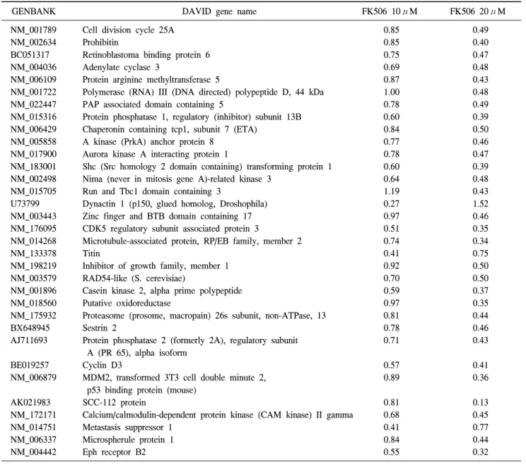
FK506에 의해 감소한 유전자는 33개였으며 이들 유전자는 FK506 처리 농도에 의존적으로 발현이 감소하였다(Table 7).
10) FK506에 의한 Jurkat 세포 사멸에서 Calcineurin 관 련 유전자의 발현 변화
cDNA microarray chip에 사용된 total gene들과 각 기능에 관련된 각각의 cluster를 모아서 분류하였다. 면역억제제 FK506은 세포내 NFAT의 인산화를 유지하여 세포의 증식 을 억제하는 serine/threonine kinase의 일종인 calcineurin의
Table 6. Negative two-fold regulated cell cycle process (32 Genes)
GENBANK DAVID gene name FK506 10μM FK506 20μM
NM_002634 Prohibitin 0.85 0.40
AF086214 Tumor necrosis factor superfacmily member 8 0.45 1.13
NM_032271 TNF receptor-associated factor 7 0.88 0.42
AB209345 Atrophin 1 0.84 0.34
NM_002575 Serpin peptidase inhibitor, member 2 0.54 0.43
NM_138622 BCL2-like 11 (apoptosis facilitator) 0.46 0.35
BC033331 Clusterin 0.37 0.25
NM_022086 Engulfment and cell motility 2 0.79 0.39
NM_015316 Protein phosphatase 1, regulatory (inhibitor) subunit 13B 0.60 0.39
NM_001455 Forkhead box O3A 0.87 0.22
NM_173824 Chromosome 3 open reading frame 38 0.81 0.47
NM_021209 Caspase recruitment domain family, member 12 0.76 0.49
BE930053 Annexin A1 0.80 0.39
NM_003901 Sphingosine-1-phosphate lyase 1 0.81 0.44
NM_002457 Mucin 2, intestinal/tracheal 0.95 0.47
NM_001737 Complement component 9 0.36 0.29
NM_003877 Suppressor of cytokine signaling 2 0.89 0.42
NM_020313 Cytokine induced apoptosis inhibitor 1 0.88 0.50
NM_032797 Hypothetical protein MGC13000 1.20 0.31
NM_000333 Ataxin 7 1.02 0.47
NM_014800 Engulfment and cell motility 1 0.74 0.42
NM_020397 Calcium/Calmodulin-dependent protein kinase ID 0.81 0.32
NM_032375 AKT1 substrate 1 (Proline-rich) 0.87 0.24
NM_139015 DKFZP586C1324 protein 0.96 0.49
NM_006282 Serine/Threonine kinase 4 0.86 0.32
NM_002371 Mal, T-cell differentiation protein 0.33 0.26
NM_002312 Ligase IV, DNA, ATP-dependent 0.89 0.42
NM_014281 Fuse-binding protein-interaction repressor 2.23 0.44
AJ711693 Protein phosphatase 2 (formerly 2A), regulatory subunit 0.71 0.43
A (PR 65), alpha isoform
NM_006384 Calcium and integrin binding 1 (Calmyrin) 1.15 0.39
NM_004309 Rho GDP dissociation inhibitor (GDI) alpha 1.26 0.37
NM_012306 Fas apoptotic inhibitory molecule 2 0.89 0.42
억제제로 잘 알려져 있다. Jurkat 세포에서 myristoylated ala- nine-rich protein kinase c substrate (MARCKS) (NM_002356) 유전자는 10μM 농도의 FK506 처리 시 0.7배의 발현을 보 였고, 20μM 농도의 FK506 처리 시에는 2.06배로 발현이 증 가하였다. 또한, transcription factor sp3 (SP3) (NM_003111) 유전자는 10μM 농도의 FK506 처리 시 1.05배, 20μM 농도 의 FK506 처리 시 2.04배로 FK506 농도 의존적으로 증가하 였다. 이들은 DAVID's를 통한 분석 결과 Keratinocyte 분화 와 관련된 유전자들임을 확인할 수 있었다(Table 8).
11) FK506에 의한 Jurkat 세포 사멸에서 G0∼S 전이 관 련 유전자의 발현 변화
T세포 활성과 증식에는 수많은 전사인자가 관련되어 있
는데 이중 G0에서 S기 l로의 전이에 관련된 전사인사인 Id 의 발현을 조사하였다. cDNA microarray 분석 결과 FK506 처리시 Jurkat 세포에 3종류의 Ids중 Id 1과 Id 3의 발현 변화 가 관찰되었다. Id 1 (NM_002165) 유전자는 10μM 농도의 FK506 처리시 2.41배, 20μM 농도의 FK506처리시 3.07배 발현이 증가하였고, Id 3 (NM_002167) 유전자는 10μM 농 도의 FK506 처리시 4.92배, 20μM 농도의 FK506 처리시 1.1배의 발현 증가를 보였다(Table 9).
12) FK506에 의한 Jurkat 세포 사멸에서 signaling path- way 관련 유전자의 발현 변화
cDNA microarray 분석 결과 FK506 처리시 Jurkat 세포에 서 thrombin, bradykinin 및 angiotensin II와 같은 bioactive
Table 7. Negative two-fold regulated cell death (33 Genes)
GENBANK DAVID gene name FK506 10μM FK506 20μM
NM_001789 Cell division cycle 25A 0.85 0.49
NM_002634 Prohibitin 0.85 0.40
BC051317 Retinoblastoma binding protein 6 0.75 0.47
NM_004036 Adenylate cyclase 3 0.69 0.48
NM_006109 Protein arginine methyltransferase 5 0.87 0.43
NM_001722 Polymerase (RNA) III (DNA directed) polypeptide D, 44 kDa 1.00 0.48
NM_022447 PAP associated domain containing 5 0.78 0.49
NM_015316 Protein phosphatase 1, regulatory (inhibitor) subunit 13B 0.60 0.39
NM_006429 Chaperonin containing tcp1, subunit 7 (ETA) 0.84 0.50
NM_005858 A kinase (PrkA) anchor protein 8 0.77 0.46
NM_017900 Aurora kinase A interacting protein 1 0.78 0.47
NM_183001 Shc (Src homology 2 domain containing) transforming protein 1 0.60 0.39
NM_002498 Nima (never in mitosis gene A)-related kinase 3 0.64 0.48
NM_015705 Run and Tbc1 domain containing 3 1.19 0.43
U73799 Dynactin 1 (p150, glued homolog, Droshophila) 0.27 1.52
NM_003443 Zinc finger and BTB domain containing 17 0.97 0.46
NM_176095 CDK5 regulatory subunit associated protein 3 0.51 0.35
NM_014268 Microtubule-associated protein, RP/EB family, member 2 0.74 0.34
NM_133378 Titin 0.41 0.75
NM_198219 Inhibitor of growth family, member 1 0.92 0.50
NM_003579 RAD54-like (S. cerevisiae) 0.70 0.50
NM_001896 Casein kinase 2, alpha prime polypeptide 0.59 0.37
NM_018560 Putative oxidoreductase 0.97 0.35
NM_175932 Proteasome (prosome, macropain) 26s subunit, non-ATPase, 13 0.81 0.44
BX648945 Sestrin 2 0.78 0.46
AJ711693 Protein phosphatase 2 (formerly 2A), regulatory subunit 0.71 0.43
A (PR 65), alpha isoform
BE019257 Cyclin D3 0.57 0.41
NM_006879 MDM2, transformed 3T3 cell double minute 2, 0.89 0.36
p53 binding protein (mouse)
AK021983 SCC-112 protein 0.81 0.13
NM_172171 Calcium/calmodulin-dependent protein kinase (CAM kinase) II gamma 0.68 0.45
NM_014751 Metastasis suppressor 1 0.41 0.77
NM_006337 Microspherule protein 1 0.84 0.44
NM_004442 Eph receptor B2 0.55 0.32
Table 8. Changes associated with calcineurin in keratinocyte differentiation by FK506
GENBANK Name FK506 10μM FK506 20μM
NM_002356 Myristoylated alanine-rich protein kinase c substrate 0.70 2.06
NM_003111 Sp3 transcription factor 1.05 2.04
Table 9. Changes of Id proteins in regulating the G0-to-S transition of the cell cycle by FK506
GENBANK Name FK506 10μM FK506 20μM
NM_002165 Inhibitor of DNA binding 1, dominant negative helix-loop-helix protein 2.41 3.07 NM_002167 Inhibitor of DNA binding 3, dominant negative helix-loop-helix protein 4.92 1.10
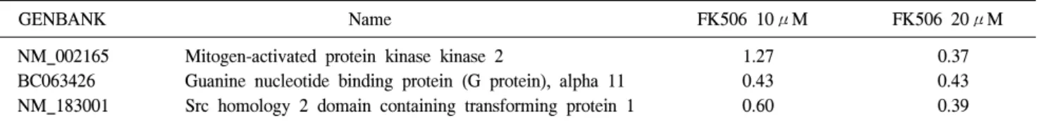
Table 10. Changes of bioactive peptide induced signaling pathway by FK506
GENBANK Name FK506 10μM FK506 20μM
NM_002165 Mitogen-activated protein kinase kinase 2 1.27 0.37
BC063426 Guanine nucleotide binding protein (G protein), alpha 11 0.43 0.43 NM_183001 Src homology 2 domain containing transforming protein 1 0.60 0.39
Table 11. Changes of gene associated with Diabetes and DRPLA pathway
GENBANK Name FK506
10μM
FK506 20μM
NM_000280 Insulin receptor 1.15 0.44
AB209345 Atrophiin 1 0.84 0.34
NM_017450 Bai1-associated protein 2 0.65 0.30
peptide에 반응하는 signaling pathway에 관여된 유전자의 발 현이 현저히 감소하였다. Mitogen-activated protein kinase kinase 2 (MAPKK) (NM_030662) 유전자는 10μM 농도의 FK506 처리시 1.27배 증가하였으나 20μM 농도의 FK506 처리시 0.37배로 발현이 감소하였고, Guanine nucleotide binding protein (G protein) alpha 11 (BC063426) 유전자는 10 μM 농도의 FK506 처리시 0.43배로 발현이 감소하였으며, 20μM 농도의 FK506 처리시에도 동일한 발현 양상을 보였 다. 또한 Shc (src homology 2 domain containing) transforming protein 1 (NM_183001) 유전자는 10μM 농도의 FK506 처리 시 0.6배, 20μM 농도의 FK506 처리 시 0.39배로 감소하였 다(Table 10).
13) FK506에 의한 Jurkat 세포 사멸에서 Diabetes와 DRPLA pathway 관련 유전자의 발현 변화 FK506 처리 시 Jurkat 세포에서 당뇨병과 연관된 유전자 의 발현을 cDNA microarray 분석결과를 통해 조사하였다.
Jurkat 세포에서 insulin receptor (NM_000208) 유전자는 10 μM 농도의 FK506 처리 시 1.15배로 발현이 증가하였으며 20μM 농도의 FK506 처리 시 0.44배로 발현이 감소하였다.
또한 Haw River syndrome (HRS)의 원인이 되는 atrophin 1 (AB209345) 유전자는 10μM 농도의 FK506 처리 시 0.84배, 20μM 농도의 FK506 처리 시 0.34배로 발현이 감소하였다.
세포의 mobility와 morphogenesis에 관련된 bai1-associated protein 2 (NM_017450) 유전자는 10μM 농도의 FK506 처리 시 0.65배, 20μM 농도의 FK506 처리 시 0.3배로 감소하였 다(Table 11).
고 찰
FK506은 calcineurin의 serine/threonine phosphatase 기능을 억제함으로서 cytokine의 전사활성에 관여하는 NFAT의 탈 인산화를 억제하여 면역억제 기능을 나타내는 것으로 알려 져 있다. 면역 반응과 관련된 초기 유전자와 IL-2, IL-3, IL-4, IL-12, granulocyte/macrophage cology-stimulating factor 및 TNF-α 등의 cytokine이 FK506에 의해서 억제됨이 보고되 었다.(7,8) 대부분은 FK506의 calcineurin 억제에 의한 전사 조절인자 NFAT의 억제와 관련된 cytokine의 발현 조절에 대한 연구가 진행되고 있고 FK506에 의한 세포자멸사와 관 련된 유전자의 발현 및 기전에 대한 연구가 미흡한 실정이 다. 따라서 본 연구에서는 cDNA microarray 분석을 통하여 FK506에 의한 Jurkat 세포의 사멸에서 관련 유전자의 발현 양상을 조사하였다. FK506은 Jurkat 세포에서 농도와 시간 의존적인 세포 독성을 야기하였다. cDNA microarray 분석 결과 FK506에 의해 2배 이상 증가하는 유전자는 348개가 도출되었으며 이중 GenBank에서 확인이 가능한 251개의 유전자를 대상으로 DAVID's Bioinformatics Resource Analysis의 웹사이트에서 gene ontology를 분석하여 수많은 biological process에 관련된 유전자의 발현이 변화하였다.
Calcineurin과 관련되어 도출된 유전자인 MARCKS는 합 성 후 plasma membrane에 위치하며 actin filament와 cross- linking하는 단백질이다. MARCKS는 calcium-calmodulin과 결합하여 plasma membrane으로부터 이탈되어 F-actin의 cross-linking 활성을 억제하며, MARCKS 단백질은 calm- odulin, actin, synapsin과 결합하여 cell motility, phagocytosis, membrane trafficking 및 mitogenesis를 조절한다. 또한 MARCKS는 세포내 calmodulin reservoir로서 작용하며 PKC 에 의해 인산화 되면 calmodulin과의 결합력이 약화되어 calmodulin을 방출한다.(9) 또한, Sp3는 isoform 특이적으로 transcription weak activator 또는 repressor로 작용하는데 con- sensus GT-와 GC- boxes promoter element와 결합하고 Sp1의 C2H2-type zinc-finger 일원으로 핵내에 존재하며 대부분의
세포에서 발현 된다.(10) 이 단백질은 하나의 zinc finger DNA-binding domain과 여러 개의 transactivation domain을 가지고 있어 수많은 유전자의 발현을 촉진 또는 억제하는 bifunctional transcription factor로 작용 한다.(11,12) 최근 E2F 계통의 전사 조절인자가 retinoblastoma-tumor-suppressor 유 전자 산물(pRB), cyclin과 cyclin dependent kinase (Cdk) 같은 cell-cycle progress에 관여하고 특히 E2F1은 DNA damage, repair, checkpoint activity 및 differentiation에 관여하는데 Sp3 는 이들을 억제 한다.(13)
G0/S cell cycle transition과 관련되어 도출된 유전자인 Id helix-loop-helix (HLH) 단백질은 Id 1∼Id 4까지 4종류가 알 려져 있으며 일반적으로 세포성장의 양성조절자(positive regulator)와 세포분화의 음성 조절자(negative regulator)로 작용 한다.(14) 이들은 다른 종류의 HLH 전사조절인자를 억제하는 dominant negative antagonist로 작용하여 진핵세포 의 세포 분화에 관여한다. 또한, Id 단백질은 Cdk와 retino- blastoma protein (Rb)에 의해 조절되는 cell-cycle-regulatory pathway에 관여한다. 최근 Id 단백질은 세포외 신호에 반응 하여 세포주기와 분화 관련 유전자 발현의 중요한 조절자 로서 주목을 받고 있다. Id 1은 특정 세포의 증식시 발현되 며 다른 HLH를 갖는 전사인자와 결합하여 DNA 결합을 억 제하며 핵내에 존재하며 cell growth, senescence 및 differ- entiation에 중요한 역할을 한다. Id 3는 basic DNA-binding domain이 결여된 HLH 단백질로서 다른 HLH 전사조절 단 백질과 반응하여 heterodimer를 형성한다. 특히 Id 3는 mus- cle creatin kinase E-box enhancer에 대한 E2A-containing 단백 질 복합체의 결합을 저해하며, phorbol 12-mystrate 13-acetate (PMA)에 의해서 발현이 유도되어 핵내에 위치하고 lung, kidney와 adrenal gland에서 발현 된다.(15,16)
Jurkat 세포에서 FK506에 의한 세포사멸에서 cDNA mi- croarray 분석 결과 2배 이상 발현이 감소된 744개 유전자 중에서 GenBank를 통해 확인 가능한 유전자 618개에 대한 gene ontology 분석 결과, 443개의 유전자를 biological proc- ess와 관련하여 분류하였다. 특히 function pathway와 관련하 여 bioactive peptide에 의한 MAP kinase의 signaling pathway 및 diabetes/DRPLA pathway 관련 유전자의 발현이 감소되었 다. Bioactive peptide에 의한 signaling pathway 유전자중 Mitogen-activated protein kinase kinase 2 (NM_030662) (MAPKK 2)는 MAP kinase kinase kinase (Raf 또는 MEKK1) 에 의해서 인산화 된다. 이들의 결여는 cardiofaciocutaneous syndrome (CFC syndrom)의 원인이 되어 heart defect 및 men-
tal retardation이 된다. 또한, MAPKK2는 ERK1과 ERK2 MAP kinase를 활성화 하며, mitogen growth factor signal transduction에서 중요한 기능을 수행한다. 이 kinase의 억제 또는 degradation은 Yersinia 와 anthrax의 질병과 관련이 있 다. Guanine nucleotide binding protein (G protein) alpha 11 (BC063426)은 다양한 transmembrane signaling system에서 modulator 또는 transducer이다. Alpha chain은 guanine nucleo- tide binding site를 가지고 있으며 phospholipase C의 activator 로 작용한다.
Shc (src homology 2 domain containing) transforming pro- tein 1 (NM_183001)은 활성화된 epidermal growth factor re- ceptor에 의해서 인산화 되고, p45 Shc, p52 Shc 및 p66 Shc의 isoform이 존재하여 insulin, hygrogen peroxide 또는 UV에 의 해서 인산화 된다. 이 단백질은 다양한 growth factor에 반응 하며 인산화된 Trk receptor와 결합하며 Ras의 활성에 관여 한다. p66 Shc은 Ras의 활성화에는 관여하지 않으나 oxida- tive stress와 life span에 관련된 signal transduction pathway에 관계한다. 이 p66 Shc는 tumor suppressor p53의 downstream target으로서 intracellular oxidants, cytochrome c release와 apoptosis의 증가에 필요한 것으로 알려져 있다.
Diabetes 및 DRPLA pathway와 관련된 유전자인 Insulin receptor (NM_000208) 유전자 발현의 감소는 insulin 저항성 에 의해 diabetes mellitus와 관련된다. Insulin receptor는 in- sulin과 반응하여 tyrosine-999 잔기가 autophosphorylated되 며 IRS1과 PI3K를 포함한 downstream mediators와 receptor와 결합을 촉진하며 최종적으로 glucose uptake를 조절하는데 관여한다.
Atrophin 1 (AB209345) 발현의 감소는 Haw River syn- drome (HRS)의 원인이 되며 HRS는 dominant neurodegene- rative disease로서 huntington disease, spinocerebella atrophy와 dentatorubropallidoluysian atropy (DRPLA)를 일으키며 neu- ron의 소실과 관련된다. DRPLA (Atrophin 1)의 생물학적 기 능은 정확하게 밝혀져 있지 않으나 세포자멸사와 관련하여 caspase-3의 기질인 nuclear repair 관련 단백질 PARP, DNA- dependent protein kinase 또는 U1-70 kDa로 절단되어 DNA degradation과 연관되어 있다.(17,18) DRPLA의 bipartite nu- clear localization signal인 N-말단은 세포자멸사시 절단되어 제거되고 핵내에서 세포질로 이동하며 절단된 DRPLA 단 백질은 세포자멸사를 유도하는 dominant negative effect를 갖는 것으로 보인다. 또한 DRPLA 단백질은 caspase protease 의 기질인 nuclear lamins, actin, fodrin, actin-associated protein
또는 Gas2처럼 microfilament 구성물과 결합하여 세포 구조 를 유지하는데 중요한 역할을 수행한다.(19-21)
Bai1-associated protein 2 (NM_017450) 유전자에 의해 생 성된 단백질은 brain-specific angiogenesis inhibitor (BAI 1)- binding 단백질로 규명되었으며 또한 insulin receptor ty- rosine kinase substrate로 작용하며 중추신경계에서 insulin, stress fiber 및 cytokine의 형성에 관여한다. BAI 1은 1,584 아마노산으로 구성된 adhesion-type G-protein coupled re- ceptor (GPCR) family의 subgroup VII에 속하는 단백질로 cell-cell 또는 cell-matrix interaction에 관여하고 뇌에서 p53 과 반응하여 angiogenesis의 neovascularization을 억제한 다.(22-24) 그러나 BAI 1에 대한 ligand는 알려져 있지 않고 yeast two-hybrid screen에 의해서 ELMO 1과 결합하는 것으 로 보고되었다. ELMO 1은 세포자멸사에 의한 세포를 en- gulfment하거나 degradation하는데 관여한다. 따라서 BAI 1 의 발현감소 또는 기능이상은 apoptotic 세포의 uptake를 억 제한다.(25) 뇌세포 이외의 세포에서 BAI 1의 발현은 human monocytes와 macrophages에서 microarray analyses에 의해 보 고된 바 있다.(26)
FK506에 의한 Jurkat 세포의 사멸에서 cDNA microarray 분석 결과 고농도 및 장시간 처리시 세포자멸사 및 세포주 기의 변화에 관련된 유전자의 발현이 증가 또는 감소되었 고 특히 MARCKS가 증가하였고 전사조절인자인 Sp3의 발 현이 증가하였다. 또한 세포주기에서 G0∼S주기로 전이하 는데 관련된 전사조절 인자의 발현을 억제하는 Id 1과 Id 3의 발현이 증가하였다. 반면에 bioactive peptide (thrimbin, bradkynin, angiotensin II)에 의한 신호경로에 관련된 Shc, MEK2 및 G protein α-11 유전자의 발현은 감소하였다. 또 한 임상실험에서 FK506의 사용에 따른 부작용으로 보고된 diabetes와 관련된 유전자로는 insulin receptor와 neuro- degeneration에 관련된 atrophin 1 및 세포의 운동성과 mor- phogeneis에 관련된 Bai 1-associated protein 2의 발현이 감소 하였다. 따라서, 장기이식 및 자가 면역 치료에 적절한 농도 및 치료 기간의 조절 등 면역 억제제 FK506의 활용 및 작용 기전에 대한 분자 생물학적 기반을 제공할 수 있을 것이다.
결 론
사람 Jurkat 세포를 대상으로 FK506에 의한 세포독성과 관련하여 cDNA microarray 분석을 통하여 세포자멸사와 관 련된 유전자의 발현 양상 및 생물학적 경로(biological path-
way) 등을 분석하여 다음과 같은 결론을 얻었다.
1) FK506은 사람 T세포인 Jurkat 세포에 농도, 시간 의존 적인 세포독성을 보였다.
2) FK506에 의한 Jurkat 세포 사멸에서 2배 이상 증가된 183개의 유전자는 기능적 gene oncology에 따라 58종류로 소분류 하였다.
3) FK506에 의한 Jurkat 세포 사멸에서 2배 이상 감소된 443개의 유전자는 기능적 gene oncology에 따라 87종류로 소분류 하였다.
4) FK506에 의한 Jurkat 세포 사멸에서 calcineurin의 활성 억제와 관련된 MARCKS의 발현과 전사조절 인자인 Sp3 유 전자의 발현은 증가하였다.
5) FK506에 의한 Jurkat 세포 사멸에서 세포주기와 관련 된 Id 1과 Id 3 유전자의 발현은 증가하였다.
6) FK506에 의한 Jurkat 세포 사멸에서 bioactive peptide (thrombin, angiotensin II) 신호경로와 관련된 Src homology 2, G protein, MEK 2 유전자의 발현은 감소하였다.
7) FK506에 의한 Jurkat 세포 사멸에서 당뇨병 발병과 관 련된 insulin receptor, DRPLA (Atrophin 1) 및 세포의 운동과 형태 유지에 관련된 Bai1-associated protein 2 유전자의 발현 은 감소하였다.
FK506은 calcineurin의 활성 억제에 따른 NFAT의 활성을 억제뿐 아니라 세포자멸사, 세포주기, 다양한 신호경로 및 임상실험에서 나타나는 당뇨병의 발현과 세포의 운동과 형 태 유지에 관련된 유전자 발현이 감소되어 장기이식 및 자 가 면역 치료에 적절한 농도 및 치료 기간의 조절 등 면역 억제제 FK506의 기전에 대한 분자 생물학적 기반을 제공할 수 있을 것이다.
REFERENCES
1) Pridohl O, Heinemann K, Hartwig T, Witzigmann H, Lamesch P, Fangmann J, et al. Low-dose immunosuppression with FK 506 and sirolimus after liver transplantation: 1-year results.
Transplant Proc 2001;33:3229-31.
2) Harding MW, Galat A, Uehling DE, Schreiber SL. A receptor for the immunosuppressant FK506 is a cis-trans peptidyl-prol- yl isomerase. Nature 1989;341:758-60.
3) Maki N, Sekiguchi F, Nishimaki J, Miwa K, Hayano T, Takahashi N, et al. Complementary DNA encoding the human T-cell FK506-binding protein, a peptidylprolyl cis-trans iso- merase distinct from cyclophilin. Proc Natl Acad Sci U S A 1990;87:5440-3.
4) Brown EJ, Albers MW, Shin TB, Ichikawa K, Keith CT, Lane WS, et al. A mammalian protein targeted by G1-arresting ra- pamycin-receptor complex. Nature 1994;369:756-8.
5) Bagnis C, Deray G, Dubois M, Adabra Y, Jacquiaud C, Jaudon MC, et al. Comparative acute nephrotoxicity of FK-506 and ciclosporin in an isolated in situ autoperfused rat kidney model. Am J Nephrol 1997;17:17-24.
6) Olyaei AJ, de Mattos AM, Bennett WM. Nephrotoxicity of immunosuppressive drugs: new insight and preventive strategies. Curr Opin Crit Care 2001;7:384-9.
7) Weiss A, Wiskocil RL, Stobo JD. The role of T3 surface mol- ecules in the activation of human T cells: a two-stimulus re- quirement for IL 2 production reflects events occurring at a pre-translational level. J Immunol 1984;133:123-8.
8) Reem GH, Cook LA, Vilcek J. Gamma interferon synthesis by human thymocytes and T lymphocytes inhibited by cyclo- sporin A. Science 1983;221:63-5.
9) Arbuzova A, Schmitz AA, Vergeres G. Cross-talk unfolded:
MARCKS proteins. Biochem J 2002;362:1-12.
10) Hagen G, Muller S, Beato M, Suske G. Sp1-mediated tran- scriptional activation is repressed by Sp3. EMBO J 1994;13:
3843-51.
11) Ge Y, Jensen TL, Matherly LH, Taub JW. Physical and func- tional interactions between USF and Sp1 proteins regulate hu- man deoxycytidine kinase promoter activity. J Biol Chem 2003;278:49901-10.
12) Ko JL, Liu HC, Loh HH. Role of an AP-2-like element in transcriptional regulation of mouse mu-opioid receptor gene.
Brain Res Mol Brain Res 2003;112:153-62.
13) Rotheneder H, Geymayer S, Haidweger E. Transcription fac- tors of the Sp1 family: interaction with E2F and regulation of the murine thymidine kinase promoter. J Mol Biol 1999;293:
1005-15.
14) Riechmann V, van Cruchten I, Sablitzky F. The expression pattern of Id4, a novel dominant negative helix-loop-helix pro- tein, is distinct from Id1, Id2 and Id3. Nucleic Acids Res 1994;22:749-55.
15) Zebedee Z, Hara E. Id proteins in cell cycle control and cel- lular senescence. Oncogene 2001;20:8317-25.
16) Yokota Y. Id and development. Oncogene 2001;20:8290-8.
17) Tewari M, Beidler DR, Dixit VM. CrmA-inhibitable cleavage of the 70-kDa protein component of the U1 small nuclear ribo- nucleoprotein during Fas- and tumor necrosis factor-induced apoptosis. J Biol Chem 1995;270:18738-41.
18) Casciola-Rosen LA, Anhalt GJ, Rosen A. DNA-dependent protein kinase is one of a subset of autoantigens specifically cleaved early during apoptosis. J Exp Med 1995;182:1625-34.
19) Lazebnik YA, Takahashi A, Moir RD, Goldman RD, Poirier GG, Kaufmann SH, et al. Studies of the lamin proteinase re- veal multiple parallel biochemical pathways during apoptotic execution. Proc Natl Acad Sci U S A 1995;92:9042-6.
20) Mashima T, Naito M, Fujita N, Noguchi K, Tsuruo T.
Identification of actin as a substrate of ICE and an ICE-like protease and involvement of an ICE-like protease but not ICE in VP-16-induced U937 apoptosis. Biochem Biophys Res Commun 1995;217:1185-92.
21) Brancolini C, Benedetti M, Schneider C. Microfilament re- organization during apoptosis: the role of Gas2, a possible sub- strate for ICE-like proteases. EMBO J 1995;14:5179-90.
22) Nishimori H, Shiratsuchi T, Urano T, Kimura Y, Kiyono K, Tatsumi K, et al. A novel brain-specific p53-target gene, BAI1, containing thrombospondin type 1 repeats inhibits ex- perimental angiogenesis. Oncogene 1997;15:2145-50.
23) Kaur B, Brat DJ, Devi NS, Van Meir EG. Vasculostatin, a proteolytic fragment of brain angiogenesis inhibitor 1, is an antiangiogenic and antitumorigenic factor. Oncogene 2005;
24:3632-42.
24) Bjarnadottir TK, Fredriksson R, Hoglund PJ, Gloriam DE, Lagerstrom MC, Schioth HB. The human and mouse reper- toire of the adhesion family of G-protein-coupled receptors.
Genomics 2004;84:23-33.
25) Park D, Tosello-Trampont AC, Elliott MR, Lu M, Haney LB, Ma Z, et al. BAI1 is an engulfment receptor for apoptotic cells upstream of the ELMO/Dock180/Rac module. Nature 2007;
450:430-4.
26) Cho HJ, Shashkin P, Gleissner CA, Dunson D, Jain N, Lee JK, et al. Induction of dendritic cell-like phenotype in macro- phages during foam cell formation. Physiol Genomics 2007;
29:149-60.