ISSN 2234-3806 • eISSN 2234-3814
214 www.annlabmed.org https://doi.org/10.3343/alm.2019.39.2.214 Ann Lab Med 2019;39:214-217
https://doi.org/10.3343/alm.2019.39.2.214
Brief Communication
Clinical Microbiology
Simultaneous Detection of Clostridioides difficile
Glutamate Dehydrogenase and Toxin A/B: Comparison of the C. DIFF QUIK CHEK COMPLETE and
RIDASCREEN Assays
In Young Yoo, M.D., Dong Joon Song, M.T., Hee Jae Huh , M.D., and Nam Yong Lee , M.D.
Department of Laboratory Medicine and Genetics, Samsung Medical Center, Sungkyunkwan University School of Medicine, Seoul, Korea
Various commercial assays have recently been developed for detecting glutamate dehy- drogenase (GDH) and/or toxin A/B to diagnose Clostridioides difficile infection (CDI). We compared the performance of two assays for the simultaneous detection of C. difficile GDH and toxin A/B, using 150 stool samples: C. DIFF QUIK CHEK COMPLETE (QCC; TechLab, Blacksburg, VA, USA) and RIDASCREEN Clostridium difficile GDH (RC-GDH) and Toxin A/B (RC-Toxin A/B; R-Biopharm, Darmstadt, Germany). For GDH detection, QCC and RC- GDH showed satisfactory sensitivity (95.7% and 94.3%, respectively) and specificity (92.5%
and 93.8%, respectively) compared with C. difficile culture. For toxin A/B detection, QCC showed higher sensitivity than RC-Toxin A/B (60.0% vs 33.3%, P <0.001) compared with toxigenic C. difficile culture. When the results of QCC or RC-GDH+RC-Toxin A/B were used as the first step of a two-step algorithm for diagnosing CDI, QCC permitted more ac- curate discrimination than RC of positive or negative results for CDI (77.3% and 65.3%, respectively). QCC is useful for the simultaneous detection of C. difficile GDH and toxin A/
B as a part of the two-step algorithm for diagnosing CDI.
Key Words: Clostridioides difficile, Glutamate dehydrogenase, Toxin A/B, Performance, Al- gorithm
Received: May 15, 2018 Revision received: July 16, 2018 Accepted: September 27, 2018
Corresponding author: Nam Yong Lee, M.D.
https://orcid.org/0000-0003-3688-0145 Department of Laboratory Medicine and Genetics, Samsung Medical Center, Sungkyunkwan University School of Medicine, 81 Irwon-ro, Gangnam-gu, Seoul 06351, Korea
Tel: +82-2-3410-2706 Fax: +82-2-3410-2719 E-mail: [email protected]
Co-corresponding author: Hee Jae Huh., M.D.
https://orcid.org/0000-0001-8999-7561 Department of Laboratory Medicine and Genetics, Samsung Medical Center, Sungkyunkwan University School of Medicine, 81 Irwon-ro, Gangnam-gu, Seoul 06351, Korea
Tel: +82-2-3410-1836 Fax: +82-2-3410-2719 E-mail: [email protected]
© Korean Society for Laboratory Medicine This is an Open Access article distributed under the terms of the Creative Commons Attribution Non-Commercial License (http://creativecom- mons.org/licenses/by-nc/4.0) which permits unrestricted non-commercial use, distribution, and reproduction in any medium, provided the original work is properly cited.
Clostridioides difficile is the major causative agent of healthcare- associated diarrhea [1-3]. Cell cytotoxicity assays and toxigenic C. difficile culture (TC) have been regarded as the gold standards for diagnosing C. difficile infection (CDI) [4, 5]. However, a mul-
tiple-step algorithm for CDI diagnosis that includes a glutamate dehydrogenase (GDH) assay and toxin A/B assay as a screening test, along with a nucleic acid amplification test (NAAT), has re- cently been recommended [6, 7].
1 / 1 CROSSMARK_logo_3_Test
2017-03-16 https://crossmark-cdn.crossref.org/widget/v2.0/logos/CROSSMARK_Color_square.svg
Yoo IY, et al.
Detection of C. difficile GDH and Toxin A/B
https://doi.org/10.3343/alm.2019.39.2.214 www.annlabmed.org 215
Several commercial assays have recently been developed for detecting GDH and/or toxin A/B. C. DIFF QUIK CHEK COMPLETE (QCC; TechLab, Blacksburg, VA, USA), a lateral flow membrane immunoassay, tests for both GDH (QCC-GDH) and toxin A/B (QCC-Toxin A/B) simultaneously in one cartridge. RIDASCREEN (RC; R-Biopharm, Darmstadt, Germany) is a widely used en- zyme immunoassay performed using a 96-well microwell plate per batch for RIDASCREEN Clostridium difficile GDH (RC-GDH) and RIDASCREEN Clostridium difficile Toxin A/B (RC-Toxin A/B) separately. We compared the performance of these two assays for the simultaneous detection of GDH and toxin A/B.
We used 150 stool samples submitted to the clinical microbi- ology laboratory at Samsung Medical Center, Seoul, Korea, from April 2017 to May 2017, including 101 consecutively collected and 49 preselected samples. This study was approved by the Institutional Review Board (IRB) of Samsung Medical Center (IRB No. SMC 2018-03-050). After routine testing, residual sam- ples were stored at 2–8°C for processing within 72 hours and frozen at -70°C for further processing and evaluation.
TC was performed to confirm the presence of C. Difficile. All stool samples were inoculated on a chromogenic agar plate (CH- ROMID C. difficile agar; BioMérieux, Marcy l’Etoile, France) and incubated anaerobically at 35°C for 24–48 hours. Gray to black colonies grown on the agar were investigated by Gram staining and tested for the production of proline-aminopeptidase using a PRO disk K1532B (Key Scientific Products, Inc., Stamford, TX, USA) [8]. Next, toxin gene PCR was performed with all C. diffi- cile isolates. Briefly, DNA was extracted using more than five col- onies by the MagNA Pure 96 kit (Roche Diagnostics, Mannheim, Germany), according to the manufacturer’s protocol. Multiplex PCR was performed targeting the tcdA (toxin A), tcdB (toxin B), and triose phosphate isomerase (tpi; a C. difficile-specific house- keeping gene) using NK2/NK3 (tcdA), NK104/NK105 (tcdB), and tpi primers, respectively [9-11].
All stool samples were tested for the presence of GDH antigen and toxin A/B by QCC and RC according to the manufacturers’
instructions. In brief, for QCC, a 500-μL mixture comprising a 25-μL stool sample with diluent and conjugate (TechLab) was transferred to the device sample well. After incubation for 15 minutes at 20–25°C, wash buffer was added, followed by addi- tion of the substrate (TechLab) to the reaction window. Results were read after 10 minutes. The presence of GDH and/or toxins was indicated by the appearance of a color bar in the appropri- ate detection zone. The RC-GDH and RC-Toxin A/B tests were carried out sequentially using separate reagents. A total of 100 μL of stool sample with biotinylated anti-GDH and toxin A/B an- tibodies was transferred to each sample well and incubated for 60 minutes at 20–25°C. After washing with washing buffer five times, streptavidin poly-peroxidase conjugates were added and then incubated for 30 minutes. A fter washing, the substrates were added, followed by 15-minute incubation and the addition of a stop reagent. The concentrations of GDH and toxin A/B were measured at a dual wavelength of 450/630 nm using an automated immunoassay system, GEMINI (STRATEC Biomedi- cal, Birkenfeld, Germany) [12].
The sensitivity and specificity of each enzyme immunoassay for GDH were calculated against the results of C. difficile cul- ture, and those for toxin A/B were calculated against the results of TC as a reference method. McNemar’s test was used to com- pare sensitivity and specificity between QCC and RC. Cohen’s kappa was computed to evaluate the inter-assay agreement be- tween QCC and RC (agreement: <0.4, poor; 0.4–0.75, fair to good; >0.75, excellent). Analyses were performed using SPSS version 24 (IBM Corp., Armonk, NY, USA). P <0.05 was consid- ered statistically significant. We also investigated the performance of two-step algorithm for diagnosis of CDI by conducting simula- tions with the results of QCC and RC-GDH and Toxin A/B.
Among the 150 stool samples, 70 C. difficile isolates were ob- tained in culture, 60 of which (85.7%) were toxigenic C. difficile.
For GDH, 73 and 71 samples were positive according to QCC- GDH and RC-GDH, respectively (Table 1). QCC-GDH and RC- GDH showed overall excellent agreement. The positive and neg- Table 1. Performance of C. DIFF QUIK CHEK COMPLETE– GDH and RIDASCREEN Clostridium difficile GDH compared with Clostridioides difficile culture
Test Result C. difficile culture Sensitivity (%)
(95% CI) Specificity (%)
(95% CI) Kappa (95% CI) Positive (N=70) Negative (N=80)
C. DIFF QUIK CHEK COMPLETE–GDH Positive 67 6 95.7 (87.2–98.9) 92.5 (83.8–96.9) 0.89 (0.82–0.97)
Negative 3 74
RIDASCREEN Clostridium difficile GDH Positive 66 5 94.3 (85.3–98.2) 93.8 (85.4–97.7)
Negative 4 75
Abbreviations: CI, confidence interval; GDH, glutamate dehydrogenase.
Yoo IY, et al.
Detection of C. difficile GDH and Toxin A/B
216 www.annlabmed.org https://doi.org/10.3343/alm.2019.39.2.214 ative percent agreement was 95.8% (95% confidence interval
[CI], 87.3–98.9%) and 93.7% (95% CI, 85.2–97.6%), respec- tively. Discordant results between QCC-GDH and RC-GDH were observed for eight samples. A total of five results were QCC-GDH- positive and RC-GDH-negative, and two of them were confirmed to be positive on C. difficile culture. Of the three QCC-GDH-neg- ative and RC-GDH-positive results, one was confirmed to be positive by C. difficile culture. In comparison with C. difficile cul- ture, QCC-GDH and RC-GDH assays showed comparable diag- nostic sensitivity and specificity (Table 1).
For toxin A/B detection, 41 and 21 samples were positive ac- cording to QCC and RC-Toxin A/B, respectively (Table 2). QCC and RC-Toxin A/B showed overall fair to good agreement. The positive and negative percent agreement was 95.3% (95% CI, 74.1–99.8%) and 83.7% (95% CI, 76.0–89.4%), respectively. In comparison with TC, the QCC-Toxin A/B assay showed higher sensitivity than the RC-Toxin A/B assay (P <0.001); however, specificity did not significantly differ between the two assays (P =0.125; Table 2).
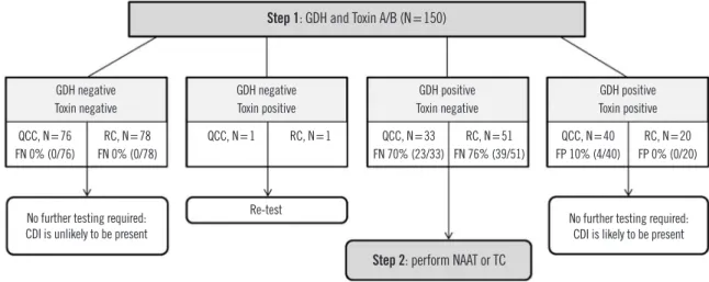
In the two-step algorithm for diagnosis (Fig. 1) [6, 13], when QCC was used as an initial screening test, no further tests were needed for 116 of 150 (77.3%) samples. When RC-GDH and Toxin A/B were used as an initial screening test, no further tests were needed in 98 of 150 (65.3%) samples.
Overall, QCC-GDH and RC-GDH showed satisfactory perfor- mance in detecting GDH, with sensitivities and specificities in the range of previous reports (81.0–100% and 82.0–94.8%, re- spectively) [14, 15]. QCC-Toxin A/B showed a sensitivity of 60%, which accords with previous reports of a wide range of sensitivity, from 29% to 79% [6, 13]. However, the sensitivity of RC-Toxin A/B was relatively low at 33.3%, below that of previous reports showing a narrow range of 48–67%, despite the same test procedure [12, 13, 16, 17]. One GDH-negative but toxin A/
B-positive sample was identified by both QCC and RC. Since this sample was determined to be negative by TC, it was desig- nated as a toxin A/B false-positive result. The most likely expla- nation for this discrepancy is cross-reactivity to toxins formed by other clostridial species, such as C. sordellii, which produce tox-
Table 2. Performance of C. DIFF QUIK CHEK COMPLETE–Toxin A/B and RIDASCREEN Clostridium difficile Toxin A/B compared with toxi- genic Clostridioides difficile culture
Test Result Toxigenic culture Sensitivity (%)
(95% CI)
Specificity (%) (95% CI)
Kappa (95% CI) Positive (N=60) Negative (N=90)
C. DIFF QUIK CHEK COMPLETE–Toxin A/B Positive 36 5 60.0 (46.5–72.2) 94.4 (86.9–97.9) 0.56 (0.41–0.71)
Negative 24 85
RIDASCREEN Clostridium difficile Toxin A/B Positive 20 1 33.3 (22.0–46.8) 98.9 (93.1–99.9)
Negative 40 89
Abbreviation: CI, confidence interval.
Fig. 1. Two-step algorithm for diagnosis of toxigenic Clostridioides difficile infection by applying QCC or RC-GDH and Toxin A/B.
Abbreviations: QCC, C. DIFF QUIK CHEK COMPLETE, QUIK-CHEK COMPLETE; RC, RIDASCREEN Clostridium difficile; FP, false positive; FN, false negative;
CDI, Clostridioides difficile infection; GDH, glutamate dehydrogenase; NAAT, nucleic acid amplification test; TC, toxigenic Clostridioides difficile culture.
Step 1: GDH and Toxin A/B (N=150)
Step 2: perform NAAT or TC GDH negative
Toxin negative QCC, N=76 FN 0% (0/76)
QCC, N=33 FN 70% (23/33)
QCC, N=40 FP 10% (4/40) RC, N=78
FN 0% (0/78)
No further testing required:
CDI is unlikely to be present No further testing required:
CDI is likely to be present Re-test
RC, N=51 FN 76% (39/51)
RC, N=20 FP 0% (0/20) QCC, N=1 RC, N=1
GDH negative Toxin positive
GDH positive Toxin negative
GDH positive Toxin positive
Yoo IY, et al.
Detection of C. difficile GDH and Toxin A/B
https://doi.org/10.3343/alm.2019.39.2.214 www.annlabmed.org 217
ins that are antigenically similar to those of C. difficile [18].
Because no single test is suitable as a stand-alone test, the use of a multiple-step algorithm for CDI diagnosis is recommended in clinical laboratories [6, 19]. Simultaneous detection of both GDH and toxin A/B is less time-consuming than conventional TC and has the advantage of being able to quickly and accu- rately discriminate negative cases as a screening test. The use of QCC and RC as the first step in a two-step algorithm elimi- nated the need for secondary testing in 77.3% and 65.3%, of the samples, respectively. In particular, QCC has the advantage that samples can be analyzed individually and do not need to be batched. However, RC is suitable for high-throughput batch testing in laboratories that require analyses of large numbers of samples.
In summary, as QCC-Toxin A/B is significantly more sensitive than RC-Toxin A/B for identifying toxin producers, QCC is useful for simultaneous detection of GDH and toxin A/B in the first step of the two-step algorithm for diagnosing CDI.
Authors’ Disclosures of Potential Conflicts of Interest
There are no potential conflicts of interest relevant to this article.
REFERENCES
1. Makristathis A, Zeller I, Mitteregger D, Kundi M, Hirschl AM. Compre- hensive evaluation of chemiluminescent immunoassays for the labora- tory diagnosis of Clostridium difficile infection. Eur J Clin Microbiol Infect Dis 2017;36:1253-9.
2. Shin BM, Yoo SM, Shin WC. Evaluation of Xpert C. difficile, BD MAX Cdiff, IMDx C. difficile for Abbott m2000, and Illumigene C. difficile as- says for direct detection of toxigenic Clostridium difficile in stool speci- mens. Ann Lab Med 2016;36:131-7.
3. Mori N and Takahashi T. Characteristics and immunological roles of sur- face layer proteins in Clostridium difficile. Ann Lab Med 2018;38:189- 95.
4. Planche T and Wilcox M. Reference assays for Clostridium difficile in- fection: one or two gold standards? J Clin Pathol 2011;64:1-5.
5. Debast SB, Bauer MP, Kuijper EJ, European Society of Clinical Microbi- ology and Infectious Diseases. European Society of Clinical Microbiology and Infectious Diseases: update of the treatment guidance document for Clostridium difficile infection. Clin Microbiol Infect 2014;20:1-26.
6. Crobach MJ, Planche T, Eckert C, Barbut F, Terveer EM, Dekkers OM, et al. European Society of Clinical Microbiology and Infectious Diseases:
update of the diagnostic guidance document for Clostridium difficile in-
fection. Clin Microbiol Infect 2016;22:S63-81.
7. McDonald LC, Gerding DN, Johnson S, Bakken JS, Carroll KC, Coffin SE, et al. Clinical practice guidelines for Clostridium difficile infection in adults and children: 2017 update by the Infectious Diseases Society of America (IDSA) and Society for Healthcare Epidemiology of America (SHEA). Clin Infect Dis 2018;66:987-94.
8. Park KS, Ki CS, Lee NY. Isolation and identification of Clostridium diffi- cile using ChromID C. difficile medium combined with Gram staining and PRO disc testing: a proposal for a simple culture process. Ann Lab Med 2015;35:404-9.
9. Terhes G, Urbán E, Sóki J, Hamid KA, Nagy E. Community-acquired Clostridium difficile diarrhea caused by binary toxin, toxin A, and toxin B gene-positive isolates in Hungary. J Clin Microbiol 2004;42:4316-8.
10. Lemee L, Dhalluin A, Testelin S, Mattrat MA, Maillard K, Lemeland JF, et al. Multiplex PCR targeting tpi (triose phosphate isomerase), tcdA (Toxin A), and tcdB (Toxin B) genes for toxigenic culture of Clostridium difficile. J Clin Microbiol 2004;42:5710-4.
11. Kato H, Kato N, Watanabe K, Iwai N, Nakamura H, Yamamoto T, et al.
Identification of toxin A-negative, toxin B-positive Clostridium difficile by PCR. J Clin Microbiol 1998;36:2178-82.
12. Vanpoucke H, De Baere T, Claeys G, Vaneechoutte M, Verschraegen G.
Evaluation of six commercial assays for the rapid detection of Clostridi- um difficile toxin and/or antigen in stool specimens. Clin Microbiol Infect 2001;7:55-64.
13. Chung HS and Lee M. Evaluation of the performance of C. DIFF QUIK CHEK COMPLETE and its usefulness in a hospital setting with a high prevalence of Clostridium difficile infection. J Investig Med 2017;65:88- 92.
14. Swindells J, Brenwald N, Reading N, Oppenheim B. Evaluation of diag- nostic tests for Clostridium difficile infection. J Clin Microbiol 2010;48:
606-8.
15. Ota KV and McGowan KL. Clostridium difficile testing algorithms using glutamate dehydrogenase antigen and C. difficile toxin enzyme immu- noassays with C. difficile nucleic acid amplification testing increase di- agnostic yield in a tertiary pediatric population. J Clin Microbiol 2012;
50:1185-8.
16. Mattner F, Winterfeld I, Mattner L. Diagnosing toxigenic Clostridium dif- ficile: new confidence bounds show culturing increases sensitivity of the toxin A/B enzyme immunoassay and refute gold standards. Scand J In- fect Dis 2012;44:578-85.
17. Eastwood K, Else P, Charlett A, Wilcox M. Comparison of nine commer- cially available Clostridium difficile toxin detection assays, a real-time PCR assay for C. difficile tcdB, and a glutamate dehydrogenase detec- tion assay to cytotoxin testing and cytotoxigenic culture methods. J Clin Microbiol 2009;47:3211-7.
18. Toltzis P, Nerandzic MM, Saade E, O’Riordan MA, Smathers S, Zaoutis T, et al. High proportion of false-positive Clostridium difficile enzyme im- munoassays for toxin A and B in pediatric patients. Infect Control Hosp Epidemiol 2012;33:175-9.
19. Cohen SH, Gerding DN, Johnson S, Kelly CP, Loo VG, McDonald LC, et al. Clinical practice guidelines for Clostridium difficile infection in adults:
2010 update by the society for healthcare epidemiology of America (SHEA) and the infectious diseases society of America (IDSA). Infect Control Hosp Epidemiol 2010;31:431-55.