a 10 cm-sized subcapsular hemorrhage connected with a multi-lobulated mass with hemorrhage and necrotic foci in the right liver. The patients underwent right hemihepatectomy with caudate lobectomy and lymphadenectomy. The oper- ation findings revealed metastatic nodules to the diaphragm and omentum. Detailed histopathological analysis through immunohistochemistry confirmed the diagnosis of sarcomatoid cholangiocarcinoma with a poorly undifferentiated sar- comatous component. The patient underwent chemotherapy. To date, the patient is doing well for 8 months after initial diagnosis. (Korean J Hepatobiliary Pancreat Surg 2012;16:70-74)
Key Words: Intrahepatic cholangicarcinoma; Sarcomatous change; Subcapsular hematoma; Chemotherapy
Received: April 30, 2012; Revised: May 14, 2012; Accepted: May 20, 2012 Corresponding author: Dong Eun Park
Department of Surgery, Wonkwang University College of Medicine, 344-2, Sinyoung-dong, Iksan 570-711, Korea Tel: +82-63-855-2389, Fax: +82-63-859-1475, E-mail: [email protected]
Copyright Ⓒ 2012 by The Korean Association of Hepato-Biliary-Pancreatic Surgery Korean Journal of Hepato-Biliary-Pancreatic Surgery ∙ pISSN: 1738-6349
INTRODUCTION
Sarcomatous change of the epithelial neoplasm is a rare condition with a poor clinical course. This condition has been observed in renal cell carcinoma and squamous cell carcinoma and originated from the esophagus, skin and lungs.1 In the liver, sarcomatous transformation has been reported in about 3.9 to 9.4% of hepatocellular carcinoma in autopsy and in about 4.5% of cholangiocarcinoma.2,3 Sarcomatous changes of cholangiocarcinoma are defined as “sarcomatous intrahepatic cholangiocarcinoma” in the World Health Organization (WHO) classification of tumor. To the best of our knowledge, intrahepatic chol- angiocarcinoma associated with intraparenchymal hemor- rhage had been rarely found. We report a patient with a large subcapsular hematoma caused by intrahepatic sarco- matoid cholangicarcinoma who underwent a hepatic re- section, through literature review.
CASE
A 59 year-old man admitted to our emergency center
complaining of pain in the right upper quadrant area which had abruptly started 1 day before. During the as- sessment, he felt dizziness and had mild fever. He had been treated for hypertension for 5 years. There was no past history of liver or biliary diseases. On physical ex- amination, mild tenderness in right upper abdomen was felt. The laboratory results were as follows: hemoglobin, 13.1 g/dl (reference range: 13-18 g/dl), white blood cell, 12.44×10³/μl (reference range: 4-10×103/μl) and plate- let, 281×103/μl (reference range: 150-450×103/μl). The results of serum chemistry and electrolyte were as fol- lows: AST, 205 IU/L (reference range: 5-35 IU/L) and ALT 325 IU/L (reference range: 5-40 IU/L). Other results were within the normal limits. Plain chest and abdomen X-rays showed no abnormal findings. Serum carbohydrate antigen (CA) 19-9 was 34.9 IU/ml (reference range: 0-33 IU/ml) and serum alpha-fetoprotein (AFP) and chorioem- bryonic antigen (CEA) were 2.58 IU/ml (reference range:
0.-5.5 IU/ml) and 7.32 IU/ml (reference range 0-4.6 IU/ml), respectively.
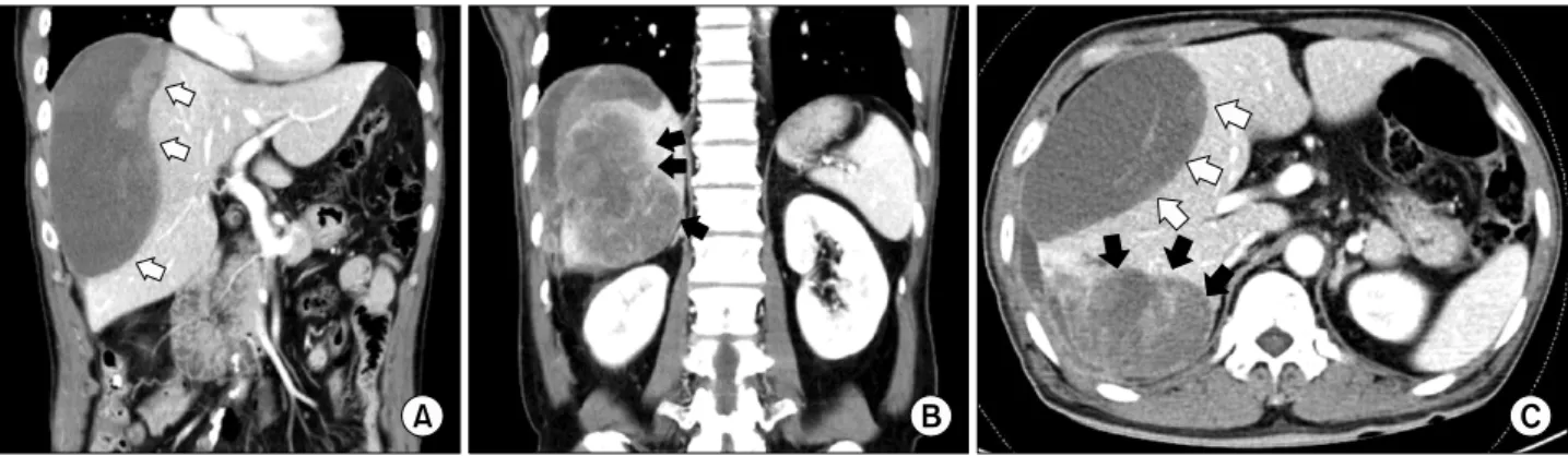
An abdominal computed tomography (CT) showed a homogenous low density lesion in the anterior section of
Fig. 1. Computed tomography (CT) images show (A) a homogenous low density lesion in the anterior section of the liver (white arrows); (B) heterogeneous enhanced mass lesion in the delayed phase with enhanced peripheral bile duct in the posterior section (black arrows); and (C) transverse CT image showing a homogenous anterior mass and heterogenous enhanced of posterior section.
Fig. 2. Magnetic resonance imaging study shows (A) the large tumor of posterior lobe in the liver show heterogeneous lower signal intensity in T1 weighted image (white arrow) and (B) heterogeneous high signal intensity with necrosis and hemorrhage on mass in T2 weighted image (black arrow). It was associated with focal dilation around the bile duct and hepatic capsular retraction of the posterior section in the liver. The large hematoma of the right anterior section was connected with the hetero- genous tumor of the right posterior section by high signal intensity in the right perihepatic space that was suspected as hemorrhage (white circle).
the liver and a heterogeneous enhanced mass lesion in the delayed phase with the enhanced peripheral bile duct in the posterior section (Fig. 1). In magnetic resonance imag- ing (MRI) study, a large tumor of the right posterior sec- tion in the liver showed heterogeneous lower signal in- tensity in T1-weighted image and heterogeneous high sig- nal intensity with necrosis and hemorrhage on mass in T2-weighted image. It was associated with focal dilation around the bile duct and hepatic capsular retraction of the posterior section in the liver. The large hematoma of the right anterior section was connected with the heterogenous tumor of the posterior section by high signal intensity in the right perihepatic space that was suspected as hemor- rhage (Fig. 2). Intrahepatic cholangiocarcinoma was high- ly suspected through MRI and CT findings. We concluded that the large subcapsular hematoma was caused by rup- tured cholangiocarcinoma.
After 1 week of conservative treatment, a follow-up CT scan showed no internal bleeding but increase in hema- toma size. The patient was discharged from the hospital for personal reasons and re-admitted after 1 month. The tumor was further increased in size compared with the previous CT scan findings.
Upon readmission, the patient underwent a laparotomy.
The intraoperative examination revealed a small amount of ascites in the peritoneal cavity. The tumor had strongly adhered to the diaphragm and a 3 cm-sized metastatic nodule was found in the diaphragm adjacent to the live dome without peritoneal seeding or lympadenopathy.
Right hemihepatectomy with cholecystectomy and radical lymph node dissection was performed. The tumor was measured 18×15×10.5 cm. On the cut section, the surface showed massive necrosis with hemorrhage and 6.5 cm of cystic change (Fig. 3). In histological examination, the tu-
Fig. 3. The gross finding of a cut surface showing a cystic change of the anterior section that was defined as a sub- capsular hematoma (red circle) and severe necrosis and hem- orrhage in the tumor with well-developed fibrous capsule (yellow circle).
Fig. 4. Microphotographs show (A) microscopic findings of the resected specimen revealing carcinomatous and sarcomatous component (H&E, ×100). (B) Sarcomatous areas are composed of poorly differentiated cells that were showed a bizarre fashion with scanty cytoplasm and without the spindle bundle (H&E, ×200). (C) There were no features of hepatocytes and carcinomatous area shows moderately differentiated adenocarcinoma (H&E, ×200).
cinoma. The sarcomatous component was positive for vi- mentin but negative for pan-CK and S-100 proteins (Fig.
5). Based on the histological and immunohistochemical findings, a diagnosis of sarcomatoid cholangiocarcinoma was made and the patient was referred to the department of Hemato-oncology for consideration of adjuvant chemo-
Fig. 5. Immunohistochemical examination showing the sarcomatous component being positive for vimentin but negative for pan- cytokeratin (A), and the carcinomatous component revealing positive staining for pan-cytokeratin stain and negative staining for vimentin (B).
therapy.
The patient was started on chemotherapy combined with 5-fluorouracil and cisplatin, which he tolerated well.
A postoperative contrast CT at 3 months after adjuvant chemotherapy confirmed disease recurrence with 2 small nodular lesions that had grown in the right subphrenic space (1 cm) and the omentum (8 mm) that was suspi- cious of peritoneal seeded metastasis. Since the follow-up CT, he received gemcitabine (1,000 mg/m2) and cisplatin (30 mg/m2) chemotherapy in a 21-day cycle. The patient received a total of six cycles and a follow up CT scan showed a partial response. At present, he is doing well for 8 months post-initial presentation.
DISCUSSION
Sarcomatous change of primary cancer of the liver is most common in hepatocellular carcinoma, especially af- ter anticancer chemotherapy or transarterial embolization therapy.2,4 However, intrahepatic cholangicarcinoma with sarcomatous changes is rarely observed and is reported only in 4.5% of surgical autopsy cases. 3 There have been no previous reports concerning the interrelationship be- tween intrahepatic sarcomatoid cholangiocarcinoma and anticancer therapy. The pathogenesis of sarcomatous change with epithelial malignancy is poorly understood.
Several theories have been suggested to explain the inter- mingling of epithelial and mesenchymal malignancies as
follows: 1) mesenchymal reaction, 2) true sarcoma (inclu- ding the collision neoplasm hypothesis), 3) malignancy proliferation of epithelial origin (including the stromal in- duction/metaplasia model), 4) an embryonic cell rest ori- gin, and 5) the totipotential stem cell hypothesis.5,6 Recently, Yoo et al.7 reported that the genetic and gene expression alterations may underlie the sarcomatous change or epithelial mesenchymal transition in cholangio- carcinoma. Diagnosis of sarcomatous change in intra- hepatic cholangiocarcinoma is very difficult before patho- logic confirmation. It appears to be difficult to differ- entiate between two carcinomas because there are no de- tailed distinguishing findings of clinical symptoms. Also, CT scans showed that the hypo-attenuated mass with pe- ripheral regional enhancement was likely reported in most cases. These features resemble those of ordinary cholagio- carcinoma. Shimada et al.8 reported the following charac- teristics of patients who had been diagnosed with chol- angicarcinoma with sarcomatous change: 1) Often demon- strating pyrexia; 2) without always demonstrating remark- able abnormal findings in the laboratory data including tu- mor markers; 3) histologically poorly differentiated ad- enocarcinoma; and 4) a very poor prognosis.
In our case, the patient admitted with severe right upper quadrant abdominal pain with fever and chills and ele- vated serum levels of CEA and CA19-9 of were detected.
Histologically, intrahepatic sarcomatoid cholangiocarci- noma showed two intermingled components, intrahepatic
vious reports showed immunoreaction to both vimentin and cytokeratin. Vimentin that is expressed in mesen- chymal cells may reflect a sarcomatous transformation process, and the cytokeratins suggested that transformed sarcomatous carcinoma cells still retain some phenotypes of carcinoma cells.10 However, our case did not show im- munoreaction of cytokeratins in the sarcomatous region, and this may be related to more dedifferentiation of tumor cells during sarcomatous transformation. It was reported that the prognosis of intrahepatic sarcomatoid chol- angiocarcinoma is worse than that of ordinary intrahepatic cholangicarcinoma. Although recurrence or metastasis of tumor rapidly occurred regardless of the resection proce- dure because the sarcomatous component in the tumor seems to have a high potential to metastasize to variety of regions, a review of previous reports proposed that rad- ical surgery is the treatment of choice in those with lim- ited disease. The survival rates of the patients with surgi- cally resected disease were significantly higher than in pa- tients without resection.10 The role of adjuvant chemo- therapy and the survival benefit to systemic chemotherapy is not well known. Malhotra et al.11 reported that the pa- tient with resected intrahepatic sarcomatoid cholangio- carcinoma achieved a partial response after chemotherapy of combination of gemcitabine and cisplatin. Our patient received this combination chemotherapy as a purpose of palliative treatment because of a microscopic positive margin (sarcomatous component) and direct invasion of the diaphragm. During chemotherapy, the patient did not show progression of the tumor until 6 months after the resection.
ACKNOWLEDGEMENTS
This case report was supported by Wonkwang Univer- sity in 2010.
REFERENCES
1. Haratake J, Horie A. An immunohistochemical study of sarcoma- toid liver carcinomas. Cancer 1991;68:93-97.
2. Kakizoe S, Kojiro M, Nakashima T. Hepatocellular carcinoma with sarcomatous change. Clinicopathologic and immunohisto- chemical studies of 14 autopsy cases. Cancer 1987;59:310-316.
3. Nakajima T, Tajima Y, Sugano I, et al. Intrahepatic cholangio- carcinoma with sarcomatous change. Clinicopathologic and im- munohistochemical evaluation of seven cases. Cancer 1993;72:
1872-1877.
4. Kojiro M, Sugihara S, Kakizoe S, et al. Hepatocellular carcinoma with sarcomatous change: a special reference to the relationship with anticancer therapy. Cancer Chemother Pharmacol 1989;23 Suppl:S4-8.
5. Kubota K, Kakuta Y, Kawamura S, et al. Undifferentiated spin- dle-cell carcinoma of the gallbladder: an immunohistochemical study. J Hepatobiliary Pancreat Surg 2006;13:468-471.
6. Diebold-Berger S, Vaiton JC, Pache JC, et al. Undifferentiated carcinoma of the gallbladder. Report of a case with immunohisto- chemical findings. Arch Pathol Lab Med 1995;119:279-282.
7. Yoo HJ, Yun BR, Kwon JH, et al. Genetic and expression alter- ations in association with the sarcomatous change of cholangio- carcinoma cells. Exp Mol Med 2009;41:102-115.
8. Shimada M, Takenaka K, Rikimaru T, et al. Characteristics of sarcomatous cholangiocarcinoma of the liver. Hepatogastroenter- ology 2000;47:956-961.
9. Kaibori M, Kawaguchi Y, Yokoigawa N, et al. Intrahepatic sarco- matoid cholangiocarcinoma. J Gastroenterol 2003;38:1097-1101.
10. Tsou YK, Wu RC, Hung CF, et al. Intrahepatic sarcomatoid chol- angiocarcinoma: clinical analysis of seven cases during a 15-year period. Chang Gung Med J 2008;31:599-605.
11. Malhotra S, Wood J, Mansy T, et al. Intrahepatic sarcomatoid cholangiocarcinoma. J Oncol 2010;70:701476.