Copyrights © 2016 by The Korean Gastric Cancer Association www.jgc-online.org This is an open-access article distributed under the terms of the Creative Commons Attribution Non-Commercial License (http://creativecommons.org/
licenses/by-nc/4.0) which permits unrestricted noncommercial use, distribution, and reproduction in any medium, provided the original work is properly cited.
Introduction
Gastric cancer is the fourth most common cancer in the world.1,2 A frequent cause of cancer-related death, it is largely responsible for the high mortality rates in East Asia. Despite de- creases in its incidence and mortality rates, it remains the most
common cancer in Korea.3
The standard surgical procedure for resectable advanced gastric cancer is D2 lymphadenectomy with radical gastrectomy. At least 15 dissected lymph nodes (LNs) are required for accurate histo- logical classification of the postoperative LN metastatic stage ac- cording to the TNM staging system of the Union for International Cancer Control/American Joint Committee on Cancer (UICC/
AJCC).4-7 In the 7th version of the TNM system, T-stages are subcategorized and N-stages are more granular.7 The efficacies of T- and N-staging are controversial, but N-stage is generally considered a much stronger prognostic indicator than is T-stage.
There are only a few reports on node-negative advanced gastric cancer, and the most recent have focused on lymphatic invasion.8,9 Correspondence to: Woo Yong Lee
Department of Surgery, Inje University Seoul Paik Hospital, Inje University College of Medicine, 9 Mareunnae-ro, Jung-gu, Seoul 04551, Korea Tel: +82-2-2270-0247, Fax: +82-2-2270-0250
E-mail: [email protected] Received June 17, 2016 Revised August 15, 2016 Accepted August 29, 2016
Prognostic Factors for Node-Negative Advanced Gastric Cancer after Curative Gastrectomy
Eun Woo Lee, Woo Yong Lee1, and Ho-Seok Koo2
Department of Surgery, Inje University Ilsan Paik Hospital, Inje University College of Medicine, Goyang,
Departments of 1Surgery and 2Internal Medicine, Inje University Seoul Paik Hospital, Inje University College of Medicine, Seoul, Korea
Purpose: Lymph node (LN) metastasis is the best prognostic indicator in non-distant metastatic advanced gastric cancer. This study aimed to assess the prognostic value of various clinicopathologic factors in node-negative advanced gastric cancer.
Materials and Methods: We retrospectively analyzed the clinical records of 254 patients with primary node-negative stage T2~4 gastric cancer. These patients were selected from a pool of 1,890 patients who underwent radical resection at Memorial Jin-Pok Kim Korea Gastric Cancer Center, Inje University Seoul Paik Hospital between 1998 and 2008.
Results: Of the 254 patients, 128 patients (50.4%), 88 patients (34.6%), 37 patients (14.6%), and 1 patient (0.4%) had T2, T3, T4a, and T4b tumors, respectively. In a univariate analysis, operation type, T-stage, venous invasion, tumor size, and less than 15 LNs significantly correlated with tumor recurrence and cumulative overall survival. In a multivariate logistic regression analysis, tumor size, venous invasion, and less than 15 LNs significantly and independently correlated with recurrence. In a multivariate Cox proportional hazards analysis, tumor size (hazard ratio [HR]: 2.926; 95% confidence interval [CI]: 1.173~7.300; P=0.021), venous invasion (HR:
3.985; 95% CI: 1.401~11.338; P=0.010), and less than 15 LNs (HR: 0.092; 95% CI: 0.029~0.290; P<0.001) significantly cor- related with overall survival.
Conclusions: Node-negative gastric cancers recurred in 8.3% of the patients in our study. Tumor size, venous invasion, and less than 15 LNs reliably predicted recurrence as well as survival. Aggressive postoperative treatments and timely follow-ups should be considered in cases with these characteristics.
Key Words: Lymph nodes; Prognosis; Stomach neoplasms
Chemotherapy is currently not indicated for gastric cancers at a stage of less than T3 without LN metastasis.10 The aim of this study was to determine the clinicopathological characteristics of and prognostic factors for LN-negative advanced gastric cancer (T2~4N0M0).
Materials and Methods
A total of 1,890 patients underwent curative gastrectomy for gastric adenocarcinoma at the Memorial Jin-Pok Kim Korea Gastric Cancer Center, Inje University Seoul Paik Hospital, be- tween September 1998 and December 2008. Of these patients, our study retrospectively enrolled 254 patients who had primary node-negative gastric cancer, stage T2~4. The exclusion criteria included metachronous gastrointestinal cancer, a previous history of surgery for gastric cancer, and palliative surgery. The patients in our study did not receive systemic chemotherapy or radio- therapy before surgery.
All patients underwent radical gastrectomy with D2 LN dis- section, as defined by the Japanese Gastric Cancer Association.6 They also received adjuvant immunochemotherapy for advanced and node-positive early gastric cancer. The regimen for adjuvant immunochemotherapy was as follows: OK-432 (Streptococcus pyogenes preparation) at 1.0 Klinische Einheit (day 5 after sur- gery, injected) and mitomycin C at 4 mg/50 kg and 5-fluorouracil (5-FU) at 800 mg/50 kg (twice per week for 2 consecutive weeks beginning day 8 after surgery and once per week after 6 weeks, injected). After completing this regimen, patients took oral 5-FU (800 mg/50 kg per day) for 2 years.
Clinicopathological data were extracted from the computerized medical records of the patients. We analyzed the following infor- mation obtained from pathological examinations: clinical stage, T-stage, tumor size, operation type, histologic type, venous inva- sion, lymphatic invasion, neural invasion, Lauren’s classification, tumor location, and the number of dissected LN.
Follow-up assessments were performed every 3 months for the first 2 years after surgery and yearly thereafter. The follow- up procedures included medical history documentation, physical examination, routine blood testing including measurement of tu- mor marker (carcinoembryonic antigen and carbohydrate antigen 19-9) levels, upper endoscopy, chest radiography, abdominal ul- trasonography, and computed tomography. Biopsy and radiologic imaging were used to confirm cancer recurrence. Recurrence was classified as locoregional, hematogenous, peritoneal dissemina-
tion, or multiple metastases. Follow-ups were performed until December 2013 or the patient was lost to follow-up. The mean duration of the follow-up period was 67 months (range, 1~162 months).
The study was reviewed and approved by the Seoul Paik Hos- pital Institutional Review Board (IIT-2016-215).
For statistical analysis, receiver operating characteristic (ROC) analysis was used to determine the optimal cutoff values for continuous variables. Univariate associations were assessed by us- ing logistic, Kaplan-Meier, and log-rank tests. Two multivariate analyses of the prognostic factors for recurrence were performed, one using logistic regression and the other using the Cox propor- tional hazards model.
The statistical analysis were performed using SPSS ver. 12.0 (SPSS Inc., Chicago, IL, USA).
Results
1. Incidence and patterns of recurrence
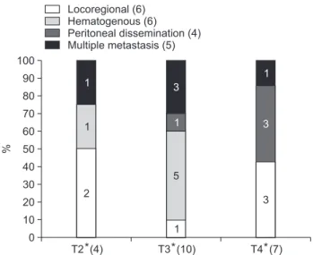
Our study included 254 patients who underwent curative sur- gery and whose pathologic diagnosis was node-negative advanced gastric cancer; 128 patients (50.4%), 88 patients (34.6%), 37 pa- tients (14.6%), and 1 patient (0.4%) had T2, T3, T4a, and T4b tumors, respectively. Tumor recurrence occurred in 21 patients (8.3%) and was locoregional in 6 patients (28.6%), hematog- enous in 6 patients (28.6%), peritoneal dissemination in 4 patients (19.0%), and multiple metastases in 5 patients (23.8%) (Fig. 1).
%
T2 (4) 100
90 80 70 60 50 40 30 20 10 0
T3 (10) T4 (7)
Locoregional (6) Hematogenous (6) Peritoneal dissemination (4) Multiple metastasis (5)
1
1
2
3
1
5
1
1
3
3
* * *
Fig. 1. Recurrence patterns according to T-stage. *Classification according to the TNM staging system of the Union for International Cancer Control/American Joint Committee on Cancer 7th edition.
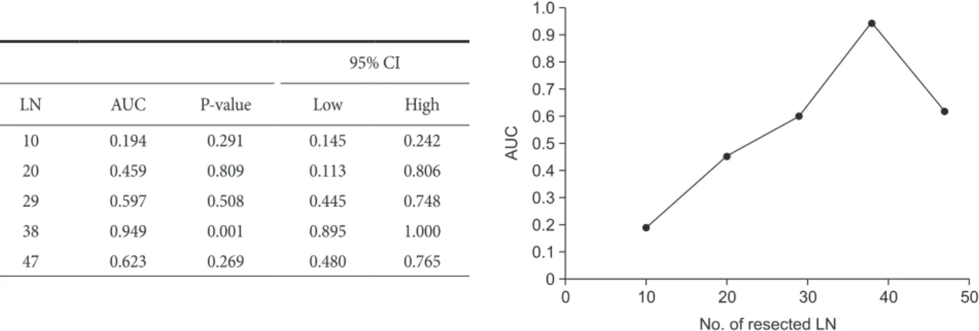
The current TNM system requires that a minimum of 15 LNs (cutoff value=15) be collected via standard gastrectomy. In our study cohort, the total number of resected LNs nodes was 11,270, and the median number of resected LNs was 43. Less than 15 LNs were resected in only 11 patients (4.3%; the LN<15 group).
Based on ROC analysis, the optimal cutoff value for dissected
Table 1. Continued
Variable No. of
patients No. of
recurrences P-value
Neural invasion 0.59
Negative 86 9
Positive 147 12
Histology 0.136
Differentiated 69 3
Undifferentiated 164 18
Signet ring cell or mucinous component 0.762
Negative 141 12
Positive 92 9
Lauren’s classification 0.944
Intestinal 97 9
Diffuse 96 9
Mixed 40 3
Tumor size (cm) 0.024
≤4.7 137 7
>4.7 96 14
Tumor location in the stomach 0.979
Lower one-third 79 7
Mid 108 10
Upper 44 4
Entire 2 0
Resected lymph nodes
<15 7 4 0.001
≥15 226 17
<38 76 8 0.609
≥38 157 13
*Classification according to the TNM staging system of the Union for International Cancer Control/American Joint Committee on Cancer 7th edition.
Table 1. Patient demographics and clinicopathologic characteristics and correlation with recurrence
Variable No. of
patients No. of
recurrences P-value
Sex 0.895
Male 152 14
Female 81 7
Age (yr) 0.538
≤60 105 8
>60 128 13
Operation type 0.021
Partial gastrectomy 158 9
Total gastrectomy 75 12
T-stage* 0.026
2 123 5
3 79 9
4a 30 7
4b 1 0
Venous invasion 0.025
Negative 213 16
Positive 20 5
Lymphatic invasion 0.958
Negative 79 7
Positive 154 14
95% CI
LN AUC P-value Low High
10 0.194 0.291 0.145 0.242
20 0.459 0.809 0.113 0.806
29 0.597 0.508 0.445 0.748
38 0.949 0.001 0.895 1.000
47 0.623 0.269 0.480 0.765
AUC
0 1.0 0.9 0.8 0.7 0.6 0.5 0.4 0.3 0.2 0.1 0
10 20 30 40 50
No. of resected LN
Fig. 2. The optimal cutoff value of LN was obtained by receiver operating characteristic analysis. CI = confidence interval; LN = lymph node; AUC
= area under curve.
LNs was 38 (Fig. 2). In our study cohort, 84 patients (33.1%) had less than 38 resected LNs (the LN<38 group) and 170 patients (66.9%) had more than 38 resected LNs (the LN≥38 group). Tu- mors recurred in 4 patients (57.1%) in the LN<15 group, 17 patients (7.52%) in the LN≥15 group, 8 patients (10.5%) in the LN<38 group, and 13 patients (8.2%) in the LN≥38 group (Table 1).
2. Analysis of clinicopathologic factors and recurrence Table 1 lists the clinicopathologic factors of the patients with node-negative advanced gastric cancer, as well as the relation- ships between these factors and recurrence. Recurrence correlated significantly with operation type (P=0.021), T-stage (P=0.026), venous invasion (P=0.025), tumor size (P=0.024), and LN number
<15 (P=0.001). A multivariate logistic regression analysis showed that tumor size (P=0.021), venous invasion (P=0.019), and LN number <15 (P=0.001) were independent predictors of recurrence.
3. Overall survival of patients with node-negative advanced gastric cancer
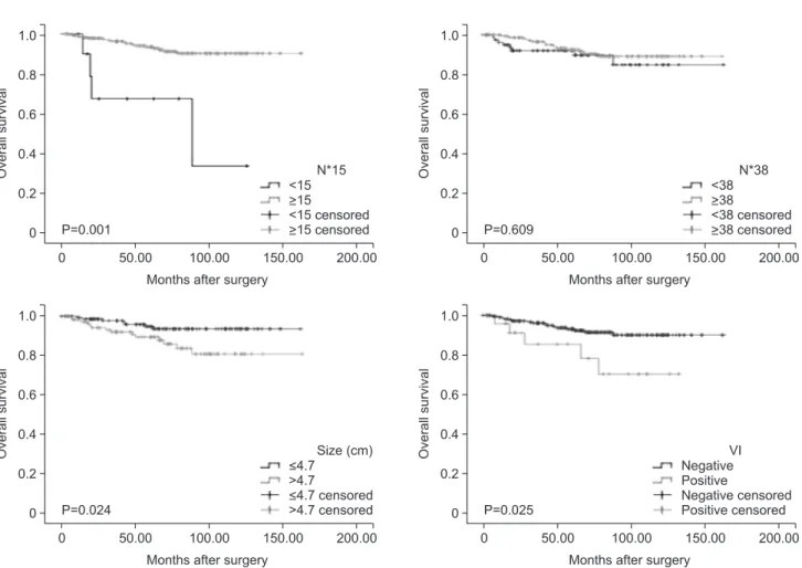
In a multivariate analysis using logistic regression, cumulative overall survival (OS) was significantly associated with operation type (P=0.043), T-stage (P=0.026), venous invasion (P=0.016), tumor size (P=0.024), and LN number<15 (P<0.001), but not LN number<38 (Fig. 3). In a multivariate analysis using the Cox pro- portional hazards model, cumulative OS was significantly associ- ated with tumor size (P=0.021), venous invasion (P=0.010), and LN number<15 (P<0.001) (Table 2).
Table 2. Multivariate Cox regression analysis for overall survival Hazard
ratio 95% confidence
interval P-value
Tumor size (cm) 2.926 1.173~7.300 0.021
Venous invasion 3.985 1.401~11.338 0.010
<15 resected lymph nodes 0.092 0.029~0.290 <0.001
Overallsurvival
0 0.8
0.6
0.4
0.2
0
50.00 100.00 150.00 200.00
Months after surgery
<15
>15
<15 censored
>15 censored N*15 1.0
P=0.001
Overallsurvival
0 0.8
0.6
0.4
0.2
0
50.00 100.00 150.00 200.00
Months after surgery
<38
>38
<38 censored
>38 censored N*38 1.0
P=0.609
Overallsurvival
0 0.8
0.6
0.4
0.2
0
50.00 100.00 150.00 200.00
Months after surgery
<4.7
>4.7
<4.7 censored
>4.7 censored Size (cm) 1.0
P=0.024
Overallsurvival
0 0.8
0.6
0.4
0.2
0
50.00 100.00 150.00 200.00
Months after surgery Negative Positive
censored Negative
Positive censored VI 1.0
P=0.025
Fig. 3. Kaplan-Meier analysis of overall survival according to significant clinicopathologic factors with node-negative advanced gastric cancer. VI = venous invasion. *Resected lymph nodes.
Discussion
Despite improvements in survival rates attributable to early detection and radical lymphadenectomy, gastric cancer remains one of the most common causes of death from cancer worldwide, as well as the fourthmost common cancer worldwide.1,2 Because concrete evidence supports its use in stage II and III gastric cancer, adjuvant chemotherapy is widely accepted as a standard treatment for gastric cancer patients with a high risk of recur- rence. Hence, N-stage is the most significant prognostic indicator in gastric cancer. However, adjuvant chemotherapy is not recom- mended for pT3N0 gastric cancers despite prognostic similarities between the stage IIA gastric cancer subgroups.10 The decision to administer adjuvant chemotherapy to patients withT3N0 gastric cancer is currently left to the clinicians’ discretion. This study, which examined T2~4N0 gastric cancers, found that T-stage was a significant prognostic factor for recurrence and OS in a univari- ate analysis, but not a multivariate analysis using the Cox propor- tional hazards model.
Among the various prognostic factors previously reported, tu- mor size is especially important in node-negative advanced gas- tric cancer. Adachi et al.11 identified gastric tumor size as a simple prognostic indicator. Kim et al.12 found that patients with node- negative gastric cancers had favorable outcomes, whereas those with relatively large tumors and serosal invasion had unfavorable outcomes. Yokota et al.13 showed that tumor size predicted sur- vival rates in patients with gastric cancer. Our study shows that gastric tumor size and prognosis are closely associated.
In the study by Zhang et al,14 the number of retrieved LNs was an independent prognostic factor in node-negative gastric can- cer, and retrieval of more than 15 LNs correlated with improved survival rates. In that study, less than 15 LNs were retrieved from most patients (74 of 106 patients, 69.8%). In our study, the average number of retrieved LNs was 44.3, and in almost all patients (243 of 254 patients, 95.7%), at least 15 were retrieved. The prognosis of patients with at least 15 retrieved LNs was significantly better than that of patients with less than 15 retrieved LNs. In the study by He et al.15 of patients with advanced node-negative cancer, survival rates improved as the number of retrieved LNs follow- ing radical gastrectomy increased. In that study, the cutoff value for retrieved LNs was 18 as determined via ROC analysis, and the prognosis of patients with more than 18 retrieved LNs was significantly better than that of patients with 18 or fewer retrieved LNs. We also used ROC analysis to determine the optimal cutoff
value for LNs in node-negative gastric cancer. We calculated the area under ROC curves at different cutoff values corresponding to different OS times and obtained a substantially greater cutoff value (38) than did He et al.15 However, there was no significant difference between the ≥38 and <38 groups in terms of recur- rence. Therefore, at least 15 LNs should be collected, as recom- mended by the UICC/AJCC cancer staging manual.
Lymphovascular invasion (LVI) is recognized as a negative prognostic factor in several malignancies, including gastric can- cer.3,5-7,16 Previous studies showing that LVI is an independent predictor of worse survival in gastric cancer include the gene ex- pression analysis by Dicken et al.17 and the comparative analysis by Lee et al.18 of patients with N0 versus N1 gastric cancer. In our study, however, there was no significant relationship between venous invasion and lymphatic invasion. Furthermore, venous invasion correlated significantly with OS and recurrence, whereas lymphatic invasion did not. These findings may reflect our study’s focus on node-negative patients only.
In conclusion, we recommend the development of compre- hensive, individualized treatment plans for patients with node- negative gastric cancer. Our analysis of these cancers showed that tumor size, venous invasion, and at least 15 retrieved LNs were independent prognostic indicators of survival. Further evaluation of prognostic factors is needed to determine whether treatments such as adjuvant therapy benefit patients with node-negative ad- vanced gastric cancer.
Conflicts of Interest
No potential conflict of interest relevant to this article was reported.
References
1. Siegel R, Ma J, Zou Z, Jemal A. Cancer statistics, 2014. CA Cancer J Clin 2014;64:9-29.
2. DeSantis CE, Lin CC, Mariotto AB, Siegel RL, Stein KD, Kramer JL, et al. Cancer treatment and survivorship statistics, 2014. CA Cancer J Clin 2014;64:252-271.
3. Jung KW, Won YJ, Kong HJ, Oh CM, Cho H, Lee DH, et al.
Cancer statistics in Korea: incidence, mortality, survival, and prevalence in 2012. Cancer Res Treat 2015;47:127-141.
4. Cuschieri SA, Hanna GB. Meta-analysis of D1 versus D2 gastrectomy for gastric adenocarcinoma: let us move on to an-
other era. Ann Surg 2014;259:e90.
5. Memon MA, Subramanya MS, Khan S, Hossain MB, Osland E, Memon B. Meta-analysis of D1 versus D2 gastrectomy for gastric adenocarcinoma. Ann Surg 2011;253:900-911.
6. Japanese Gastric Cancer Association. Japanese classification of gastric carcinoma: 2nd English edition. Gastric Cancer 1998;1:10-24.
7. Washington K. 7th edition of the AJCC cancer staging manual:
stomach. Ann Surg Oncol 2010;17:3077-3079.
8. Kim SS, Choi BY, Seo SI, Jung MY, Choi HS, Ahn SM, et al.
The comparison between 6th and 7th International Union against Cancer/American Joint Committee on Cancer clas- sification for survival prognosis of gastric cancer. Korean J Gastroenterol 2011;58:258-263.
9. Kim JH, Kim CW, Choi NK, Kwak JH, Choi KM, Jang HJ, et al. The comparison between 6th and 7th UICC/AJCC N Stage for prognostic significance in gastric cancer. J Korean Surg Soc 2010;79:202-206.
10. Imamura T, Komatsu S, Ichikawa D, Kubota T, Okamoto K, Konishi H, et al. Poor prognostic subgroup in T3N0 stage IIA gastric cancer, suggesting an indication for adjuvant chemo- therapy. J Surg Oncol 2015;111:221-225.
11. Adachi Y, Oshiro T, Mori M, Maehara Y, Sugimachi K. Tumor size as a simple prognostic indicator for gastric carcinoma.
Ann Surg Oncol 1997;4:137-140.
12. Kim DY, Seo KW, Joo JK, Park YK, Ryu SY, Kim HR, et al.
Prognostic factors in patients with node-negative gastric car- cinoma: a comparison with node-positive gastric carcinoma.
World J Gastroenterol 2006;12:1182-1186.
13. Yokota T, Ishiyama S, Saito T, Teshima S, Yamada Y, Iwamoto K, et al. Is tumor size a prognostic indicator for gastric carci- noma? Anticancer Res 2002;22:3673-3677.
14. Zhang BY, Yuan J, Cui ZS, Li ZW, Li XH, Lu YY. Evaluation of the prognostic value of the metastatic lymph node ratio for gastric cancer. Am J Surg 2014;207:555-565.
15. He H, Shen Z, Wang X, Qin J, Sun Y, Qin X. Survival benefit of greater number of lymph nodes dissection for advanced node- negative gastric cancer patients following radical gastrectomy.
Jpn J Clin Oncol 2016;46:63-70.
16. Rausei S, Dionigi G, Ruspi L, Proserpio I, Galli F, Tirotta F, et al. Lymph node staging in gastric cancer: new criteria, old problems. Int J Surg 2013;11 Suppl 1:S90-S94.
17. Dicken BJ, Graham K, Hamilton SM, Andrews S, Lai R, List- garten J, et al. Lymphovascular invasion is associated with poor survival in gastric cancer: an application of gene-expression and tissue array techniques. Ann Surg 2006;243:64-73.
18. Lee JH, Kim MG, Jung MS, Kwon SJ. Prognostic significance of lymphovascular invasion in node-negative gastric cancer.
World J Surg 2015;39:732-739.