Copyright © 2019 The Korean Society for Bone and Mineral Research
This is an Open Access article distributed under the terms of the Creative Commons Attribution Non-Commercial Li- cense (http://creativecommons.org/licenses/by-nc/4.0/) which permits unrestricted non-commercial use, distribu- tion, and reproduction in any medium, provided the original work is properly cited.
The Function of the Vitamin D Receptor and a Possible Role of Enhancer RNA in Epigenomic
Regulation of Target Genes: Implications for Bone Metabolism
Shun Sawatsubashi1, Koichi Nishimura2,3, Jinichi Mori2,3, Alexander Kouzmenko3, Shigeaki Kato2,3
1Department of Molecular Endocrinology, Fujii Memorial Institute of Medical Sciences, Institute of Advanced Medical Sciences, Tokushima University, Tokushima;
2Center for Regional Cooperation, Iwaki Meisei University, Iwaki;
3Research Institute of Innovative Medicine, Tokiwa Foundation, Jyoban Kamiyunagayamachi, Iwaki, Japan
Vitamin D (VD) is essential for bone health, and VD or its analogues are widely used in clinics to ameliorate bone loss. The targets and mode of VD anti-osteoporotic actions appear to be different from those of other classes of drugs modulating bone remodel- ing. VD exerts its biological activities through the nuclear VD receptor (VDR)-mediated transcriptional regulation of target mRNA and non-coding RNA genes. VD-induced gene regulation involves epigenetic modifications of chromatin conformation at the target loci as well as reconfiguration of higher-order chromosomal organization through VDR- mediated recruitment of various regulatory factors. Enhancer RNAs (eRNA), a class of non-coding enhancer-derived RNAs, have recently emerged as VDR target gene candi- dates that act through reorganization of chromatin looping to induce enhancer-pro- moter interaction in activation of mRNA-encoding genes. This review outlines the mo- lecular mechanisms of VD actions mediated by the VDR and suggests novel function of eRNAs in VDR transactivation.
Key Words: Chromatin reorganization · Enhancer RNA · Non-coding RNA · Vitamin D · Vi- tamin D receptor
INTRODUCTION
Vitamin D (VD) is a pivotal hormone/vitamin controlling calcium homeostasis, but also regulating a number of biological processes.[1] VD exerts a wide variety of biological actions through nuclear VD receptor (VDR)-mediated transcriptional regulations of the target genes, and the target gene products facilitate the bio- logical effects.[2-4] Clinically, VD as well as the related compounds is successful as an anti-osteoporotic agent, drawing huge attention to the molecular basis of the VD actions in the bone field.[5] Ligand-dependent transcriptional by VDR is cou- pled with chromatin reconfiguration/epigenetic alteration.[3,4] Recent progress of whole human genome analysis has uncovered the presence of numerous of non-coding RNAs (ncRNAs) and tertiary structure of chromatin and their signifi- Corresponding author
Shigeaki Kato
Center for Regional Cooperation, Iwaki Meisei University, 5-5-1 Chuodai Iino, Iwaki City, Fukushima 970-8551, Japan
Tel: +81-246-29-5394 Fax: +81-246-29-5414
E-mail: [email protected];
[email protected] Received: September 21, 2018 Revised: December 18, 2018 Accepted: January 15, 2019
No potential conflict of interest relevant to this article was reported.
cance in gene regulation.[6] As the certain group of the non-coding RNA genes are transcribed by RNA polymerase II,[7] such ncRNAs could be VD/VDR target genes. In this short review, the role of enhancer RNA (eRNA), a newly emerged class of ncRNAs, in the gene regulations is re- viewed, and possible contribution of eRNA to the VDR-me- diated epigenetic regulation is discussed.
BIOLOGICAL ROLE OF THE NUCLEAR VDR) 1. The VDR mediates the biological actions of VD
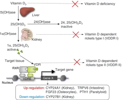
VD is a fat-soluble vitamin normally present as a nutri- ent in balanced diets; it can also be biosynthesized in the body in response to ultraviolet light exposure. In verte- brates, VD exerts a wide variety of biological actions and plays a pivotal role in calcium homeostasis.[1] Owing to its significance in physiological processes, an excess or a defi- ciency of VD leads to severe pathological conditions, in- cluding diseases like rickets.[2] The biologically active form of VD, 1α,25-dihydroxy-vitamin D3 (1α,25[OH]2D3), is pro- duced through 2 hydroxylation steps in the liver and then the kidney. Hepatic CYP24B1 hydroxylates the precursor of VD into 25(OH)D3 followed by its conversion into the active VD form through the second (1α-hydroxylation) by the re-nal enzyme CYP27B1.[1,8] Loss-of-function of CYP27B1 by genetic mutations causes type I hereditary rickets, howev- er these patients can be rescued by administration of pre- cursor 1α(OH)D3 or 1α,25(OH)2D3.[9] Excess VD activates kidney CYP24A1, resulting in the conversion of 1α,25(OH)2D3 into 24,25(OH)2D3, a biologically inactive form of VD (Fig. 1).[1,8]
Most of the known biological actions of VD are mediated by the nuclear VDR.[3,4,10] The VDR belongs to the nuclear receptor (NR) gene superfamily comprised of 48 currently known members.[11] Similar to other family members, the VDR acts as a ligand-dependent DNA-binding transcrip- tional factor. For DNA binding, the VDR requires coopera- tion with one of the 3 subtypes (α, β, or γ) of another NR superfamily member, the retinoid X receptor (RXR) and for- mation of VDR-RXR heterodimers.[3,4] A consensus DNA binding site for VDR-RXR is composed of 2 AGGTCA direct repeats (DR) spaced by 3 nucleotides (a so-called DR3-type motif sequence), 5’-AGGTCANNNAGGTCA-3’. This DNA se- quence serves as an efficient VD-responsive transcriptional enhancer named the VD response element (VDRE).[3,4]
ChIP-sequencing (ChIP-seq) analyses have shown that the numbers and locations of genomic VDR binding sites differ significantly between different cell types. Surprising-
Fig. 1. Schematic representation of vitamin D signaling and its related disease. Vitamin D is converted into an active form as a vitamin D receptor (VDR) ligand for gene regulation. Deficiency of dietary vitamin D as well as genetic mutations inducing malfunction of 1α-hydroxylase and VDR causes rachitic abnormality. VDDRI, vitamin D-dependent rickets type 1; VDDRII, vitamin D-dependent rickets type 2; RXR, retinoid X receptor;
VDRE, vitamin D response element; 1α,25(OH)2D3, 1α,25-dihydroxy-vitamin Dw.
Vitamin D3
25(OH)ase
Liver
25(OH)D3 24(OH)ase
Kidney
24, 25(OH)2D3 inactive 1α(OH)ase
1α, 25(OH)2D3 active
Target tissue VDR
Up-regulation: CYP24A1 (Kidney)、TRPV6 (Intestine) FGF23 (Osteocytes)、PTH1 (Paratyloid) Down-regulation: CYP27B1 (Kidney)
Target gene
Vitamin D deficiency
Vitamin D dependent rickets type I (VDDR I)
Vitamin D dependent rickets type II (VDDR II)
ly, only a minority of identified genomic VDR binding sites contain functional variants of the VDRE consensus sequence, [12] suggesting that VDRE-independent chromatin bind- ing of VDR might be mediated by various cell-specific reg- ulatory factors. It has been shown that for chromatin bind- ing, sex steroid receptors (androgen or estrogen receptor homodimers) require an association with so-called pioneer- ing factors, such as FOXA1.[13,14] Therefore, it is likely that similar, yet to be identified regulatory factors modulate (i.e., facilitate or inhibit) VDR binding to chromatin regard- less of genomic VDRE motif sequences. In this regard, as a candidate, co-localization of PU.1 with VDR on chromatin was reported.[15]
2. VDR is a VD-dependent transcription factor
Although the VDR may associate with chromatin and specific DNA elements even in the absence of VD, the li- gand is required for the VDR to regulate target gene tran- scription (Fig. 2).[3] VD-induced transcription requires the assembly of multi-subunit protein complexes at the target gene promoters.[16] These complexes include the transcrip- tion initiation complex (RNA polymerase II plus basal tran- scription factors) and a mediator complex (MED) bridging the promoter-bound VDR with the transcription initiationcomplex.[17,18]
In the absence of ligand, the VDR in the nucleus appears to associate with transcriptional co-repressors. For VD-in- duced modulation of gene expression, functionally oppos- ing VDR transcriptional co-regulators sequentially dissoci- ate and associate upon ligand biding. In transactivation, co-repressors that are associated with the VDR dissociate and are replaced by co-activator complexes. In VD-depen- dent transrepression, dissociation of co-repressors appears to be aborted and/or different types of co-repressors are recruited by the VDR in a ligand-dependent manner.[19,20]
However, in comparison with transactivation, there is un- certainty regarding the molecular events that occur during ligand-induced transrepression.
3. The VDR is essential for bone growth and health
Lack-of-function VDR mutations lead to development of severe skeletal abnormalities known as hereditary type II rick- ets.[2,10] In contrast to type I rickets,[9] neither 1α,25(OH)2D3
nor 1α(OH)D3 can rescue severe rachitic phenotypes in type II rickets patients.[2,10]
Type II rickets phenotypes can be experimentally reca- pitulated in mice by experimental VDR gene knock-out
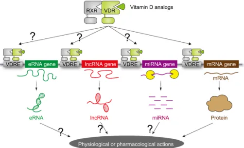
Fig. 2. Schematic representation of vitamin D target genes. The target genes for vitamin D receptor (VDR) were considered to be limited to the mRNA genes encoding proteins. However, recent progress in human genome by means of the next generation DNA sequencers have uncovered that more than 80% of the human genome encode non-coding RNAs (ncRNA). As some classes of the ncRNAs are transcribed by RNA polymerase II, VDR is assumed to transcribe a set of ncRNAs as target genes. RXR, retinoid X receptor; VDRE, vitamin D response element; eRNA, enhancer RNA; IncRNA, long non-coding RNA; miRNA, microRNA.
(KO), and animals with genetic VDR deficiency display se- vere skeletal abnormalities.[10] However, rachitic pheno- types in VDR KO mice develop after weaning.[10] This sug- gests that VD-VDR signaling is dispensable in nursing ani- mals, but becomes essential after the transition to a nor- mal diet. Remarkably, severe skeletal defects in the adult VDR KO mice can be ameliorated simply by feeding with a high calcium diet.[21] Thus, high calcium intake from nurs- ing milk appears to protect the mutant neonates from ra- chitic disorders. This suggests that the VDR-mediated ac- tions of VD in bone growth and metabolism do not target bone tissue directly, but rather mediate serum calcium con- centrations sufficient for bone formation.[22,23] Indeed, an elegant study on mice with selectively ablated VDR in the intestine has shown that calcium mobilization from bone is enhanced when a hypocalcemic state was induced.
[24,25]
4. Synthetic VD compounds are beneficial for bone health in hypervitaminosis
Osteomalacia is attributed to insufficient calcium intake, similar to VD deficiency,[1] as postulated from the results obtained with VDR mutant mice.[10,21-25] VD dietary sup- plementation is widely recommended to prevent a hypo- calcemic state, and in the US, calcium is widely distributed through milk. Due to low levels of calcium in water in Ja- pan and in other countries, calcium intake from the diet tends to be insufficient, particularly for the elderly who lose the ability to absorb calcium. This could explain why VD is clinically effective for bone mass increases for osteo- porotic patients in Japan. Use of eldecalcitol (ELD) has been a clinically successful approach to preventing bone fracture and increasing bone mass. However, it has less of a calce- mic effect than does calcitriol.[5,26] Furthermore, supple- mentation of ELD appears beneficial in any anti-osteopo- rotic drug treatment in terms of target tissues and skeletal cell types.[5,23] The other anti-osteoporotic drugs on the market modulate bone metabolism by attenuating bone resorption and/or stimulating bone formation for bone fracture prevention. In contrast, the major target tissue for ELD appears to be the intestine and kidney.[23,26] This dif- ference in the target tissues/cells for ELD and the other drugs warrants the use of ELD as a co-treatment with other anti- osteoporotic drugs.
MOLECULAR MECHANISMS OF
TRANSCRIPTIONAL AND EPIGENETIC CONTROL BY THE VDR
1. Pleiotropic tissue-specific action of VDR
The impaired growth and maintenance of bone in type II rickets patients and VDR KO mutant mice can be corrected by high levels of calcium supplementation.[14,17] Howev- er, other notable phenotypic abnormalities such as alope- cia cannot be reversed by any currently known treatment.Since type I rickets patients do not exhibit hair loss in early childhood, infant alopecia is a clinical indication distinguish- ing type II hereditary rickets from type I.[4,17] None of the available natural or synthetic VDR agonists has been able to prevent hair loss in these patients or mutant mice.[10]
However, targeted expression of a VDR transgene in the skin of VDR KO mice rescued the alopecia phenotype,[27]
suggesting that epithelial follicular cells are direct target of the VDR. These and similar observations indicate that the VDR targets tissue-specific sets of genes (Fig. 1).[3,4]
Initially, the VD-induced transcription of protein-encod- ing genes was assumed to account for all pleiotropic ac- tions of VDR. However, recent mammalian transcriptome studies demonstrated that a significant fraction of the RNA polymerase II transcripts represent non-protein coding RNA species,[6,28] and ChIP-seq experiments showed that the VDR was also bound to genomic sites remote from protein encoding genes.[12] These results suggest that non-cod- ing RNA genes might potentially be regulated by VD-VDR signaling (Fig. 2), though this idea still awaits experimental confirmation.
2. VDR-mediated transactivation involves
chromatin reorganization at target loci
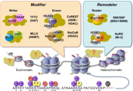
The VDR regulates cell type-specific sets of genes that reflect differences in the state of chromatin at the target loci between cells of different tissues. Arrays of target genes also change during cell differentiation. Such spatial and temporal expression of genes targeted by the VDR is achieved through highly regulated chromatin remodeling and his- tone modification.[29,30] The ordered alterations of chro- matin are controlled by a range of nuclear protein factors that support fundamental steps of gene regulation com- mon for all kinds of DNA-binding transcriptional factors, including the VDR.[29,30]Regulated chromatin remodeling [31,32] is directed by post-translational modification (PTM) of histone proteins (Fig. 3) known as the “histone code”.[33-35] Histone PTMs can be reversed through the action of functionally oppos- ing enzymes (histone modifiers). Their activities on chro- matin are finely tuned by intracellular and extracellular sti- muli that direct the transitions between active and repressed states of chromatin at specific genomic loci.[33,34]
Initially, histone acetylation and deacetylation were thou- ght to direct the transition between active and repressed chromatin states.[33,34] However, in signaling cascades, several histone PTMs were identified upstream that impact- ed chromatin conformation.[33,35,36] Currently, specific patterns of histone methylations are regarded as pivotal determinants for downstream histone PTMs.[33,34,37] A lysine (K) residue NH3+ group can be mono-, di- or tri-meth- ylated, and the arginine (R) NH2+side chain group can be mono- or dimethylated. Patterns of methylation at particu- lar histone H3 lysine residues appear to be crucial in defin- ing chromatin’s state. Methylations at H3K4 and H3K36 rep- resent reliable markers of activated chromatin, whereas methylations at H3K9 and H3K27 are markers of repressed genomic regions. The degree of methylation, or the num-
ber of methyl moieties on specific H3 lysine residues ap- pears to vary between different functional genomic regions.
Di- and tri-methylated H3K4 (H3K4me2/3) are closely asso- ciated with activated chromatin at mRNA-encoding gene loci, including their promoters.[33,34,37] Whereas H3K27m2/3 are more prevalent at silent enhancer regions of chroma- tin, acetylated H3K27 is associated with activated enhanc- er genomic regions. Apparently, repressing lysine methyla- tion residues, like H3K27 or H3K9, can be either acetylated or methylated, thereby providing a mechanism of revers- ing opposing states of chromatin.[33,36] Interestingly, acet- ylation of activating methylation residues has not been de- tected and would most likely be functionally redundant.
Thus, the effects of histone lysine methylation on chroma- tin organization are more complex than that of acetylation.
Thus, histone acetylation and deacetylation invariably pro- duce opposing results. Moreover, chromatin activation can be accomplished by both methylation (at H3K4 and H3K36) and demethylation (methylated H3K9 and H3K27); and in reverse, depending on the substrate, histone methylation (H3K9 and H3K27) and demethylation (methylated H3K4 and H3K36) would likely result in transcriptional repression.
[33,34,37]
Fig. 3. Schematic representation of chromatin reorganization and epigenetic regulators. Chromatin reorganization is achieved through histone modification and chromatin remodeling. The specific combination of histone modifications direct the state of chromatin activation. The chromatin remodelers and histone modifiers form in general multi-subunit complexes. TFTC, T cell transcription factor; TRRAP, transformation/transcription domain-associated protein; GCN5, general control of amino acid synthesis 5; OGT, O-GlcNAc transferase; MLL5, mixed lineage leukemia 5; LSD1, lysine-specific demethylase 1; RetCoR, RNA helicase DHX30; CoREST, repressor element 1 silencing transcription factor corepressor; HDM, his- tone demethylase; HDAC, histone deacetylase; SWI, switching defective; SNF, sucrose nonfermenting; BRG, Brahma-related gene; BRM, Brahma;
NuRD, nucleosome remodelling and deacetylase.
3. Histone modifiers and chromatin remodelers are transcriptional co-regulators of the VDR
The VDR exerts its regulatory effects on chromatin in co- operation with various histone modifiers and ATPase-de- pendent chromatin remodelers that function as transcrip- tional co-regulators of the VDR (Fig. 3).[29,30] Generally, his- tone modifying and chromatin remodeling enzymes do not act alone, but function as catalytic subunits in large multi- subunit nuclear protein complexes often exceeding 2 MDa in size (Fig. 3),[17,18,38] by aid of histone chaperones.[39-41]Histone acetyltransferases (HATs) and histone deacety- lases (HDACs) were the first cofactors shown to co-activate and co-repress the VDR and other NRs through ε-N lysine acetylation and ε-N-acetyl lysine deacetylation, respective- ly.[3,12,17-19,29] During VDR transactivation, HDAC com- plexes dissociate from ligand-receptor complexes followed by VDR recruitment of HAT activities. This basic mechanism accounts only for the relatively fast activation by NRs of eu- chromatic loci that were being slowly transcribed. Howev- er, the dynamics of induction of VDR target genes appear to correlate with the degree of their chromatin repression.
The transactivation of repressed target loci requires more extensive multistep rewriting of histone code markers (i.e., erasing of repression and introduction of activation PTMs) leading to more profound changes in chromatin configu- ration. As an upstream signal, histone methylation is be- lieved to direct the rewriting of histone PTMs through co- ordinated action of various histone methyltransferase (HMT) and histone demethylase (HDM) activities.[33,36,37] Guid- ed by de novo introduced histone PTMs, recruited chroma- tin remodeling factors reconfigure chromatin into confor- mations permissive for transcriptional activation.[33,34,36]
In vitro assays showed that some, but not all HMTs and HDMs directly interact with the VDR and other NRs.[20,36,42] In contrast, chromatin remodeling ATPases interact with NRs indirectly, through bridging proteins of multi-subunit re- modeler complexes.[17,18]
A POSSIBLE ROLE OF ERNA IN
CHROMOSOMAL RECONFIGURATION BY THE VDR
1. Dynamic chromatin reorganization supports gene regulation
Efforts to understand cis- and trans-regulatory factors
and their impact on gene activities are being extended to higher orders of chromosomal organization. Studies of in- teractions between genomic segments by various chromo- some conformation capture techniques (Hi-C, ChIP-loop, chromatin interaction analysis with paired-end tag sequenc- ing and related methods) have revealed that individual chromosomes are organized into topologically associating domains (TADs) made distinctive by increased intra-do- main chromatin contacts.[43,44] Structurally, each TAD is flanked by TAD boundary elements and contains multiple subdomains of DNA loops. The molecular basis of TAD for- mation and subdomain organization and dynamics remains largely unclear. However, it has been established that the TAD DNA loops are formed and stabilized by interactions between cohesins, ring-like protein complexes better known for holding sister chromatid together, and CCCTC-binding factor (CTCF), a DNA binding protein initially identified as a transcriptional repressor.[6,44,45] The observed correlation between the distribution of specific epigenetic markers or bound RNA polymerase II and boundaries of individual TADs along the chromosome suggests that individual TADs may have different functionalities (active, repressed or inert), and that genes within the same TAD are transcribed toge- ther. The CTCF binding was indeed observed in the pro- moters of the VD target genes.[46] Furthermore, genome- wide studies of long-range chromatin interactions indicate that chromatin interactions are restricted within TADs, and that DNA loop subdomains provide spatial interaction be- tween distal enhancers and promoters of regulated gene (Fig. 4).[6,45]
2. A possible role of eRNA in chromatin looping and enhancer-mediated gene activation by the VDR
TADs contribute to gene regulation by modulating the interaction between DNA regulatory elements, such as en- hancers, and their target genes through intra-domain DNA looping dynamics. Various nuclear proteins and their com- plexes have been implicated in bridging distal DNA enhanc- ers with proximal promoters of regulated target genes. Re- cently, eRNA (Fig. 5) has been identified as a factor that fa- cilitates DNA looping and intra-loop sequence interaction (Fig. 4).[6,7,45,47] Interestingly, as a trans-mechanism of regulation, eRNA has been shown to recruit cohesin to tar- geted gene promoters located on different chromosomes.
eRNAs are transcribed by RNA polymerase II from clustered, or super enhancers (Fig. 4).[45,47] Ligand-dependent in- duction of eRNAs and their involvement in gene regulations were shown in the target mRNA genes for estrogen recep- tor α (ERα).[48,49] It is therefore, conceivable that eRNA genes may target genes for the VDR, similar to other class- es of ncRNAs shown to be transcribed by RNA polymerase II, like miRNAs (Fig. 6), although no report is yet available about eRNAs’ involvement in gene regulations by the VDR.
Our group is currently testing the above hypothesis us- ing human 24-hydroxylase (CYP24A1) as a model gene, the transcription of which can be activated by VD.[4,8] In hu-
man keratinocyte HaCaT cells, we observed strong induc- tion of endogenous CYP24A1 expression after treatment with VD.[50] Public database records show that the inter- genic region downstream from the structural CYP24A1 gene harbors a super enhancer. The presence of several function- al VDREs has been reported in this super enhancer region.
[51]
In a preliminary screening of transcripts, we identified several eRNA candidates transcribed from the 3’-intergenic enhancer region. It has been reported that eRNAs are ex- pressed at low levels and their half-lives are quite short due to instability.[47] Furthermore, distinct from the single start site used in mRNA transcription, eRNA genes are transcribed from multiple start sites and from both DNA strands. In our experiments, the expression levels of eRNAs transcribed from the intergenic enhancer downstream from CYP24A1 were very low. We also confirmed the presence of multiple start sites of these eRNA genes. Knock-down of one of the candidate transcripts by siRNA resulted in a significant de- crease in CYP24A1 gene induction by VD in HaCaT cells. Con- sistent with earlier reports,[29,46,51] chromosome interac- tion analysis has shown a ligand-induced reconfiguration in chromatin looping of this downstream intergenic super enhancer region. Though the studied eRNAs appear to fa- cilitate chromatin looping,[47,52] their properties and in- volvement in chromatin reconfiguration remain to be de- Fig. 4. Schematic representation of enhancer RNAs (eRNAs) transcription from enhancer. One class of non-coding RNAs is eRNA, that is tran- scribed from strong enhancers (not all enhancers) like super enhancers. The expression levels of eRNAs are very low and the transcription start sites are multiple and ambiguous. TF, transcription factor; TAD, topologically associating domain; CTCF, CCCTC-binding factor.
Fig. 5. Property of enhancer RNAs (eRNAs). The currently known in- formation of eRNAs is shown.
What is enhancer RNA ?
I . eRNA is one class of several non-coding RNA species.
II. eRNA is transcribed from super enhancers, that define and govern cell fate.
III. eRNA is a short RNA (50 – 1000 nt?) with shorter half-life than that of mRNA.
VI. eRNA facilitates chromatin looping for efficient transcription of target mRNA gene to mediate function of
super enhancer.
termined. Taken together, our findings (Nishimura et al., unpublished data) support the hypothesis that eRNAs tran- scribed from the intergenic super enhancer downstream from the CYP24A1 locus facilitate the induction of genes by VD (Fig. 6).
CONCLUSIONS AND PERSPECTIVES
Involvement of enhancers is a common feature in mech- anisms of transactivation, including by NRs. It now appears that enhancers produce eRNA transcripts that contribute to the cis-action of DNA enhancers through trans-action regu- latory mechanisms.[6,7,47] Furthermore, as RNA Poly- merase II products, the expression of at least some eRNA genes may be regulated by extracellular signals, including hormones. In this review we introduced our preliminary data that suggest that eRNAs cooperate with the VDR in the transactivation of target genes, probably through in- duction of chromatin looping favorable for target promot- er-enhancer interaction, as the chromatin looping was al- ready seen in the promoters of the VDR target genes.[29, 30,46,51] However, the exact mechanisms of eRNA action remain to be identified. Some key questions to be resolved include the following: (1) How do eRNAs induce reconfigu- ration of higher-order chromosomal structure? (2) What
protein factors cooperate with eRNA in gene regulation?
(3) Can eRNAs affect not only intra-TAD chromatin loop in- teraction, but also bridge inter-TAD subdomains? In this respect, the functional interaction of such eRNAs with T cell transcription factor [46] and Yin Yang 1 (YY1) [52] is in- teresting to be assessed in details.
Recent reports indicate that recruitment of cohesin may represent a common mechanism in the induction of chro- mosomal DNA looping.[6,45] Cohesin recruitment has been shown for a muscle-specific eRNA in trans-regulation of the myogenin gene,[52] and for the ubiquitous transcrip- tion factor YY1 protein in loop formation to facilitate en- hancer-promoter interaction.[53] Therefore, regarding the eRNAs that we propose to be involved in CYP24A1 gene transactivation by the VDR, it would be interesting to in- vestigate whether these eRNAs interact with cohesin to fa- cilitate the looping and whether these eRNAs interact with VDR co-factors. This notion is based on the previous re- ports,[48,49] in which estrogen-induced eRNAs facilitate transcriptional regulation of the ERα target mRNA genes.
Pleiotropic actions of the VDR may also depend on cooper- ation with cell-type specific eRNAs. Clearly, VDR target gene regulation involves novel epigenomic mechanisms at the level of chromatin conformation and TAD subdomain or- ganization. This mechanism is expected to account for at Fig. 6. Schematic representation of chromatin looping by enhancer RNAs (eRNAs) in the vitamin D receptor (VDR) target mRNA genes. Chromatin looping is stabilized by cohesion complex by aid of eRNAs for efficient transcription of the VDR target mRNA genes. POL, polymerase; RXR, reti- noid X receptor; VDRE, vitamin D response element.
least in part, the complicated but highly regulated process of bone cell proliferation and differentiation.
ACKNOWLEDGMENTS
We thank all of the former laboratory members and col- leagues contributed to this study. This study is supported by Tokiwa Foundation.
REFERENCES
1. Morris HA, Anderson PH. Autocrine and paracrine actions of vitamin D. Clin Biochem Rev 2010;31:129-38.
2. Feldman D, Malloy PJ. Mutations in the vitamin D receptor and hereditary vitamin D-resistant rickets. Bonekey Rep 2014; 3:510.
3. Kato S. The function of vitamin D receptor in vitamin D ac- tion. J Biochem 2000;127:717-22.
4. Haussler MR, Whitfield GK, Haussler CA, et al. The nuclear vitamin D receptor: biological and molecular regulatory properties revealed. J Bone Miner Res 1998;13:325-49.
5. Matsumoto T, Ito M, Hayashi Y, et al. A new active vitamin D3 analog, eldecalcitol, prevents the risk of osteoporotic fractures-a randomized, active comparator, double-blind study. Bone 2011;49:605-12.
6. Andersson R, Gebhard C, Miguel-Escalada I, et al. An atlas of active enhancers across human cell types and tissues.
Nature 2014;507:455-61.
7. Djebali S, Davis CA, Merkel A, et al. Landscape of transcrip- tion in human cells. Nature 2012;489:101-8.
8. Takeyama K, Kitanaka S, Sato T, et al. 25-Hydroxyvitamin D3 1alpha-hydroxylase and vitamin D synthesis. Science 1997;277:1827-30.
9. Kitanaka S, Takeyama K, Murayama A, et al. Inactivating mutations in the 25-hydroxyvitamin D3 1alpha-hydroxy- lase gene in patients with pseudovitamin D-deficiency rickets. N Engl J Med 1998;338:653-61.
10. Yoshizawa T, Handa Y, Uematsu Y, et al. Mice lacking the vitamin D receptor exhibit impaired bone formation, uter- ine hypoplasia and growth retardation after weaning. Nat Genet 1997;16:391-6.
11. Mangelsdorf DJ, Thummel C, Beato M, et al. The nuclear receptor superfamily: the second decade. Cell 1995;83:
835-9.
12. Pike JW. Genome-wide principles of gene regulation by
the vitamin D receptor and its activating ligand. Mol Cell Endocrinol 2011;347:3-10.
13. Lupien M, Eeckhoute J, Meyer CA, et al. FoxA1 translates epigenetic signatures into enhancer-driven lineage-spe- cific transcription. Cell 2008;132:958-70.
14. Zaret KS, Mango SE. Pioneer transcription factors, chroma- tin dynamics, and cell fate control. Curr Opin Genet Dev 2016;37:76-81.
15. Seuter S, Neme A, Carlberg C. Epigenomic PU.1-VDR cross- talk modulates vitamin D signaling. Biochim Biophys Acta Gene Regul Mech 2017;1860:405-15.
16. Rachez C, Suldan Z, Ward J, et al. A novel protein complex that interacts with the vitamin D3 receptor in a ligand-de- pendent manner and enhances VDR transactivation in a cell-free system. Genes Dev 1998;12:1787-800.
17. Rosenfeld MG, Lunyak VV, Glass CK. Sensors and signals: a coactivator/corepressor/epigenetic code for integrating signal-dependent programs of transcriptional response.
Genes Dev 2006;20:1405-28.
18. Kato S, Yokoyama A, Fujiki R. Nuclear receptor coregula- tors merge transcriptional coregulation with epigenetic regulation. Trends Biochem Sci 2011;36:272-81.
19. Seuter S, Heikkinen S, Carlberg C. Chromatin acetylation at transcription start sites and vitamin D receptor binding regions relates to effects of 1alpha,25-dihydroxyvitamin D3 and histone deacetylase inhibitors on gene expression.
Nucleic Acids Res 2013;41:110-24.
20. Battaglia S, Karasik E, Gillard B, et al. LSD1 dual function in mediating epigenetic corruption of the vitamin D signal- ing in prostate cancer. Clin Epigenetics 2017;9:82.
21. Masuyama R, Nakaya Y, Tanaka S, et al. Dietary phospho- rus restriction reverses the impaired bone mineralization in vitamin D receptor knockout mice. Endocrinology 2001;
142:494-7.
22. Yamamoto Y, Yoshizawa T, Fukuda T, et al. Vitamin D recep- tor in osteoblasts is a negative regulator of bone mass con- trol. Endocrinology 2013;154:1008-20.
23. Uenishi K, Tokiwa M, Kato S, et al. Stimulation of intestinal calcium absorption by orally administrated vitamin D3 compounds: a prospective open-label randomized trial in osteoporosis. Osteoporos Int 2018;29:723-32.
24. Bouillon R, Carmeliet G, Verlinden L, et al. Vitamin D and human health: lessons from vitamin D receptor null mice.
Endocr Rev 2008;29:726-76.
25. Lieben L, Masuyama R, Torrekens S, et al. Normocalcemia
is maintained in mice under conditions of calcium malab- sorption by vitamin D-induced inhibition of bone miner- alization. J Clin Invest 2012;122:1803-15.
26. Nakamura T, Takano T, Fukunaga M, et al. Eldecalcitol is more effective for the prevention of osteoporotic fractures than alfacalcidol. J Bone Miner Metab 2013;31:417-22.
27. Sakai Y, Kishimoto J, Demay MB. Metabolic and cellular analysis of alopecia in vitamin D receptor knockout mice.
J Clin Invest 2001;107:961-6.
28. Esteller M. Non-coding RNAs in human disease. Nat Rev Genet 2011;12:861-74.
29. Meyer MB, Benkusky NA, Lee CH, et al. Genomic determi- nants of gene regulation by 1,25-dihydroxyvitamin D3 dur- ing osteoblast-lineage cell differentiation. J Biol Chem 2014;
289:19539-54.
30. Seuter S, Pehkonen P, Heikkinen S, et al. Dynamics of 1alpha, 25-dihydroxyvitamin D3-dependent chromatin accessibil- ity of early vitamin D receptor target genes. Biochim Bio- phys Acta 2013;1829:1266-75.
31. Luger K, Dechassa ML, Tremethick DJ. New insights into nucleosome and chromatin structure: an ordered state or a disordered affair? Nat Rev Mol Cell Biol 2012;13:436-47.
32. Allis CD, Berger SL, Cote J, et al. New nomenclature for chro- matin-modifying enzymes. Cell 2007;131:633-6.
33. Suganuma T, Workman JL. Signals and combinatorial func- tions of histone modifications. Annu Rev Biochem 2011;
80:473-99.
34. Tan M, Luo H, Lee S, et al. Identification of 67 histone marks and histone lysine crotonylation as a new type of histone modification. Cell 2011;146:1016-28.
35. Fujiki R, Hashiba W, Sekine H, et al. GlcNAcylation of his- tone H2B facilitates its monoubiquitination. Nature 2011;
480:557-60.
36. Baba A, Ohtake F, Okuno Y, et al. PKA-dependent regulation of the histone lysine demethylase complex PHF2-ARID5B.
Nat Cell Biol 2011;13:668-75.
37. Greer EL, Shi Y. Histone methylation: a dynamic mark in health, disease and inheritance. Nat Rev Genet 2012;13:
343-57.
38. Chen T, Dent SY. Chromatin modifiers and remodellers:
regulators of cellular differentiation. Nat Rev Genet 2014;
15:93-106.
39. Kato S, Ishii T, Kouzmenko A. Point mutations in an epi- genetic factor lead to multiple types of bone tumors: role
of H3.3 histone variant in bone development and disease.
Bonekey Rep 2015;4:715.
40. Burgess RJ, Zhang Z. Histone chaperones in nucleosome assembly and human disease. Nat Struct Mol Biol 2013;20:
14-22.
41. Sawatsubashi S, Murata T, Lim J, et al. A histone chaper- one, DEK, transcriptionally coactivates a nuclear receptor.
Genes Dev 2010;24:159-70.
42. Metzger E, Wissmann M, Yin N, et al. LSD1 demethylates repressive histone marks to promote androgen-receptor- dependent transcription. Nature 2005;437:436-9.
43. Ren B, Dixon JR. A CRISPR connection between chromatin topology and genetic disorders. Cell 2015;161:955-7.
44. Hnisz D, Shrinivas K, Young RA, et al. A phase separation model for transcriptional control. Cell 2017;169:13-23.
45. Kim TK, Shiekhattar R. Architectural and functional com- monalities between enhancers and promoters. Cell 2015;
162:948-59.
46. Neme A, Seuter S, Carlberg C. Vitamin D-dependent chro- matin association of CTCF in human monocytes. Biochim Biophys Acta 2016;1859:1380-8.
47. Li W, Notani D, Rosenfeld MG. Enhancers as non-coding RNA transcription units: recent insights and future perspec- tives. Nat Rev Genet 2016;17:207-23.
48. Li W, Notani D, Ma Q, et al. Functional roles of enhancer RNAs for oestrogen-dependent transcriptional activation.
Nature 2013;498:516-20.
49. Lam MT, Cho H, Lesch HP, et al. Rev-Erbs repress macro- phage gene expression by inhibiting enhancer-directed transcription. Nature 2013;498:511-5.
50. Sawatsubashi S, Joko Y, Fukumoto S, et al. Development of versatile non-homologous end joining-based knock-in module for genome editing. Sci Rep 2018;8:593.
51. Meyer MB, Goetsch PD, Pike JW. A downstream intergenic cluster of regulatory enhancers contributes to the induc- tion of CYP24A1 expression by 1alpha,25-dihydroxyvita- min D3. J Biol Chem 2010;285:15599-610.
52. Tsai PF, Dell'Orso S, Rodriguez J, et al. A muscle-specific enhancer RNA mediates cohesin recruitment and regu- lates transcription in trans. Mol Cell 2018;71:129-41.e8.
53. Weintraub AS, Li CH, Zamudio AV, et al. YY1 Is a structural regulator of enhancer-promoter loops. Cell 2017;171:1573- 88.e28.