114 서 론
1980년대 이후 세포주기 조절의 양성인자인 다양한 cyclin 유전자들이 동정된 후, 그들의 합성, 인산화 및 분
해 연구를 통하여 이들이 전 세포주기에 걸쳐 매우 다양 하게 존재한다는 것이 알려지게 되었다.1,2) 그 후 세포주 기 특이적인 cyclin-dependent kinases (Cdks)의 기능이 세포 주기 진행에 필수적임이 밝혀졌고, 이것이 cyclins에 의해 활성화된다는 것도 알려지게 되었다.1,3) 또한 세포주기
인체 백혈병 세포에서 Histone Deacetylase Inhibitor Trichostatin A에 의한 세포주기 G1 Arrest 유발에 관한 연구
동의대학교 한의과대학 생화학교실 및 대학원 바이오물질제어학과 우 현 주․최 영 현
G1 Phase Arrest of the Cell Cycle by Histone Deacetylase Inhibitor Trichostatin A in U937 Human Leukemic Cells
Hyun Joo Woo and Yung Hyun Choi
Department of Biochemistry, Dongeui University College of Oriental Medicine and Department of Biomaterial Control, Dongeui University Graduate School, Busan 614-052, Korea
Histone deacetylase (HDAC) inhibitors inhibit cell proliferation, induce differentiation and/or apoptotic cell death, and exhibit potent antimetastatic and antiangiogenic properties in cancer cells in vitro and in vivo. Although they are emerging as a promising new treatment strategy in malignancy, how they exert their effect on human leukemic cells is as yet unclear. The present study was undertaken to investigate the underlying mechanism of a HDAC inhibitor trichostatin A (TSA)-induced growth arrest and its effect on the cell cycle control gene products in a human leukemic cell line U937. TSA treatment induced the growth inhibition and morphological changes in a concentration-dependent manner. Treatment of U937 cells with TSA resulted in a concentration-dependent increased G1 cell population of the cell cycle as determined by flow cytometry. Moreover, 75 ng/ml TSA treatment significantly induced the population of apoptotic sub-G1 cells (10.9 fold of control). This anti-proliferative effect of TSA was accompanied by a inhibition of cyclins and proliferating cell nuclear antigen (PCNA), positive regulators of cell cycle progression, and cyclin-dependent kinases (Cdks) expression and concomitant induction of Cdk inhibitors such as p16, p21 and p27. Although further studies are needed, these findings provide important insights into the possible molecular mechanisms of the anti-cancer activity of TSA in human leukemic cells. (Cancer Prev Res 11, 114-122, 2006)
ꠏꠏꠏꠏꠏꠏꠏꠏꠏꠏꠏꠏꠏꠏꠏꠏꠏꠏꠏꠏꠏꠏꠏꠏꠏꠏꠏꠏꠏꠏꠏꠏꠏꠏꠏꠏꠏꠏꠏꠏꠏꠏꠏꠏꠏꠏꠏꠏꠏꠏꠏꠏꠏꠏꠏꠏꠏꠏꠏꠏꠏꠏꠏꠏꠏꠏꠏꠏꠏꠏꠏꠏꠏꠏꠏꠏꠏꠏꠏꠏꠏꠏꠏꠏꠏꠏꠏꠏꠏꠏꠏꠏꠏꠏꠏꠏꠏꠏꠏꠏꠏꠏ Key Words: Histone deacetylase inhibitor, Trichostatin A, U937 leukemic cells, Cell cycle
Correspondence to:Yung Hyun Choi
Department of Biochemistry, Dongeui University College of Oriental Medicine, San 45 Yangjeong-dong, Busanjin-gu, Busan 614-052, Korea Tel: +82-51-850-7413, Fax: +82-51-853-4036
E-mail: [email protected] 책임저자:최영현, ꂕ 614-052, 부산시 부산진구 양정동 산 45번지
동의대학교 한의과대학 생화학교실 Tel: 051-850-7413, Fax: 051-853-4036 E-mail: [email protected]
접수일:2006년 5월 2일, 게재승인일:2006년 6월 7일
조절에서 중요한 역할을 하는 Cdks의 활성은 세포주기 음성 조절인자에 해당되는 Cdk inhibitors에 의해서도 조 절되는데, 이들은 선택적인 Cdks와의 결합으로 cyclin/ Cdk 복합체의 활성 억제를 통하여 세포주기 특이적인 증식 의 억제 및 apoptosis에 의한 세포사를 유도하는 것으로 알려져 있다.4,5) 따라서 세포주기 조절관점에서 특정 후 보물질의 암세포 증식 억제 현상의 규명은 항암제의 개 발이나, 후보물질의 항암작용 규명에 필수적인 과정으 로 인식되고 있다.
한편 DNA와 복합체를 이루는 histone 단백질의 기능 은 acetylation, methylation, 인산화, ubiquitination 및 ADP- ribosylation 여부 등에 의하여 이루어진다.6,7) 그중 histone 단백질의 N-말단 부위에 존재하는 lysine을 acetylation 시 킴으로서 chromatin의 구조를 변화시키는 데 관여하는 것이 histone acetyltransferase (HAT) 복합체이다.8) 특히 HAT에 의하여 histone 복합체를 이루는 4가지 histone의 lysine 잔기가 acetylation 되지만, N-말단 부위는 histone의 상호작용에 의한 nucleosome 구조 유지에 중요한 영향을 미치지 않는 곳이며 그곳이 hyper-acetylation 되어도 nu- cleosome 구조의 변화에도 큰 영향이 없다. 그러나 histone 의 acetylation에 의하여 chromatin의 구조적 folding이 불안 전화되어 DNA에 RNA polymerase II의 접근이 쉽게 될 수 있도록 하여 전사가 활성화될 수 있도록 하여 준다. 따라 서 histone deacetylases (HDACs)에 의한 histone의 deacetyla- tion은 전사활성을 억제한다는 의미가 된다.9,10) 따라서 HDAC 억제제의 개발은 항암치료제 개발의 수단으로서 매우 중요한 의미를 가지고 있으며, 현재까지 알려진 HDAC 억제제는 약 20여 가지 이상이고,7) HDAC 억제제 의 대부분은 인체 암세포의 세포주기 교란 및 apoptosis 유발과 동물 모델에서 암세포의 증식억제 효과가 있는 것으로 보고되고 있다. HDAC 억제제 중, Streptomyces에 서 유래된 trichostatin A (TSA)는 항균제로서 처음 개발된 것이었으나 저농도 처리 조건에서 포유동물 세포 histone 단백질의 acetylation 유도효과가 매우 높았으며, HDAC의 활성 또한 효과적으로 억제하는 것으로 알려진 후 대표 적인 HDAC 억제제로 사용되는 물질이다.11) 선행연구들 에서 TSA는 세포의 증식과 생존을 억제하는 유전자들의 전사활성을 유도하여 다양한 암세포의 세포주기 교란 및 apoptosis를 유발하는 것으로 알려져 왔다.12∼17) 그리고 TSA 및 TSA의 hydroxamate 유도체들은 현재 임상시험에 사용 중이지만,16,18) 아직 인체 혈구암세포에 관한 연구는 상대적으로 매우 미비하게 이루어져 있다. 따라서 본 연 구에서는 대표적인 HDAC 억제제인 TSA가 U937 백혈병 세포의 성장에 미치는 영향을 세포주기 조절인자 중심
으로 조사하였다.
재료 및 방법
1. 암세포배양 및 HDAC 억제제 TSA의 처리
본 연구에 사용된 U037 인체 백혈병 세포는 한국생명 공학연구소에서 분주 받아 사용하였으며, 90%의 RPMI- 1640 (Gibco BRL, Grand Island, NY, USA)에 10% fetal bo- vine serum (FBS), 1%의 penicillin 및 streptomycin (Biofluids, Rockville, MD, USA)이 포함된 배지를 사용하여 배양하였 다. TSA는 Sigma Chemical Co. (St. Luis, MO, USA)에서 구 입하였으며, dimethyl sulfoxide (DMSO, Sigma)에 용해하여 -20oC에 보관하였고, 매회 처리 전 배지에 희석 후 사용 하였다.
2. MTT assay를 이용한 세포 성장률의 측정 및 세포형 태 변화의 관찰
분주된 U937 세포를 24시간 동안 안정화시킨 후, TSA 를 적정 농도별로 처리하고 48시간 동안 배양하였다. 그 후, 배지를 제거하고 tetrazolium bromide salt (MTT, Amres- co, Solon, Ohio, USA) 시약(0.5 mg/ml)을 성장배지로 희석 하여 분주하고 3시간 동안 배양 후 MTT 시약을 제거하 고 DMSO를 첨가하여 well에 생성된 formazin을 모두 녹 인 후 ELISA reader (Molecular Devices, Sunnyvale, CA, USA) 로 540 nm에서 흡광도를 측정하여 정상 및 TSA가 처리 된 배지에서 배양된 세포들의 성장률을 비교하였다. 세 포형태 변화 관찰을 위해서는 세포배양용 petridish에 세 포를 24시간 동안 안정화시킨 다음 TSA를 농도별로 처 리하여 48시간 동안 배양한 후, 위상차 현미경하에서 각 농도에 따른 변화를 관찰하였다.
3. DNA flow cytometry에 의한 세포주기의 분석
세포주기 변화에 미치는 TSA의 영향을 조사하기 위하 여 Cycle TEST PLUS kit (Becton Dickinson, San Jose, CA, USA)를 사용하였으며, TSA 미함유 및 TSA가 함유된 배 지에서 48시간 배양된 세포들을 kit에서 제공된 buffer solution을 이용하여 씻어내고, Cycle TEST PLUS solution A 및 B를 상온에서 각각 10분씩 처리한 후 Cycle TEST PLUS solution C를 처리하여 4oC에서 30분 동안 염색하였 다. 이를 nylon mesh로 세포를 하나씩으로 분리시킨 후 DNA flow cytometry (Becton Dickinson, San Jose, CA, USA) 에 적용시켜 형광반응에 따른 histogram을 ModiFit LT (Becton Dickinson) 프로그램으로 분석하였다.
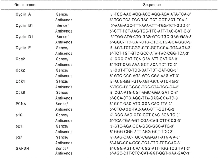
4. RT-PCR에 의한 mRNA 발현의 분석
TSA 미함유 및 TSA가 함유된 배지에서 48시간 배양된 세포들에 TRIzol reagent (Invitrogen Co., Carlsbad, CA, USA) 를 4oC에서 1시간 동안 처리하여 total RNA를 분리하였 다. 분리된 RNA를 정량한 후, oligo dT primer와 AMV reverse transcriptase (RT)를 이용하여 2μg의 RNA에서 ss cDNA를 합성하였다. 이 cDNA를 template로 사용하여 관 찰 대상 유전자(Table 1)를 polymerase chain reaction (PCR) 방법으로 증폭하였다. 이때 housekeeping 유전자인 glyce- raldehyde-3-phosphate dehydrogenase (GAPDH)를 internal control로 사용하였다. 각 PCR 산물들을 1% agarose gel을 이용하여 전기영동하고 ethidium bromide (EtBr, Sigma)를 이용하여 염색한 후 ultra violet (UV)하에서 확인하였다.
5. Western blot analysis에 의한 단백질 발현의 분석 TSA 미처리 및 TSA가 처리된 배지에서 자란 세포들을
수집하여 적당량의 lysis buffer (40 mM Tris, pH 8.0, 120 mM NaCl, 0.5% Nonidet P-40, 0.1 mM sodium orthovana- date, 2μg/ml aprotinin, 2μg/ml leupeptin, and 100μg/ml PMSF)를 첨가하여 4oC에서 30분간 반응시킨 후, 14,000 rpm으로 15분간 원심분리하여 그 상층액을 취하였다. 상 층액의 단백질 농도는 Bio-Rad 단백질 정량 시약(Bio-Rad, Hercules, CA, USA)을 이용하여 측정하였으며, 동량의 단 백질을 sodium dodecyl sulphate (SDS)-polyacrylamide gel을 이용하여 전기영동으로 분리하였다. 분리된 단백질을 함유한 acrylamide gel을 nitrocellulose membrane (Schleicher and Schuell, Keene, NH, USA)으로 electroblotting에 의해 전 이시킨 후, 10% skim milk를 함유한 PBS-T (0.1% Tween 20 in PBS)에 4oC에서 1시간 이상 배양하면서 비특이적인 단백질들에 대한 blocking을 실시하였다. 그리고 특정 단 백질에 대한 항체를 membrane에 적용시켜 항원 항체 반 응을 일으킨 후, PBS-T로 씻어내고 특정 항체에 대한 이 차 항체 반응을 실시한 후 enhanced chemiluminoesence (ECL)
Table 1. Sequences of primers used for RT-PCR
ꠏꠏꠏꠏꠏꠏꠏꠏꠏꠏꠏꠏꠏꠏꠏꠏꠏꠏꠏꠏꠏꠏꠏꠏꠏꠏꠏꠏꠏꠏꠏꠏꠏꠏꠏꠏꠏꠏꠏꠏꠏꠏꠏꠏꠏꠏꠏꠏꠏꠏꠏꠏꠏꠏꠏꠏꠏꠏꠏꠏꠏꠏꠏꠏꠏꠏꠏꠏꠏꠏꠏꠏꠏꠏꠏꠏꠏꠏꠏꠏꠏꠏꠏꠏꠏꠏꠏꠏꠏꠏꠏꠏꠏꠏꠏꠏꠏꠏꠏꠏꠏꠏꠏꠏꠏꠏꠏꠏꠏꠏꠏꠏꠏꠏꠏ
Gene name Sequence
ꠏꠏꠏꠏꠏꠏꠏꠏꠏꠏꠏꠏꠏꠏꠏꠏꠏꠏꠏꠏꠏꠏꠏꠏꠏꠏꠏꠏꠏꠏꠏꠏꠏꠏꠏꠏꠏꠏꠏꠏꠏꠏꠏꠏꠏꠏꠏꠏꠏꠏꠏꠏꠏꠏꠏꠏꠏꠏꠏꠏꠏꠏꠏꠏꠏꠏꠏꠏꠏꠏꠏꠏꠏꠏꠏꠏꠏꠏꠏꠏꠏꠏꠏꠏꠏꠏꠏꠏꠏꠏꠏꠏꠏꠏꠏꠏꠏꠏꠏꠏꠏꠏꠏꠏꠏꠏꠏꠏꠏꠏꠏꠏꠏꠏꠏ
Cyclin A Sence/ 5’-TCC-AAG-AGG-ACC-AGG-AGA-ATA-TCA-3’
Antisence 5’-TCC-TCA-TGG-TAG-TCT-GGT-ACT-TCA-3’
Cyclin B1 Sence/ 5’-AAG-AGC-TTT-AAA-CTT-TGG-TCT-GGG-3’
Antisence 5’-CTT-TGT-AAG-TCC-TTG-ATT-TAC-CAT-G-3’
Cyclin D1 Sence/ 5’-TGG-ATG-CTG-GAG-GTC-TGC-GAG-GAA-3’
Antisence 5’-GGC-TTC-GAT-CTG-CTC-CTG-GCA-GGC-3’
Cyclin E Sence/ 5’-AGT-TCT-CGG-CTC-GCT-CCA-GGA-AGA-3’
Antisence 5’-TCT-TGT-GTC-GCC-ATA-TAC-CGG-TCA-3’
Cdc2 Sence/ 5’-GGG-GAT-TCA-GAA-ATT-GAT-CA-3’
Antisence 5’-TGT-CAG-AAA-GCT-ACA-TCT-TC-3’
Cdk2 Sence/ 5’-GCT-TTC-TGC-CAT-TCT-CAT-CG-3’
Antisence 5’-GTC-CCC-AGA-GTC-CGA-AAG-AT-3’
Cdk4 Sence/ 5’-ACG-GGT-GTA-AGT-GCC-ATC-TG-3’
Antisence 5’-TGG-TGT-CGG-TGC-CTA-TGG-GA-3’
Cdk6 Sence/ 5’-CGA-ATG-CGT-GGC-GGA-GAT-C-3’
Antisence 5’-CCA-CTG-AGG-TTA-GAG-CCA-TC-3’
PCNA Sence/ 5’-GCT-GAC-ATG-GGA-CAC-TTA-3’
Antisence 5’-CTC-AGG-TAC-AAA-CTT-GGT-G-3’
p16 Sence/ 5’-CGG-AAG-GTC-CCT-CAG-ACA-TC-3’
Antisence 5’-TCA-TGA-AGT-CGA-CAG-CTT-CCG-3’
p21 Sence/ 5’-CTC-AGA-GGA-GGC-GCC-ATG-3’
Antisence 5’-GGG-CGG-ATT-AGG-GCT-TCC-3’
p27 Sence/ 5’-AAG-CAC-TGC-CGG-GAT-ATG-GA-3’
Antisence 5’-AAC-CCA-GCC-TGA-TTG-TCT-GAC-3’
GAPDH Sence/ 5’-CGG-AGT-CAA-CGG-ATT-TGG-TCG-TAT-3’
Antisence 5’-AGC-CTT-CTC-CAT-GGT-GGT-GAA-GAC-3’
ꠏꠏꠏꠏꠏꠏꠏꠏꠏꠏꠏꠏꠏꠏꠏꠏꠏꠏꠏꠏꠏꠏꠏꠏꠏꠏꠏꠏꠏꠏꠏꠏꠏꠏꠏꠏꠏꠏꠏꠏꠏꠏꠏꠏꠏꠏꠏꠏꠏꠏꠏꠏꠏꠏꠏꠏꠏꠏꠏꠏꠏꠏꠏꠏꠏꠏꠏꠏꠏꠏꠏꠏꠏꠏꠏꠏꠏꠏꠏꠏꠏꠏꠏꠏꠏꠏꠏꠏꠏꠏꠏꠏꠏꠏꠏꠏꠏꠏꠏꠏꠏꠏꠏꠏꠏꠏꠏꠏꠏꠏꠏꠏꠏꠏꠏ
용액(Amersham Life Science Corp., Arlington Heights, IL, USA)을 적용시킨 다음 X-ray film에 감광시켜 특정 단백 질의 양을 분석하였다. 본 실험에 사용된 항체들은 Santa Cruz Biotechnology Inc. (Santa Cruz, CA, USA) 및 Calbiochem (Cambridge, MA, USA)에서 구입하였으며, 이차 항체로 사 용된 horseradish peroxidase-labeled donkey anti-rabbit im- munoglobulin 및 peroxidase-labeled sheep anti-mouse immu- noglobulin은 Amersham Corp.에서 구입하였다.
결과 및 고찰
1. U937 세포의 증식에 미치는 TSA의 영향
U937 백혈병 세포의 증식에 미치는 TSA의 영향을 MTT assay에 준하여 실시한 결과는 Fig. 1A에 나타낸 바와 같 다. 48시간 동안 TSA 미처리 배지에서 배양한 U937 세포 에 비하여 TSA가 함유된 배지에서 배양한 세포는 TSA의 처리 농도에 의존적으로 증식이 감소하였음을 알 수 있 었다(Fig. 1A). 즉 45 ng/ml 농도의 TSA 처리군의 경우 대 조군에 비하여 40% 이상 세포증식이 억제되었으며, 75 ng/ml 농도의 처리군에서는 80% 정도의 세포증식 억제 현상을 관찰할 수 있었다. 이러한 결과들은 다양한 인체 암세포에서 관찰된 최근의 선행연구들과 유사한 경향성 을 보여주는 것으로,12,19∼22) TSA는 백혈병 세포에서도 다 른 고형암세포에서처럼 유사한 처리 농도 조건에서 비 슷한 암세포 증식억제 효능이 있음을 알 수 있었다. 아울 러 TSA의 처리가 백혈병 세포의 형태에 미치는 영향을 Fig. 1B에 나타내었다. TSA 처리 농도 의존적으로 현저한
세포밀도의 감소현상과 다양한 형태적인 변화를 관찰 할 수 있었다. 이러한 현상은 인체 자궁경부암 및 간암세 포 등에서 관찰된 최근의 선행연구 결과와 유사한 경향 성을 보여주는 것이었다.12,23∼25)
2. 세포주기 분포에 미치는 TSA의 영향
세포의 증식과 연관된 세포주기 조절은 G1, S 및 G2/M 기 각 주기별 관여하는 다양한 유전자들에 의해 조절되 는데, 기본적으로 세포주기 checkpoint 각 시기에 요구되 는 양성 조절인자인 cyclins에 의하여 Cdks의 연속적인 활 성과 불활성이 결정된다.1,2) 세포주기 조절의 관점에서 암세포는 세포주기의 비정상화에 기인된 질병으로 정의 될 수 있으며, 특정 시기의 세포주기 억제는 세포주기 조절 양성인자의 발현 저하 또는 음성 조절 인자의 과발 현과 관계가 있다고 요약될 수 있다.1∼3) 따라서 TSA의 처리에 의한 U937 세포의 증식억제가 세포주기 특정 시 기의 교란과 연관성을 지니는지의 여부를 조사하기 위 하여 세포주기 분포에 미치는 TSA의 영향을 조사한 flow cytometry 결과를 Fig. 2에 나타내었으며, 이 중 sub-G1기 를 제외한 나머지 세포군을 대상으로 세포주기 빈도를 산출한 결과는 Table 2에 나타낸 바와 같다. 결과에서 알 수 있듯이 TSA가 함유되지 않은 배지에서 자란 암세포 의 경우 전체 조사 대상 세포 중 G1, S 및 G2/M기에 해당 되는 세포의 빈도는 각각 약 61.10%, 17.50% 및 21.40%
정도였다. 그리고 45 ng/ml 및 75 ng/ml 농도의 TSA 처리 군의 경우 G1기는 각각 53.60% 및 81.39%였으며, S기나 G2/M기에 해당되는 세포의 빈도는 상대적으로 TSA 처 Fig. 1. Anti-proliferative effect and morphological changes of human leukemic U937 cells following incubation with TSA. (A) Cells were seeded as described in materials and methods, and treated with various concentrations of TSA. After 48 h incubation with TSA, MTT assay was performed. Results are expressed as average from two separate experiments. (B) Exponentially growing U937 cells were incubated with TSA for 48 h. Cell morphology was visualized by light microscopy. Magnification, X200.
0 15 30 45 60 75
Relative Growth (%)
Trichostatin A (nM) 0
20 40 60 80 100
A B
0 15 30
45 60 75
Trichostatin A (nM)
리 농도 의존적으로 감소되었다. 그러나 Fig. 2의 결과에 서처럼 TSA의 처리 농도가 증가될수록 sub-G1기에 해당 되는 세포의 빈도가 대조군에서 2.40%였던 것이 45 ng/ml 농도의 TSA 처리군에서는 6.61%, 그리고 75 ng/ml 농도의 TSA 처리군에서는 26.05%로 크게 증가되어 TSA 처리 농 도 의존적으로 U937 세포의 증식 억제는 apoptosis 유발 과 직접적인 관련이 있음을 알 수 있었다. 이와 같이 U937 세포에서 관찰된 TSA 농도에 따른 세포주기 G1기 의 증가 및 apoptosis 유발 현상은 다양한 고형암 세포주 에서 관찰된 결과들과도 유사하였다.10,12,19-22)
3. Cyclins 및 Cdks의 발현에 미치는 TSA의 영향
진핵세포에서 세포주기의 진행을 위해서는 세포주기 특이적 양성조절인자인 cyclin의 발현 증가가 우선적으 로 이루어져야 한다. 즉 mid G1에서 D-type cyclin (cyclin
D1, D2 및 D3)의 발현을 필요로 하며, late G1에서 S기로 의 진입을 위해서는 cyclin E의 발현이 증가되어야 한다.
그리고 S기에서 G2기로의 진입 및 G2기와 M기 진행 동 안에는 각각 cyclin A 및 B-type cyclin (cyclin B1 및 B2)의 발현이 증가되어야 한다. 그리고 이들 cyclin들은 특정 Cdks와 특이적인 결합에 의하여 cyclin/Cdk 복합체를 형 성하여 세포주기진행을 조절한다.1-3) G1기에서는 D-type cyclin이 Cdk4/6와 복합체를 이루고, Cdk2는 cyclin A와 결 합하여 S기와 G2기 동안 역할을 하는 반면 Cdc2는 cyclin B1과 결합하면 핵막의 histone H1과 lamin이 인산화에 의 해 kinase 활성이 증가하여 핵막의 붕괴 및 염색체의 재 배열이 일어나서 M기로 진행이 된다.1-3) 따라서 U937 세 포에서 TSA의 처리에 따른 세포주기 조절관련 기전을 조사하기 위하여 cyclins 및 Cdks의 발현에 미치는 TSA의 영향을 RT-PCR 및 Western immunoblotting 법으로 조사하 였다. Cyclins의 경우, Fig. 3A 및 B의 결과에서 알 수 있듯 이 조사된 cyclins 단백질의 대부분이 TSA 처리 농도 의존 적으로 발현이 감소되었음을 알 수 있었다. 특히 cyclin A 및 E의 경우 전사 및 번역 수준에서 동시에 감소되었 으나, 전사 수준에서 나머지 cyclins의 발현은 큰 변화가 없는 것으로 보아 TSA 처리에 의한 cyclins 단백질의 발현 감소는 전사 수준보다는 단백질의 half-life의 감소나 번 역 수준에서 더 조절되는 것으로 추정된다. 그리고 Cdks 의 발현에 미치는 TSA의 영향을 조사한 결과는 Fig. 4A 및 B에 나타낸 바와 같이, Cdc2, Cdk2 및 Cdk6의 경우 TSA 처리 농도 의존적으로 전사 및 번역 수준에서 발현 이 모두 감소되었으나, Cdk4는 mRNA 및 단백질 발현 수 준에서 모두 큰 변화가 없었다. 한편 세포증식 전반에 걸쳐 양성조절인자로서 작용하는 proliferating cell nuclear Fig. 2. DNA-flourescence histogram of human leukemic U937 cell nuclei after treatment with TSA. Exponentially growing cells at 50% confluency were treated for 48 h with indicated concentrations of TSA. Cells were trypsinized and pellets were collected. The cells were fixed and treated by RNase, and then cellular DNA was stained with PI, and analyzed by flow cytometry. Arrows mean the apoptotic sub-G1 populations.
2.40%
0 15
2.92% 3.58%
30
6.61%
45
15.25%
60 75
26.05%
DNA content
Trichostatin A (nM)
Cell number
Table 2. Fractions of each cell cycle phase of U937 cells cultured in the presence or absence of various concentrations of TSA. Each phase was analyzed by flow cytometry after 48 h treatment with TSA
ꠏꠏꠏꠏꠏꠏꠏꠏꠏꠏꠏꠏꠏꠏꠏꠏꠏꠏꠏꠏꠏꠏꠏꠏꠏꠏꠏꠏꠏꠏꠏꠏꠏꠏꠏꠏꠏꠏꠏꠏꠏꠏꠏꠏꠏꠏꠏꠏꠏꠏꠏꠏꠏꠏꠏ
% of cells
TSA (nM) ꠏꠏꠏꠏꠏꠏꠏꠏꠏꠏꠏꠏꠏꠏꠏꠏꠏꠏꠏꠏꠏꠏꠏꠏꠏꠏꠏꠏꠏꠏꠏꠏꠏꠏꠏꠏꠏꠏꠏꠏꠏ
G1 S G2/M
ꠏꠏꠏꠏꠏꠏꠏꠏꠏꠏꠏꠏꠏꠏꠏꠏꠏꠏꠏꠏꠏꠏꠏꠏꠏꠏꠏꠏꠏꠏꠏꠏꠏꠏꠏꠏꠏꠏꠏꠏꠏꠏꠏꠏꠏꠏꠏꠏꠏꠏꠏꠏꠏꠏꠏ
0 61.10 17.50 21.40
15 61.60 16.00 22.40
30 63.70 13.60 22.70
45 63.60 13.10 23.30
60 51.40 26.00 22.60
75 81.39 14.11 4.50
ꠏꠏꠏꠏꠏꠏꠏꠏꠏꠏꠏꠏꠏꠏꠏꠏꠏꠏꠏꠏꠏꠏꠏꠏꠏꠏꠏꠏꠏꠏꠏꠏꠏꠏꠏꠏꠏꠏꠏꠏꠏꠏꠏꠏꠏꠏꠏꠏꠏꠏꠏꠏꠏꠏꠏ
antigen (PCNA) 발현26,27)의 경우도 TSA 처리 농도 증가에 따라 전사 및 번역 수준에서 점차 감소되었다. 따라서 TSA 처리에 따른 U937 세포의 증식억제는 세포주기 양 성 조절인자인 cyclins뿐만 아니라 Cdks 및 PCNA 등의 발 현 감소와도 직접적인 연관이 있음을 알 수 있었다.
4. Cdk inhibitors의 발현에 미치는 TSA의 영향
Cdk inhibitors는 cyclin/Cdk 복합체와 결합하여 그 활성 을 억제하는 것으로 알려져 있는데, 특히 CIP/KIP 군에 속하는 p21은 종양 억제유전자인 p53에 의하여 활성화 되어 G1기뿐만 아니라 G2/M기를 포함한 전체 세포주기 의 진행을 억제하는 주요한 조절인자이다.1∼3) Cdk inhibi- Fig. 3. Effects of TSA treatment on the levels of cyclins in human leukemic U937 cells. (A) Cells were incubated with TSA for 48 h and total RNAs were isolated and RT-PCR was performed using indicated primers. GAPDH was used as a house-keeping control gene. (B) Cells were incubated with TSA for 48 h, lysed and cellular proteins were separated by 10% SDS-polyacrylamide gels and transferred onto nitrocellulose membranes. The membranes were probed with the indicated antibodies. Proteins were visualized using ECL detection system. Actin was used as a loading control.
A B
Trichostatin A (nM)
0 15 30 45 60
Cyclin A
Cyclin B1
Cyclin D1
Trichostatin A (nM)
0 30 45 60 75
Cyclin A
Cyclin B1
Cyclin D1
Cyclin E Cyclin E
GAPDH 75
Actin 15
Fig. 4. Effects of TSA treatment on the levels of Cdks and PCNA in human leukemic U937 cells. (A) Cells were incubated with TSA for 48 h and total RNAs were isolated and RT-PCR was performed using indicated primers. GAPDH was used as a house-keeping control gene. (B) Cells were incubated with TSA for 48 h, lysed and cellular proteins were separated by 10%
SDS-polyacrylamide gels and transferred onto nitrocellulose membranes. The membranes were probed with the indicated antibodies.
Proteins were visualized using ECL detection system. Actin was used as a loading control.
A B
Trichostatin A (nM)
0 15 30 45 60
Cdk2
Cdk4
Cdk6
Trichostatin A (nM)
0 30 45 60 75
Cdk2
Cdk4
Cdk6
Cdc2 Cdc2
PCNA 75
PCNA 15
GAPDH Actin
tor p21의 전사활성에는 p53이 관여하는 것이 일반적이 지만 암세포의 종류나 항암제 및 후보물질의 종류에 따 라서 p53 비의존적인 경로를 통하여 p21이 활성화된다 고도 알려져 있다.4,28) 그러나 p16의 경우 일반적으로 G1 기 arrest에만 관여하는 것으로 알려져 있으며, p27 역시 G1기 arrest에 중요하지만 부분적으로 G2/M기 arrest에도 관여할 수 있는 것으로 보고되고 있다.4) 따라서 본 연구 에서는 TSA의 처리에 의한 U937 세포의 증식억제가 Cdk inhibitors의 발현 증가에 의한 것인지의 여부를 조사하였 다. Fig. 5A 및 B의 결과에서 알 수 있듯이 조사된 3가지 Cdk inhibitor 모두 단백질 수준에서 TSA가 처리된 배지 에서 배양된 세포에서 전체적으로 처리 농도 의존적으 로 높게 나타났다. 특히 TSA에 의한 p21의 전사 및 번역 수준에서의 발현 증가 현상은 다양한 인체 암세포주를 대상으로 한 선행 연구의 결과12,14,21,22,25,29,30,33)
와도 유사 하였으며, 전사 수준에 관한 연구의 결과들에서 아마도 p21의 promoter 영역 중 전사조절인자 Sp-1의 결합부위가 중요한 조절영역으로 추정된다.14,29,30,33) 아울러 다양한 유전자의 promoter에 존재하는 Sp-1 결합부위의 활성은 세포주기 교란뿐만 아니라 apoptosis 유발에도 중요한 영 향을 줄 것으로 추정된다.31,34) 또한 U937 세포에서 TSA 처리에 의한 p16 및 p27의 발현 증가 역시 대장암 및 간 암세포 등에서 관찰된 것과 유사한 결과였다.12,35.36) 따라 서 TSA 처리에 의한 인체암세포의 증식 억제는 세포주 기 전반에 걸쳐 세포증식에 관여하는 유전자들의 발현 을 억제시키면서 Cdk inhibitors와 같은 세포증식 억제 유 전자들의 발현 증가를 통하여 암세포의 증식 억제를 유 도하는 것으로 생각된다.
결 론
Histone deacetylase (HDAC) 억제제가 새로운 항암치료 제 후보물질로서 유용성이 높은 것으로 평가되지만, 아 직까지 백혈병 세포에 관한 연구는 상대적으로 미미한 실정이다. 따라서 본 연구에서는 인체 백혈병세포에 미 치는 HDAC 억제제의 항암작용 기전을 조사하기 위하여 U937 세포주를 대상으로 그들의 증식에 미치는 대표적 인 HDAC 억제제인 trichostatin A (TSA)에 의한 영향을 세 포주기 조절관련 인자 중심으로 조사하였다. TSA의 처 리에 의하여 U937 세포의 증식은 처리 농도 의존적으로 억제되었으며, 심한 형태적 변형을 동반하였으며, 이는 세포주기 G1기의 빈도 증가와 연관성이 있었다. 또한 apoptosis 유발의 간접적인 지표가 되는 sub-G1기에 속하 는 세포 집단의 빈도 역시 TSA 처리 농도 의존적으로 매우 증가되었다. 이러한 TSA의 U937 세포 증식억제 효 과는 cyclins 및 Cdks의 발현 억제, 그리고 Cdk inhibitor인 p16, p21과 p27 등의 발현 증가와도 연관성이 있었다.
TSA의 항암 기전을 규명하기 위해서는 더 많은 연구가 부가적으로 필요하겠지만, 본 연구의 결과들에 의하면 TSA는 강력한 백혈병 세포의 증식 억제 및 항암작용이 있음을 시사하여 준다고 할 수 있다.
감사의 글
이 논문은 정부(교육인적자원부)의 재원으로 한국학 술진흥재단의 지원을 받아 수행된 연구의 일부임(KRF- Fig. 5. Induction of Cdk inhibitors by TSA treatment in human leukemic U937 cells. (A) Cells were incubated with TSA for 48 h and total RNAs were isolated and RT-PCR was performed using indicated primers. GAPDH was used as a house-keeping control gene. (B) Cells were incubated with TSA for 48 h, lysed and cellular proteins were separated by 10% or 12% SDS- polyacrylamide gels and transferred onto nitrocellulose membranes. The membranes were probed with anti-p16, anti-p21 and anti-p27 antibodies.
Proteins were visualized using ECL detection system. Actin was used as a loading control.
A B
Trichostatin A (nM)
0 15 30 45 60
p16
p21
p27
Trichostatin A (nM)
0 30 45 60 75
p16
p21
p27
GAPDH Actin
75 15
2004-202-E00039).
참 고 문 헌
1) Sherr CJ. The Pezcoller lecture: cancer cell cycles revisited.
Cancer Res 60, 3689-3695, 2000.
2) Weinberg RA. The retinoblastoma protein and cell cycle control. Cell 81, 323-330, 1995.
3) Elledge SJ, Harper JW. Cdk inhibitors: on the threshold of checkpoints and development. Curr Opin Cell Biol 6, 847- 852, 1994.
4) Harper JW. Cyclin dependent kinase inhibitors. Cencer Surv 29, 91-107, 1995.
5) Li Y, Jenkins CW, Nichols MA, Xiong Y. Cell cycle expression and p53 regulation of the cyclin-dependent kinase inhibitor p21. Oncogene 9, 2261-2268, 1994.
6) Gerber M, Shilatifard A. Transcriptional elongation by RNA polymerase II and histone methylation. J Biol Chem 278, 26303-26306, 2003.
7) Marks PA, Richon VM, Miller T, Kelly WK. Histone de- acetylase inhibitors. Adv Cancer Res 91, 137-168, 2004.
8) Kuo MH, Allis CD. Roles of histone acetyltransferases and deacetylases in gene regulation. Bioessays 20, 615-626, 1998.
9) Aksan I. Chromatin goes global. Trends Biochem Sci 27, 7-8, 2002.
10) Yoshida M, Furumai R, Nishiyama M, Komatsu Y, Nishino N, Horinouchi S. Histone deacetylase as a new target for cancer chemotherapy. Cancer Chemother Pharmacol 48 (Suppl 1), S20-26, 2001.
11) Yoshida M, Kijima M, Akita M, Beppu T. Potent and specific inhibition of mammalian histone deacetylase both in vivo and in vitro by trichostatin A. J Biol Chem 265, 17174- 17179, 1990.
12) Herold C, Ganslmayer M, Ocker M, Hermann M, Geerts A, Hahn EG, Schuppan D. The histone-deacetylase inhibitor trichostatin a blocks proliferation and triggers apoptotic pro- grams in hepatoma cells. J Hepatol 36, 233-240, 2000.
13) Kim YB, Ki SW, Yoshida M, Horinouchi S. Mechanism of cell cycle arrest caused by histone deacetylase inhibitors in human carcinoma cells. J Antibiot (Tokyo) 53, 1191-200, 2000.
14) Sowa Y, Orita T, Minamikawa S, Nakano K, Mizuno T, Nomura H, Sakai T. Histone deacetylase inhibitor activates the WAF1/Cip1 gene promoter through the Sp1 sites.
Biochem Biophys Res Commun 241, 142-150, 1997.
15) Suzuki T, Yokozaki H, Kuniyasu H, Hayashi K, Naka K, Ono S, Ishikawa T, Tahara E, Yasui W. Effect of trichostatin A on cell growth and expression of cell cycle- and apopto- sis-related molecules in human gastric and oral carcinoma cell lines. Int J Cancer 88, 992-997, 2000.
16) Vanhaecke T, Papeleu P, Elaut G, Rogiers V. Trichostatin A-like hydroxamate histone deacetylase inhibitors as thera-
peutic agents: toxicological point of view. Curr Med Chem 11, 1629-1643, 2004.
17) Yamashita Y, Shimada M, Harimoto N, Rikimaru T, Shirabe K, Tanaka S, Sugimachi K. Histone deacetylase inhibitor trichostatin induces cell-cycle arrest/apoptosis and hepa- tocyte differentiation in human hepatoma cells. Int J Cancer 103, 572-576, 2003.
18) Bouchain G, Delorme D. Novel hydroxamate and anilide derivatives as potent histone deacetylase inhibitors: synthesis and antiproliferative evaluation. Curr Med Chem 10, 2359- 2372, 2003.
19) Bordonaro M, Mariadason JM, Aslam F, Heerdt BG, Aug- enlicht LH. Butyrate-induced apoptotic cascade in colonic carcinoma cells: modulation of the β-catenin-Tcf pathway and concordance with effects of sulindac and trichostatin A but not curcumin. Cell Growth Differ 10, 713-720, 1999.
20) Joung KE, Kim DK, Sheen YY. Antiproliferative effect of trichostatin A and HC-toxin in T47D human breast cancer cells. Arch Pharm Res 27, 640-645, 2004.
21) Noh EJ, Lee JS. Functional interplay between modulation of histone deacetylase activity and its regulatory role in G2-M transition. Biochem Biophys Res Commun 310, 267-273, 2003.
22) Roh MS, Kim CW, Park BS, Kim GC, Jeong JH, Kwon HC, Suh DJ, Cho KH, Yee SB, Yoo YH. Mechanism of histone deacetylase inhibitor Trichostatin A induced apop- tosis in human osteosarcoma cells. Apoptosis 9, 583-589, 2004.
23) Papeleu P, Vanhaecke T, Elaut G, Vinken M, Henkens T, Snykers S, Rogiers V. Differential effects of histone deace- tylase inhibitors in tumor and normal cells-what is the toxicological relevance? Crit Rev Toxicol 35, 363-378, 2005.
24) Lopatina NG, Poole JC, Saldanha SN, Hansen NJ, Key JS, Pita MA, Andrews LG, Tollefsbol TO. Control mechanisms in the regulation of telomerase reverse transcriptase expres- sion in differentiating human teratocarcinoma cells. Biochem Biophys Res Commun 306, 650-659, 2003.
25) Li H, Wu X. Histone deacetylase inhibitor, Trichostatin A, activates p21WAF1/CIP1 expression through downregulation of c-myc and release of the repression of c-myc from the promoter in human cervical cancer cells. Biochem Biophys Res Commun 324, 860-867, 2004.
26) Prasanth SG, Mendez J, Prasanth KV, Stillman B. Dynamics of pre-replication complex proteins during the cell division cycle. Philos Trans R Soc Lond B Biol Sci 359, 7-16, 2004.
27) Tachibana KE, Gonzalez MA, Coleman N. Cell-cycle-depen- dent regulation of DNA replication and its relevance to can- cer pathology. J Pathol 205, 123-129, 2005.
28) Xiong Y, Hannon GJ, Zhang H, Casso D, Kobayashi R, Beach D. p21 is a universal inhibitor of cyclin kinases. Nature 366, 701-704, 1993.
29) Hirsch CL, Bonham K. Histone deacetylase inhibitors regu- late p21WAF1 gene expression at the post-transcriptional level in HepG2 cells. FEBS Lett 570, 37-40, 2004.
30) Lagger G, Doetzlhofer A, Schuettengruber B, Haidweger E,
Simboeck E, Tischler J, Chiocca S, Suske G, Rotheneder H, Wintersberger E, Seiser C. The tumor suppressor p53 and histone deacetylase 1 are antagonistic regulators of the cyclin- dependent kinase inhibitor p21/WAF1/CIP1 gene. Mol Cell Biol 23, 2669-2679, 2003.
31) Margueron R, Duong V, Castet A, Cavailles V. Histone deacetylase inhibition and estrogen signalling in human breast cancer cells. Biochem Pharmacol 68, 1239-1246, 2004.
32) Moore PS, Barbi S, Donadelli M, Costanzo C, Bassi C, Palmieri M, Scarpa A. Gene expression profiling after trea- tment with the histone deacetylase inhibitor trichostatin A reveals altered expression of both pro- and anti-apoptotic genes in pancreatic adenocarcinoma cells. Biochim Biophys Acta 1693, 167-176, 2004.
33) Xiao H, Hasegawa T, Isobe K. Both Sp1 and Sp3 are
responsible for p21waf1 promoter activity induced by histone deacetylase inhibitor in NIH3T3 cells. J Cell Biochem 73, 291-302, 1999.
34) Salminen A, Tapiola T, Korhonen P, Suuronen T. Neuronal apoptosis induced by histone deacetylase inhibitors. Brain Res Mol Brain Res 61, 203-206, 1998.
35) Chen JS, Faller DV. Histone deacetylase inhibition-mediated post-translational elevation of p27KIP1 protein levels is re- quired for G1 arrest in fibroblasts. J Cell Physiol 202, 87-99, 2005.
36) Chen YX, Fang JY, Lu J, Qiu DK. Regulation of histone acetylation on the expression of cell cycle-associated genes in human colon cancer cell lines. Zhonghua Yi Xue Za Zhi 84, 312-317, 2004.