Introduction
Obesity has become the most dangerous public health problem in many societies of the world. It is a complex metabolic disorder that is thought to result from an imbalance of energy intake and energy expenditure leading to the excess accumulation of fat in various adipose
tissues and organs; that is, at least, 20 % heavier than ideal weight
1).
Recently there are a lot of attempts to treat obesity through the energy expenditure. Espe- cially Uncoupling Protein (UCP) and Peroxisome proliferator-activated receptors (PPAR) are known to play a key role for energy dissipation through the increasing thermogenesis.
Uncoupling Proteins (UCPs) are mitochondrial inner membrane proteins sustaining an inducible proton conductance. They weaken the proton electrochemical gradient built up by the mitoc- hondrial respiratory chain, and induce the energy dissipation through the thermogenesis
2).
Peroxisome proliferator-activated receptors
received : 21 November 2007
received in revised from : 22 November 2007 accepted : 7 December 2007
Correspondence to : Beom-Joon Lee
ADDRESS; East-West medical department of The Graduate School of East-West Medical Science, Kyung-Hee University, Suwon, Korea
(Tel : +82-2-3457-9006 / Fax : + 82-2-958-9212 E-mail : [email protected])
The Anti-obesity Effects of Gambi-hwan Extract on Obese Rats Induced by High-fat Diet through the
Expression of UCP-1 and PPAR-δ δ δ δ
Beom-Joon Lee
1, Jae-Hwan Ryu
1, Jae-Wan Kim
1, Jong-Hun Park
1, Jae-Woo Park
21The Graduate School of East-West Medical Science, Kyung-Hee University
2the 3rd Internal medicine, college of Oriental medicine, Kyung-Hee University Original Article
Objective : Recently there are a lot of attempts to treat obesity through energy expenditure. Especially UCP-1 and PPAR-δ is known to play a key role for energy dissipation through the increasing thermogenesis. Gambi-hwan extract is a traditional medicine made of herbs containing the polyunsaturate fatty acids related to the energy expenditure. It is expected to reduce the weight by means of the expression of UCP-1 and PPAR-δ.
Meterial and Method : We divided 21 rats into 3 groups and assigned 8 rats respectively. The normal group was administered normal diet, the control group was administered high-fat diet, and the G50 group was administered high-fat diet with Gambi-hwan extracts50 mg/kg. And then the weights of body, food intake, the changes of lipids in blood stream, and the expressions of UCP-1 and PPAR-δ on adipose tissues were measured respectively.
Result : The reduction of body weight and the increasing tendency of expression of UCP-1 and PPAR-δ mRNA were shown in G50 group. In the G50 group the Triglyceride level is decreased and the HDL-cholesterol level and the expression of PPAR-δ and UCP-1 protein on Visceral adipose tissue were significantly increased.
Conclusion : This result indicates that Gambi-hwan Extract upregulate the expression of UCP-1 and PPAR-δ in adipose tissue, which may contribute to reducing the weight of adipose tissue.
Key Words : Antiobesity, UCP, PPAR, Croton tiglium, buthus martensi karsch
(PPARs) are the ligand-activated transcription factors belonging to the nuclear receptor superfamily
3). Among PPARs, PPAR- δ plays an important role as a powerful regulator of fatty acid catabolism and energy dissipation
4).
In oriental medicine, there are several investigations about increasing energy expen- diture such as inducing the expression of UCP administering sobium
5), cheongpesagan-tang
6), SBY- Ⅲ
7)and so on. Gambi-hwan extract is a traditional medicine made of herbs, croton tiglium (Badou) and Buthus martensi karsch (Chinese-Scorpion), which are regarded to move qi and free stagnation with pungency
8). It is expected to contain the polyunsaturate fatty acids which were related to increase the energy expenditure.
We measured the weight of body, food intake and plasma lipid level to show the anti-obesity effects in many fields. Specially we focused on the previous study which showed the relatio- nship among obesity and UCP-1 and PPAR- δ in the field of energy metabolism
4,25,26). Therefore we tried to carry out this research to demonstrate the anti-obesity effects of Gambi-hwan Extract through the energy dissipation.
Materials and Methods 1. Materials
1) Animals
Male Copenhagen rats, 5 weeks of age and weighing from 230 to 250g, were purchased
from an animal breeder (Orient Bio, Seoul, Korea) and were housed at 24±1 ℃ and at 50 % relative humidity with 12/12-h light/dark cycle.
Animals had free access to drinking water and were fed diet with AIN-76A for 1 week in order for acclimation. After acclimation, animals were randomly divided into each 5 groups of 8 mice and each group was separated into two cages. All the rats were given high fat diet and experim- ental diet.
2) Preparation of oriental herbs and their extracts
Gambi-hwan is a mixture of herbal drugs. It is made of buthus martensi kirsch (Chinese Scorpion) and croton tiglium (Badou). Buthus martensi karsch and croton tiglium were purch- ased from the Korean Pharmacy (Kyunghee herb phamacy Ltd). When we used croton tiglium, we made it to the croton tiglium powder. At first, we removed the nutshell of croton tigliumand smashed it into the pulp. During that period, we removed the oil from the pulp with paper and applied heat to it a little in order to volatilize the oil
8). Finally we made it to the powder containing 18-20 % of the oil contents. In case of buthus martensi karsch, we grinded the dried it into the buthus martensi karschpowder. Croton tiglium dried powder were mixed the same amount of buthus martensi karschdried powder. Mixed powder were dissolved in 200 ml 80 % ethanol and boiled for 6 hours in water bath. The supernatants were collected and concentrated with vacuum evaporator (EYELA CA-1500,
Scientific Name Weight (g)
Buthus martensi karsch 10
Croton tiglium 10
Total amount 20
Table 1. The Composition of Gambi-hwan
Rikakikai, Japan) to 60 ml and then the residue was freeze-dried in a freezing drier and was stored in a refrigerator. The Gambi-hwan ethanol extract was dissolved in distilled water before use (Table 1).
2. Methods
1) Administration of materials
We divided 21 rats into 3 groups and assigned 8 rats respectively. Rats were categorized as normal, control and G50 groups. Normal group was administered normal diet (AIN-76A feed
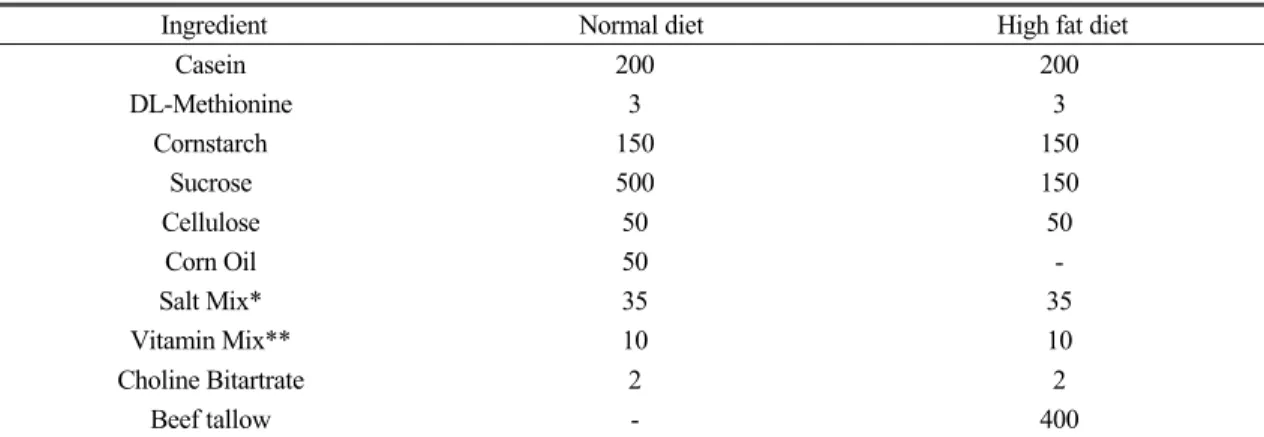
#100000, Dyets Inc, Bethlehm, PA, USA), Control group was administered high-fat diet (AIN-76A+40 % beef Tallow#101556, Dyets Inc, Bethlehm, PA, USA), and G50 group was administered high-fat diet with Gambi-hwan extracts (Table 2).
Food and water were provided at libitum and we change food every other day for preventing insufficiency. We administered normal saline as placebo to the normal and control group, on the other hand we administered Gambi-hwan extract
50 mg/kg to G50 group once in a day for 10 weeks.
2) Measurement of Weight and Food intake We measured the daily food intake and body weight and at 10 am on Monday every week for 10 weeks. At the 10th week, rats were killed with ethyl ether and we measured the weight of visceral adipose tissue and liver.
3) Blood sampling and plasma assay
Blood was withdrawn from the tail venous plexus, using a heparinized capillary tube wit- hout anesthesia. The blood samples were placed on ice, centrifuged at 1,100 g for 15 minutes, and then stored at -80 ℃ until assay. Triglycerides, Total cholesterol, LDL-cholesterol, and HDL- cholesterol were enzymatically analyzed using a commercial kit based on the cholesterol oxidase method. Adipose tissues were collected and weighed, immediately frozen inliquid nitrogen, placed in 1.5-mL Eppendorf tubes, and stored at -80°C until analysis.
Ingredient Normal diet High fat diet
Casein 200 200
DL-Methionine 3 3
Cornstarch 150 150
Sucrose 500 150
Cellulose 50 50
Corn Oil 50 -
Salt Mix* 35 35
Vitamin Mix** 10 10
Choline Bitartrate 2 2
Beef tallow - 400
* AIN 76 salt mixture: Calcium Phosphate Dibasic 500 g, Sodium Chloride 74 g, Potassium Citrate H2O 220 g, Potassium Sulfate 52 g, Magnesium Oxide 24 g, Manganous Carbonate 3.5 g, Ferric Citrate U.S.P. 6 g, Zinc Carbonate 1.6 g, Cupric Carbonate 0.3 g, Potassium Iodate 0.01 g, Sodium Selenite 0.01 g, Chromium Potassium Sulfate 12H2O 0.55 g, Sucrose, finely powdered 118.03 g
** AIN 76 vitamin mixture: Thiamine HCl 0.6 g,, Riboflavin 0.6 g,, Pyridoxine HCl 0.7 g,, Niacin 3 g,, Calcium Pantothenate 1.6 g,, Folic Acid 0.2 g,, Biotin 0.02 g,, Vitamin B12 (0.1%) 1 g,, Vitamin A Palmitate (500,000 IU/g) 0.8 g,, Vitamin D3 (400,000 IU/g) 0.25 g,, Vitamin E Acetate (500 IU/g) 10 g,, Menadione Sodium Bisulfite 0.08 g,, Sucrose finely powdered 981.15 g
Table 2. The composition of Normal Diet and High Fat Diet (g/kg)
4) RNA Preparation and RT-PCR
Total RNA was isolated from the white adipose tissue using the TriZol reagent (Life Technologies, Inc) and isopropanol precip- itation. The RNA was reverse transcribed into cDNA using the Moloney murine leukemia virus transcriptase system. mRNA expression was determined by polymerase chain reaction (PCR) using the following PCR primer sequences:
forward 5'-GCTTCGTCACCCATGAGTTCTT-3' and reverse 5'-GATCTGGCCCTTTTCATTG-3' to amplify PPAR- δ; forward 5'-CGGCAGCCT- TTTTCAAAGG-3' and reverse 5'-ACATAGG- CGACTTGGAGAAAGG-3' to amplify UCP-1.
In case of PPAR- δ the PCR cycling conditions were 94 ℃ for 30sec, 62℃ for 1 min, and 25 cycles of 72 ℃ for 1 min. In case of UCP-1, they were 92 ℃ for 30sec, 60℃ for 1 min, and 35 cycles of 72 ℃ for 1 min. The RT-PCR products were electrophorosed in 2 % agarose gels under 100 V and was stained with 0.5 ㎍/ml ethidium bromide. The density of the PCR product was measured using a GS-700 imaging densitometer.
5) Western blotting
White adipose tissue of rats was homogenize using ELB buffer(50 mM HEPES pH 7.0, 250 mM NaCl, 5 mM EDTA, 0.1 % Nonidet P-40, 1 mM phenylmethylsulfonyl fluoride, 0.5 mM dithiothreitol, 5 mM NaF, 0.5 mM sodium orthovanadate) containing protease inhibitor cocktail. After centrifugation at 10,000 g for 5 min, the fat cake discarded, and the infranatant was quantified by Bradford method and used for Western blot analysis of UCP1.
The supernatants were separated by 10 % SDS-polyacrylacide gel electrophoresis. Proteins were transferred to nitrocellulose membrane for 1 hour at 100 mA (semi-dry system). The mem-
branes were blocked at 5 % skim milk solution and was incubated with an primary antibody for 4 hour and then was incubated with secondary antibody conjugated horseradish peroxidase for 1 hour. The membranes were treated with the reagents in the chemiluminescene detection kit (ECL system) according to the manufacturer's instructions.
3. Statistical analysis
Data are expressed as means ± standard error.
Statistical analysis between groups were deter- mined by ANOVA. Statistical comparisons were made by Dunnett's test. Differences with P<0.05 were considered significant.
Result 1. Body Weight
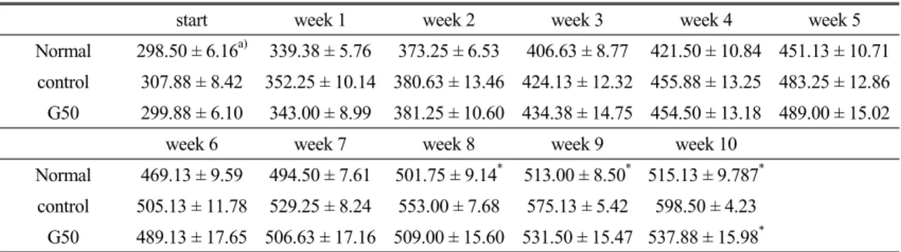
We administered normal diet, high-fat diet and high-fat diet with Gambi-hwan extract respectively for 10 weeks. The body weight of the control group significantly increased than that of normal group from the 8th week to 10th week. The body weight of the G50 group decreased than that of the control group in 10th week (Table 3, Fig 1).
2. Food intake
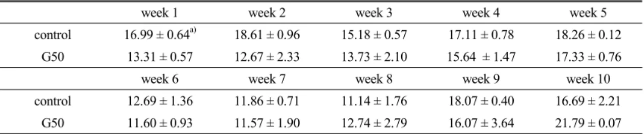
There was no significant difference in food intake between groups, but we observed the tendency of decrease in the G50 group compared with the control group from 1st week to 6th week (Table 4).
3. Plasma lipid level
There were significant decreases in Trigly-
ceride and LDL-Cholesterol level between the
normal group and control group. We also obse-
rved the HDL-Cholesterol level of the G50 group significantly increased more than that of control group (Table 5).
4. The Weight of Visceral Adipose Tissue The weight of visceral adipose tissue of the G50 group decreased more than that of the control group, and there was no significant difference in the weight of liver (Table 6, Fig 2).
5. The Expression of PPAR-δ and UCP-1 in the Visceral Adipose Tissue
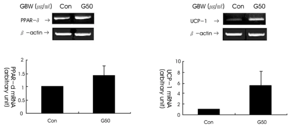
We observed the expression of mRNA and protein of UCP-1 and PPAR- δ in Visceral adipose tissue. A trend toward decrease in the
mRNA expression of UCP-1 and PPAR- δ of the G50 group compared with the control group (Fig 3). The expression of UCP-1 and PPAR- δ protein of experimental group significantly increased than that of control group (Fig 4).
Discussion
Obesity has become a global health epidemic and its prevalence continues to increase at a rapid rate in our society. Moreover, it often gives rise to complications related to the metabolic disorderssuch as dyslipidemia, diabetes, hypert- ension and so on. In the developed countries, it is attributed to changes in dietary and lifestyle
start week 1 week 2 week 3 week 4 week 5
Normal 298.50 ± 6.16a) 339.38 ± 5.76 373.25 ± 6.53 406.63 ± 8.77 421.50 ± 10.84 451.13 ± 10.71 control 307.88 ± 8.42 352.25 ± 10.14 380.63 ± 13.46 424.13 ± 12.32 455.88 ± 13.25 483.25 ± 12.86 G50 299.88 ± 6.10 343.00 ± 8.99 381.25 ± 10.60 434.38 ± 14.75 454.50 ± 13.18 489.00 ± 15.02
week 6 week 7 week 8 week 9 week 10
Normal 469.13 ± 9.59 494.50 ± 7.61 501.75 ± 9.14* 513.00 ± 8.50* 515.13 ± 9.787* control 505.13 ± 11.78 529.25 ± 8.24 553.00 ± 7.68 575.13 ± 5.42 598.50 ± 4.23
G50 489.13 ± 17.65 506.63 ± 17.16 509.00 ± 15.60 531.50 ± 15.47 537.88 ± 15.98* a) Values represent Mean ± Standard Error
* ; significantly different (p<0.05), ANOVA followed by Dunnett's test from high-fat diet group
Normal; Normal diet group, Control; High-fat diet group, G50; Gambi-hwan Extract 50 mg with high fat diet group Table 3. The Change of Body Weight in Each Group.
Period of Experiment 650
600 550 500 450 400 350 300 250
Body weight (g)
start week1 week2 week3 week4 week5 week6 week7 week8 week9 week10
Normal Control G50
Fig. 1. Body weight gain in each group.
habits, such as rapidly changing diets, increased availability of high-energy foods, and reduced physical activity of people
27).
In the ancient text Yellow Emperor's Inner Canon, huang-di-nei-ding written in the 3rd centry BC, there are comments about cause of obesity. “Both obese and noble people have diseases that result from their rich, fatty diet,”
and “Obese people often eat sweet foods”. As an etiology of obesity, it said “Obese persons have too much blood and qi and their skin is too thick.
In this situation, they are easy to be retained by evil-qi. So they often have phlegm-damp obstruction in their body. As a result it induced stagnation of qi flow and various illness occur due to the stagnation.”
9)Therefore, in oriental
medical point of view, it is an important key whether we can remove that phlegm-damp obstruction or not
10).
In this study, we use croton tiglium and buthus martensi karsch for the solution. Croton tigliu- mis pungent herb which free phlegm-damp obstruction and usually have been used laxatives. Croton tiglium contains oil, that is croton oil, from 34 % to 57 %. Croton oil is very toxic, so we removed the oil down to the 18 % and then we use it. It also contains various fatty acids - that is palmitic acid, stearic acid, oleic acid, tiglic acid, linolenic acid, myristic acid, arachidonic acid, glycerides, and phorbol- 12,13-esters and so on
8,11). Buthus martensi karschis also pungent herb which move qi and
Normal Control G50
Triglyceride 68.00 ± 8.66 *a) 116.38 ± 4.91 99.38 ± 2.63
Total Cholesterol 82.00 ± 3.85 78.50 ± 2.99 70.88 ± 3.29
HDL-Cholesterol 23.75 ± 2.01 22.88 ± 1.04 28.63 ± 0.91*
LDL-Cholesterol 9.50 ± 0.93* 15.50 ± 2.03 13.00 ± 1.32
Phospholipid 148.63 ± 15.29 131.13 ± 7.26 138.00 ± 4.86
Free Fatty Acid 969.88 ± 87.68 539.25 ± 32.91 620.75 ± 86.81
a) Values represent Mean ± Standard Error
* ; significantly different (p<0.05), ANOVA followed by Dunnett's test from high-fat diet group
Normal; Normal diet group, Control; High-fat diet group, G50; Gambi-hwan Extract 50 mg with high fat diet group
Table 5. Effects of Gambi-hwan Extract on Plama lipid level of Normal Rat and Obese Rat Induced of High-fat Diet (mg/dl)
week 1 week 2 week 3 week 4 week 5
control 16.99 ± 0.64a) 18.61 ± 0.96 15.18 ± 0.57 17.11 ± 0.78 18.26 ± 0.12 G50 13.31 ± 0.57 12.67 ± 2.33 13.73 ± 2.10 15.64 ± 1.47 17.33 ± 0.76
week 6 week 7 week 8 week 9 week 10
control 12.69 ± 1.36 11.86 ± 0.71 11.14 ± 1.76 18.07 ± 0.40 16.69 ± 2.21 G50 11.60 ± 0.93 11.57 ± 1.90 12.74 ± 2.79 16.07 ± 3.64 21.79 ± 0.07 a) Values represent Mean ± Standard Error
* ; significantly different (p<0.05), ANOVA followed by Dunnett's test from high-fat diet group
Normal; Normal diet group, Control; High-fat diet group, G50; Gambi-hwan Extract 50 mg with high fat diet group Table 4. Effects of Gambi-hwan Extract on Food Intake of Rat fed on High-fat Diet
dissipate stagnation. It contains toxin and various fatty acids - that is palmitic acid, stearic acid, oleic acid, linolenic acid, gammalinolenic acid, and behenic acid and so on
8). So both medicines contain a variety of ingredients - polyunsaturated fatty acids, toxins, vitamins and so on
11). From the oriental medical point of view, as mentioned above, obesity is induced by the phlegm-damp obstruction and stagnation of qi.
Therefore we expected they can remove the cause of obesity and then we made Gambi-hwan of them.
Nowadays there are a lot of attempts to treat obesity through energy expenditure. Especially Uncoupling Protein (UCPs) and Peroxisome proliferator-activated receptors (PPARs) are known to play a key role for energy dissipation through the increasing thermogenesis
12,13).
Uncoupling Proteins (UCPs), inner mitoch- ondrial membrane proteins, have five members with different purported functions. UCP-1, UCP-2, and UCP-3 arethought to be related to obesity. Among them, UCP-1 is the first to be discovered and it is the main mediator of adaptive thermogenesis. UCP-1 can dissipate energy as heat by uncoupling oxidative phosph- orylation. Because UCP-2 and UCP-3 have the similar to UCP-1 molecular composition, they were thought that they were related to the energy expenditure
2,14). However, there are some exper- iments that have questioned the relationships between obesity and UCP-2, UCP-3. That is obesity wasnot induced in the UCP-2
15)and UCP-3
16)knock-out mouse. Thus it is unclear whether UCP-2 and UCP-3 are related to the obesity and energy expenditure or not. Apart
Normal Control G50
Visceral 2.19 ± 0.13a) 2.82 ± 0.11 2.04 ± 0.31*
Liver 3.61 ± 0.28 3.36 ± 0.22 3.41 ± 0.18
a) Values represent Mean ± Standard Error
* ; significantly different (p<0.05), ANOVA followed by Dunnett's test from high-fat diet group
Normal; Normal diet group, Control; High-fat diet group, G50; Gambi-hwan Extract 50 mg with high fat diet group
Table 6. Effects of Gambi-hwan Extract on Weight of Visceral Adipose Tissue and Liver of Normal Rat and Obese Rat Induced of High-fat Diet (mg)
4.5 4 3.5 3 2.5 2 1.5 1 0.5 0
Weight (mg)
Visceral Liver
Normal Control G50
*
Fig. 2.Effects of Gambi-hwan Extract on Weight of Visceral Adipose tissue and Liver of Normal Rat and Obese Rat Induced of High-fat Diet (mg)
* ; significantly different (p<0.05), ANOVA followed by Dunnett's test from high-fat diet group Normal; Normal diet group, Control; High-fat diet group, G50; Gambi-hwan Extract 50 mg with high fat diet group
from UCP-2 and UCP-3, it is certain that UCP-1 can increase energy expenditure through ada- ptive thermogenesis
17).
UCP-1 was originally regarded as an important factor of thermogenesis and mainly found in Brown Adipose Tissue in rodents. But there is scarcely brown adipose tissue in human.
So upregulation of UCP-1 in white adipose
tissueis more important practically. So some researchers recently reported that they found UCP-1 and could make the expression of UCP-1 increase in white adipose tissue and decrease the weight of white adipose tissue though the extracts from sea food
17). Thus we investigated the expression of UCP-1 in white adipose tissue.
Peroxisome proliferator-activated receptors
UCP-1 → β-actin → PPAR-δ →
β-actin →
GBW (㎍/㎖) Con G50
4 3.5
0
G50 Con
PPAR-d(arbitrary unit)
GBW (㎍/㎖) Con G50
*
3 2.5 2 1.5 1 0.5
4 3.5
0
G50 Con
UCP-1(arbitrary unit)
*
3 2.5 2 1.5 1 0.5
Fig. 4.Expression of PPAR-δ and UCP-1 Protein in Visceral White Adipose Tissue in Rat fed the Gambi-hwan Extract
PPAR-δ or UCP-1 was detected by western blot analysis. β-actin was used as an equal loading control. Densitometric analysis shows relative PPAR-δ or UCP-1 expression levels (mean ±SE). * p < 0.05 vs. control group; significance of differences between treatment groups was evaluated using the ANOVA with Dunnett's test.
UCP-1 → β-actin → PPAR-δ →
β-actin →
GBW (㎍/㎖) Con G50
2
1.5
`1
0.5
0
G50 Con
PPAR-d mRNA(arbitrary unit)
GBW (㎍/㎖) Con G50
10
0
G50 Con
UCP-1 mRNA(arbitrary unit)
8 6 4 2
Fig. 3.Expression of PPAR-δ and UCP-1 mRNA in Visceral White Adipose Tissue in Rat fed the Gambi-hwan Extract
PPAR-δ or UCP-1 was detected by western blot analysis. β-actin was used as an equal loading control. Densitometric analysis shows relative PPAR-δ or UCP-1 expression levels (mean ± SE). * p < 0.05 vs. control group; significance of differences between treatment groups was evaluated using the ANOVA with Dunnett's test.
(PPARs) form obligate heterodimers with the retinoid X receptor and bind to defined PPAR elements in the promoter region of target genes.
The PPAR subgroup comprises three closely related members - PPAR α, γ, and δ. They are activated by a variety of fatty acids, fatty acid derivatives, and synthetic compounds
18). Among PPARs, PPAR- δ plays an important role as a powerful regulator of fatty acid catabolism and energy dissipation
4).
PPAR- δ is located in 6p21.1-21.2 of human gene. Recent studies have shown that this was closely related to lipid metabolism. There are several recent studies about PPAR- δ. Synthetic PPAR- δ agonists increase serum HDL-Chole- sterol while lowering triglyceride levels in obese mice
19). Acute treatment of (Lepr db/db) mice, genetically predisposed obese mice, with a PPAR- δ agonist depletes lipid accumulation. In parallel, PPAR- δ-deficient mice challenged with high-fat diet show reduced energy uncoupling and are prone to obesity
4). In rhesus monkey model, activation of PPAR- δ induced decreasing triglyceride level and increasing HDL-Choles- terol level. Also there are other reports that activation of PPAR- δ induced fatty acid β -oxidation in skeletal muscle and reduced the metabolic syndrome
20).
In vivo study, the selective expression of PPAR- δ in adipose tissue induced decreasing lipid accumulation in both adipose tissue and serum, and the activation of PPAR- δ stimulated β-oxidation and triglyceride utilization in adipocytes and myocytes. Short-term treatment of obese mice with PPAR- δ agonist causes a dramatic lipid depletion in tissues. Furthermore, PPAR- δ-deficient mice fed with high-fat diet display reduced energy uncoupling and are prone to obesity
4).
Thus PPAR- δ plays an important role as a powerful regulator of fatty acid catabolism and energy dissipation. As stated above, there are several PPAR- δ agonists; synthetic PPAR-δ agonist (GW501516), fatty acids, fatty acids derivates. Among them, polyunsaturated fatty acids (PUFA), particularly those of the n-3 family are strong activators of PPAR- δ
21).
In this study, we administered to rats normal diet, high-fat diet and high-fat diet with Gambi-hwan extract respectively for 10 weeks.
The body weight of the control group signifi- cantly increased more than that of normal group from 8th week to 10th week, so we proved obesity was induced by high-fat diet. The body weight of G50 group decreased than that of control group in 10th week, so we also found Gambi-hwan extract reduced the body weight.
In plasma lipid level, there were significant decreases in Triglyceride and LDL-Cholesterol level between the normal group and control group. We also observed the HDL-Cholesterol level of G50 group significantly increased more than that of control group. HDL-Cholesterol act as reverse cholesterol transport, so improve the complication related to hyperlipidemia such as atherosclerosis
19). Therefore it appeared that Gambi-hwan extract reduced lipid level and would prevent hyperlipidemia and complic- ations.
The weight of visceral adipose tissue of the G50 group decreased more than that of the control group. The expression of UCP-1 and PPAR- δ protein of the G50 group significantly increased more than that of the control group.
We also observed a trend toward decrease in the
mRNA expression of UCP-1 and PPAR- δ of
G50 group compared with control group. These
results demonstrated that Gambi-hwan extract
can reduce the weight of white adipose tissue thorough the upregulation of genes relate to energy dissipation in white adipose tissue.
The results are in agreement with the previous reports
4,24)in aspect of the expression of PPAR- δ and the level of HDL-cholesterol and also consistent with the previous studies
5,6,7)incre- asing energy expenditure. But this study is different from the previous studies in aspect of the expression of UCP in white adipose tissue.
On the contrary, the previous studies
5,6,7)showed the expression of UCP in brown adipose tissue.
It is more important whether genes are upregulated in white adipose tissue than in brown adipose tissue because humans scarcely have brown adipose tissue in their bodies.
Moreover this is the first study that increased the expression of PPAR- δ in adipose tissue.
In oriental medicine, there are the five flavors of oriental medicinal herbs. Among them, pungent herbs act as promoting the flow of qi and blood, resolving dampness, dispelling wind, resolving phlengm, diminishing stagnation and so on
28). There are several studies of some elements extracted from pungent herbs. For example Caffein
22)and Capsiate
23)can increase energy dissipation through the upregulation of UCPs. Therefore we speculated that Gambi- hwan, made of pungent herbs resolving phlengm and diminishing stagnation, can increase energy dissipation, too.
On the other hand, from a western medical point of view fatty acids generally increase the expression of UCP
21). There are some report that polyunsaturated fatty acids (PUFA), especially n-3 PUFA increase the expression of UCPs or PPARs, which result in preventing obesity
21). As mentioned above, Gambi-hwan extract contains a great variety of fatty acids. So we postulate that
some fatty acids in Gambi-hwan extract might play a key role to induce the same effect through increasing of the expression of UCP-1 and PPAR- δ.
Future studies will be required which PUFA of Gambi-hwan extract played a key role in energy dissipation in white adipose tissue and whether other pungent herbs have the same effect on energy dissipation or not.
Conclusion
In this study, we administered to rats normal diet, high-fat diet and high-fat diet with Gambi- hwan extract respectively for 10 weeks.
1. The body weight of control group signif- icantly increased than that of the normal group from 8th week to 10th week. The body weight of the G50 group decreased more than that of the control group in 10th week.
2. There were significant decreases in Trigly- ceride and LDL-Cholesterol level between the normal group and control group. We also observed the HDL-Cholesterol level of G50 group significantly increased more than that of the control group
3. The weight of visceral adipose tissue of the G50 group decreased more than that of the control group.
4. The expression of UCP-1 and PPAR- δ protein of the G50 group significantly increased more than that of the control group and the mRNA expression of UCP-1 and PPAR- δ of the G50 group compared with the control group tends to decrease.
This result indicated that Gambi-hwan Extract
upregulate the expression of UCP-1 and PPAR- δ
in adipose tissue, which may contribute to
reducing the weight of adipose tissue.
Reference
1. Burton B.T, Foster W.R. Hirsch J. Van Itallie TB.: Health impliation of obesity. an NIH Consensus Development Conference 2. Jezek P. Possible physiological roles of
mitochondria uncoupling proteins-UCPn. Int.
J. Biochem. Cell Biol. 2002; 34: 1190-206 3. Kersten S, Desvergne B, and Wahli W.
Roles of PPARs in health and disease.
Nature 2000; 405: 421-4.
4. Wang Y.X, Lee C.H, Tiep S, Yu R.T, Ham J, Kang H, Evans R.M: peroxisome prolif- erator-activated receptor delta activates fat metabolism to prevent obesity. Cell, 2003;
113: 159-70.
5. Song M.Y, Chung S.H, Lee J.S, Kim S.S, Shin H.D.: Effects of Sobieum (Xiaofeiyin) on Weight, Plasma, and UCP mRNA Expressions in Zucker Rats. Journal of Oriental Rehabilitation Medicine. 2001;
11(2): 75-84
6. Bae J.H, Lee J.S, Jung S.H, Kim S.S, Shin H.D. Effect of Cheongpesagan-tang on the changes of the weight, plasma and UCP mRNA expression in obese SD rats induced high fat feed. J Oriental Rehab Med, 2002;
12(1): 133-56
7. Yang J.H, Kim S.S, Lee J.S, Shin H.D. The study for the prohibitive effect of SBY- on obesity formation. J Oriental Rehab Med.
2004; 14(2): 55-73
8. An D.K, Kim H.C The textbook of Herbal Foundation. Yunglimsa. 1991; 255,446 9. Hong WS. The Yellow Emperor's Inner
Cannon. Juntongmoonhwa yunkuhoi. 1992;
78-81, 174-80, 280-6.
10. Kim J.Y, Song Y.S. East and west medical theory about obesity. J Oriental Rehab Med.
1993; 3(1); 299.
11. Wu X, ZHAO Y.M. Advance on chemical composition and pharmacological action of
croton L. Natural product research and development. 2004; 16(5): 467-72
12. George A. B, Louis A. Tartaglia: Medical strategies in the treatment of obesity. Nature 2000; 404; 672-7
13. Dale A.S The importance of clinical research: the role of thermogenesis in human obesity. Am J Clin Nutr 2001; 73: 511-6.
14. Gonzalez M.M, Ricquier D, Cassard A.M.:
human uncoupling protein-1 gene (UCP-1):
present status and perspectives in obesity research. Obesity review 2004; 1: 61-72 15. Arsenijevic D, Onuma H, Pecqueur C,
Raimbault S. Disruption of the uncoupling protein-2 gene in mice reveals a role in immunity and reactive oxygen species production, Nat. Genet. 2000; 26: 435-9.
16. Vidal-Puig A.J, Grujic D, Zhang C.Y, Hagen T, Boss O, Ido Y, Szczepanik A, Wade J, Mootha V, Cortright R, Lowell B.B. Energy metabolism in uncoupling protein 3 gene knockout mice, J. Biol. Chem. 2000; 275:
16258-66.
17. Hayato M, Masashi H, Tokutake S, Katsura F, Kazuo M. Fucoxanthin from edible seaweed, Undaria pinnatifida, shows antio- besity effect through UCP-1 expression in white adipose tissues; Biochemical and Bio- physical Research Communications 2005;
332: 392-7.
18. Joel Berger, David E. Moller. The Mecha- nisms of action of PPARs. Annu. Rev. Med.
2002; 53: 409-35
19. Oliver, W.R., Shenk, J.L., Snaith, M.R., Russell, C.S., Plunket, K.D., Bodkin, N.L., Lewis, M.C., Winegar, D.A., Sznaidman, M.L., Lambert, M.H., et al. A selective peroxisome proliferator-activated receptor δ agonist promotes reverse cholesterol trans- port. Proc. Natl. Acad. Sci 2001; 98: 5306- 11.
20. Winegar, D.A: Effects of fenofibrate on lipid
parameters in obese rhesus monkeys. J.
Lipid. Res. 2001; 42: 1543-51.
21. Clarke. S.D. Polyunsaturated fatty acid regulation of gene transcription: a mechanism to improve energy balance and insulin resistance, Br. J. Nutr. 2000; 83: 59-66.
22. Kogure. A, Sakane. K, Y. Takakura, T.
Umekawa, K. Yoshioka, H. Nishino, T.
Yamamoto, T. Kawada, T. Yoshikawa, T.
Yoshida, Effects of caffeine on the uncou- pling protein family in obese yellow KK mice, Clin. Exp. Pharm. Physiol. 2002; 29:
391-4.
23. Masuda Y, Haramizu S, Oki K, Ohnuki K, Watanabe T, Yazawa S, Kawada T, Hashizume S, Fushiki F. Upregulation of uncoupling proteins by oral administration of
capsiate, a nonpungent capsaicin analog, J.
Appl. Physiol. 2003; 95: 2408-15.
24. Leibowitz MD, Catherine Fievet, Nathalie Hennuyer et. al Activation of PPAR-δ alters lipid metabolism in db/db mice FEBS Lett 2000; 473: 333-6.
25. Bruce M, Jefferey S. Flier. Obesity and the Regulation of Energy Balance Cell, 2001;
104: 531-43
26. Pecqueur C. Couplan E. Genetic and physi- ological analysis of the role of uncoupling proteins in human energy homeostasis. J.
Mol. Med 2001; 79: 48-56
27. Peter G. Kopelman Obesity as a medical problem. Nature 2000; 404: 635-43 28. Liu Shijue. On Pungent Herbs. Journal of
Zhejiang College of Tcm 2004; 05: 3-5