Corresponding Author : Jekal, Seung-Joo, Department of Clinical Laboratory Science, Wonkwang Helath Science University, Jeonbuk 570-750, Korea
Tel : +82-63-840-1215 Fax : +82-63-840-1219 Mobile +82-10-3084-4275
E-mail : [email protected]
Received : 29 November 2012
Return for modification : 5 December 2012 Accepted : 14 December 2012
본 연구는 2012년도 원광보건대학교 연구지원에 의하여 연구되었으며 이에 감사드립니다.
Korean J Clin Lab Sci. 2012, 44(4) : 229 - 238 ISSN 1738-3544
서 론
Cyclooxygenase (COX)는 arachidonic acid로부터 염증 중개제로 중요한 Prostaglandin2 (PG2), prostacyclin 그리 고 thromboxane 등의 전구체인 prostaglandin H2 (PGH2) 로 전환시키는데 관여하는 속도제한효소로서 COX-1과 COX-2의 두가지 isoform이 존재한다(Dubois 등, 1998).
COX-1은 정상적으로 많은 세포에서 발현되며 PG의 항상 성 기능에 관여하는 반면에, COX-2는 정상 상태에서는 존 재하지 않지만 시토카인이나 지질다당체(LPS)와 같은 다양 한 염증유발 자극에 의해 합성된다(Appleton 등, 1996; Si- monin 등, 2002). 이 중 COX-2는 염증, 섬유화 그리고 발
Cyclooxygenase-2 over-expression is associated with increased mast cells in CCl
4-induced hepatic fibrosis
Seung-Joo Jekal1, Jae-Hyoung Lee2, and Seung-Teack Park3
Department of Clinical Laboratory Science, Wonkwang Health Science University, Jeonbuk, 570-750, Korea1 Department of Physical Therapy, Wonkwang Health Science University, Jeonbuk, 570-750, Korea2
Department of Anatomy, College of Medicine, Wonkwang University, Jeonbuk, 570-749, Korea3
Cyclooxygenase(COX-2) is an inducible enzyme that catalyzes the synthesis of prostaglandins (PGs) from arachidonic acid. Over-expression of COX-2 has been reported to be associated with progressive hepatic fibrosis in chronic hepatic C infection and rat liver fibrosis induced by carbon tetrachloride(CCl4).
Recently, it is well known that mast cell products can stimulate the proliferation of hepatic stellate cells and key players in liver fibrosis. But little is known regarding their role in CCl4-induced liver fibrosis in rat. Our aim was to investigate the relation between COX-2 expression and mast cells during liver fibrosis after CCl4 treatment. Thirty Wistar rats were divided into five groups (non-treated 0, 2, 4, 6 and 8-week after CCl4-treatment). Reverse transcription polymerase chain reaction (RT-PCR) and immuno- histochemistry were used to assess the expression of α-smooth muscle actin (α-SMA), collagen-1 and COX-2 in liver tissue from CCl4-treated rats. The density of collagen and mast cells were determined using a computerized image analysis system in liver sections stained with picrosirius red and toluidine blue, respectively. The expression levels of α-SMA, collagen-1 and COX-2 mRNA were significantly higher at 2 wk in CCl4-treated groups than non-treated group. The number of mast cells in liver tis- sues increased gradually from 2 wk to 6 wk depending on the fibrosis severity but decreased abruptly at 8 wk. The significant increase of collagen-1 and α-SMA mRNA expression in CCl4-treated rats was continued until 6 wk while the COX-2 mRNA was significantly decreased at 8 wk. These results suggest that increased mast cells are closely associated with COX-2 over-expression during hepatic fibrogenesis of CCl4-treated rats.
Key Words : Hepatic fibrosis, Cyclooxygenase-2, Mast Cells
증 군은 2, 4, 6, 8주 간격으로 희생시켰고 나머지 CCl4를 처 리하지 않은 한 군은 비처리 대조(0 주)로 사용하였다. 각 군으로부터 채취한 간조직에서 일부는 즉시 10% 중성완충 포르말린에 24시간 고정하여 조직학적, 면역조직학적 검사 에 사용하였고, 나머지 조직은 나중에 RNA 추출을 위해 잘 게 썰어 RNAlaterTM(Ambion, Inc.,Texas, U.S.A)에 담아 4℃
에서 1일간 두었다가 -20℃에 옮겨 보관하였다. 동물 실 험은 원광보건대학 동물윤리심의위원회에서 정한 동물실험 관리 및 사용에 관한 규정에 따라 수행하였다.
2. 조직학적 분석
각 군으로부터 얻은 포르말린에 고정된 간조직은 일반적 인 방법에 따라 탈수, 침투과정을 거쳐 파라핀 포매 한 다 음 4 ㎛ 두께로 연속절편을 만들어 hematoxylin-eosin(HE) 염색, 콜라겐 침착을 보기 위해 picrosirius red염색(López- De León과 Rojkind 등, 1985) 그리고 비만세포를 보기 위해 toluidine blue염색(Enerbäck, 1966)을 하였다.
3. 면역조직화학 분석
면역염색은 DAKO EnVision 킷트를 사용하여 습윤챔버 에서 손으로 실온에서 수행하였다. 4μm 두께의 파라핀 절 편을 제작하여 ProbeOnTM Plus slide(Fisher Scientific, Pitts- burgh, U.S.A)에 붙이고 58℃의 슬라이드 건조기에서 충 분히 건조시킨 후 자일렌에 탈파라핀, 하강 계열알코올에 함수한 다음 면역조직화학염색을 습윤챔버를 사용하여 손 으로 시행하였다. 그 과정을 간단히 요약하면 절편을 수세 후 α-SMA의 경우 항원부활 없이, COX-2는 0.01 M citrate buffer(pH 6.0)에 넣어 압력솥(Cell marque, Austin, TX, USA)에서 15분간 항원부활을 하였다. 이어 조직 내인성 과 산화효소의 활성을 차단하기 위해 3% hydrogen peroxide로 5분간 작용시킨 후, Tris buffer saline(ScyTex laboratories, Utah, USA)으로 세척한 다음 비특이적 반응을 차단하기 위 해 protein block(Dako, A/S; Grostrup, Denmark)에 옮겨 20분간 작용시켰다. 이어 일차 항체로 mouse anti-human α-SMA antibody(1:400; Dako, Denmark)와 rabbit antimu- rine/rat Cox-2 antibody(1:500; Cayman Chemical, USA)를 각각 30분간 작용시킨 다음 Tris buffer saline로 적당히 세척 암과정에 밀접하게 관련되어 있다고 알려져 왔고, 특히 간
에서는 COX-2의 상향조절이 염증유발 및 유사분열 촉진 자극에 반응하여 PGs의 생성을 증가시키는 것으로 추정하 고 있다(Friedman, 2000; Shattuck-Brandt 등, 2000; Kwon 등, 2012). 최근 몇몇 연구에서도 간암종 주위 간경변조직, 만성 C형 간염환자에서 간섬유화 그리고 사염화탄소(CCl4) 에 의한 간섬유화 과정에서 COX-2 상향조절이 있음을 보 여 주었다(Cheng 등, 2004; Mohammed 등, 2004; Hui 등, 2006).
비만세포는 염증 중개제의 주요 생성세포로서 분비과립 내에 중개제의 일부를 미리 만들어 저장하고 있으며, 시토 카인과 지질유래 eicosanoid와 같은 것들은 세포내에서 생 합성하기도 한다(Levi-Schaffer와 Kupietzky, 1990). 또한 비만세포는 간별모양세포의 증식을 자극하여 간섬유화 과 정에서 주요 연출자로서 역할을 하고 있으나 사염화탄소 유 도 간섬유화 과정에서의 비만세포 역할에 대해서는 거의 알 려져 있지 않다. 최근 사람비만세포(HMC)주와 사람 섬유모 세포 공생배양에서 섬유모세포에서의 PGs 합성과 PGE2 생 성에 COX-2의 상향조절이 있으며 이 작용에는 비만세포로 부터 생성된 IL-4가 관여한다고 보고하였다. 그리고 이때 생성된 PGD2를 포함한 PGs는 섬유모세포의 증식과 I형 콜 라겐 생성을 조절할 수 있음도 보여주었다(Smith 등, 1999).
따라서 본 연구는 CCl4로 유도한 간섬유화 과정에서 COX-2 과발현이 비만세포 수의 증가와 상호관련성이 있는 지를 조사하기 위해 시도하였다.
재료 및 방법
1. 실험동물 및 처치
130-150g의 Wistar 흰쥐 숫컷 30마리를 구입하여 실험실 에서 2~3일간 적응시킨 후 사용하였다. 모든 쥐는 19-22℃
의 실내에서 12시간 명암주기로 상품화된 사료(카길 애그퓨 리나, 한국)와 수돗물을 자유롭게 공급하면서 사육하였다.
간섬유증 유도를 위해 올리브기름에 1:1로 희석한 CCl4를 8 주 동안 1주에 3번씩 체중 100g 당 0.2ml를 복강 내로 주사 하였다. 실험쥐는 각 군당 6마리씩 5군으로 나누어 간섬유
하고 Dako Envision Detection kit를 사용하여 30분간 작용 시켰다. 그 후 DAB로 5분간 발색시킨 후 Gill’s hematoxylin 으로 대조염색하고 탈수, 투명을 거쳐 permount로 봉입하 였다.
4. 영상분석
간섬유화 정도와 α-SMA-양성세포 면적을 정량적으로 분석하기 위하여 picrosirius와 면역염색표본을 광학현미경 (Olymphus BX 50, Olymphus Optical Lts., Japan) 100배에 서, 비만세포를 보기 위한 toluidine blue 염색의 경우는 400 배에서 전체 시야를 디지털 카메라(Olympus DP72, Japan) 로 촬영하여 JPG 파일로 저장한 후, 영상분석 프로그램(Im- age-Pro® Plus ver 4.5, Media Cybernetics Inc., Georgia, USA)을 사용하여 콜라겐과 α-SMA-양성세포의 경우는 양 성면적부위의 백분율을 산출하였고, 비만세포는 1mm2 당 수를 세어 비만세포밀도를 구하였다.
5. RT-PCR에 의한 collagen α1(I), α-SMA 및 COX-2 mRNA의 발현률 분석
-20℃에 보관되어 있는 동결 간조직 약 50mg을 RNA zol(Tel-Test Inc, U.S.A)에 2ml에 옮겨 조직분쇄기(ho- mogenizer)를 사용하여 균질화한 후 chloroform 0.2ml을 첨 가하여 4℃에서 12,000 × g로 15분간 원심분리한 후 상층 액을 취하여 isopropanol 0.5 ml을 넣어 잘 섞고 실온에서 10분간 두었다가 4℃에서 12,000 x g로 10분간 원심분리한 후 상층액을 버리고 80% ethanol로 세척한 다음 건조시켰 다. 추출한 RNA는 agarose gel 전기영동하여 RNA 질을 확 인하고 Qubit fluorometer(invitrogen corp, USA)로 RNA 양 을 측정하였다.
총RNA는 Reverse trascription kit(Bioneer, Korea)를 사용
하여 cDNA를 합성하였고 합성된 cDNA 1 ul를 취하여 각 PCR을 시행하였다. 정상 표준 유전자로는 β-actin house- keeping 유전자를 사용하였다. 프라이머는 Bioneer회사에 의뢰하여 제작하였고, 프라이머 염기서열과 PCR 산물크기 는 Table 1과 같다.
β-actin, collagen α1(I), α-SMA, COX-2의 PCR은 β-actin의 경우 94℃에서 30초간 변성, 59℃에서 30초간 어닐링, 72℃에서 30초간 연장하여 30회 반복 시행, col- lagen α1(I)은 94℃에서 60초간 변성, 59℃에서 60초간 어 닐링, 72℃에서 60초간 연장하여 30회 반복 시행, α-SMA 은 94℃에서 30초간 변성, 59℃에서 30초간 어닐링, 72℃
에서 30초간 연장하여 30회 반복 시행, COX-2는 72℃
에서 30초간 변성, 94℃에서 60초간 어닐링, 59℃에서 60 초간 연장하여 35회 반복 시행하였다. 증폭산물은 50bp DNA ladder(Invitrogen Co., California, USA)와 함께 2 % agarose gel에서 85 V로 45분간 전기영동한 후 Gel Doc 2,000TM(Bio-Rad Laboratories Inc., USA)를 사용하여 RNA 크기를 확인하였다. 획득한 전기영동 밴드 영상은 영상분 석 프로그램인 Quantity One 1-D image analysis software ver 4.1(Bio-Rad Laboratories Inc., USA)을 사용하여 RNA 정점 밀도를 측정하였다. DNA ladder의 밀도를 기준으로 β-actin, collagen α1(I), α-SMA 그리고 COX-2 mRNA의 밀도를 비율(ratio)로 환산한 후, β-actin의 밀도를 기준으로 하여 collagen α1(I), α-SMA 그리고 COX-2 mRNA의 밀도 비율을 각각 산출하였다.
6. 자료 통계 분석
각 군 사이의 차이를 비교하기 위하여 일원분산분석 (one-Way ANOVA test)를 하였으며, 사후검정을 위해 터 어키 다중 검사(Turkey multiple range test)를 시행하였다.
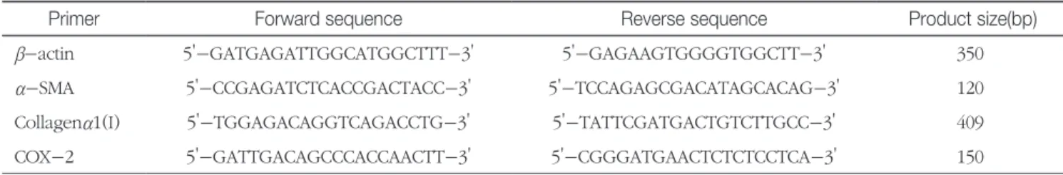
Table 1. Primer sequences and PCR product sizes
Primer Forward sequence Reverse sequence Product size(bp)
β-actin 5'-GATGAGATTGGCATGGCTTT-3' 5'-GAGAAGTGGGGTGGCTT-3' 350
α-SMA 5'-CCGAGATCTCACCGACTACC-3' 5'-TCCAGAGCGACATAGCACAG-3' 120
Collagenα1(I) 5'-TGGAGACAGGTCAGACCTG-3' 5'-TATTCGATGACTGTCTTGCC-3' 409
COX-2 5'-GATTGACAGCCCACCAACTT-3' 5'-CGGGATGAACTCTCTCCTCA-3' 150
모든 결과는 평균과 표준편차로 표현하였으며, p값이 0.05 이하이면 통계학적으로 유의한 것으로 판정하였다. 분석에 는 SPSS ver 10.0 패키지 프로그램을 사용하였다.
결 과
CCl4-투여 쥐의 간조직 HE염색표본에서는 2주 후부터 중심정맥 주변으로 지방변성, 염증세포 침윤, 간세포의 풍 선화 그리고 괴사세포들이 띠모양으로 형성하기 시작하여 4주 후에는 문맥부위까지 뻗어나가 중심정맥과 문맥부위의 연결이 관찰 되었으며, 6주 후에는 중심정맥과 문맥부위, 문 맥부위와 문맥부위 사이에 가교가 만들어졌으며 가교를 따
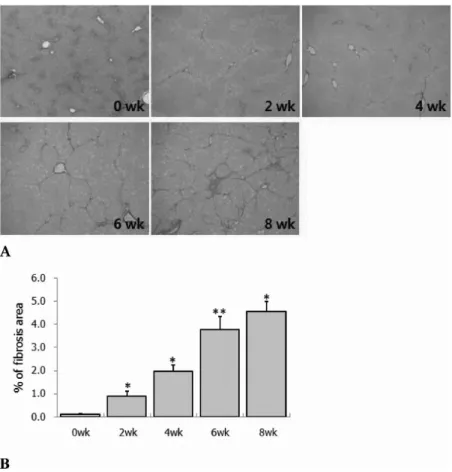
라 방추형의 세포들이 뚜렷이 증가하였다. 8주 후에는 가교 를 따라 방추형의 세포들이 더 많이 증가하여 굵고 진한 띠 로 관찰되었으며 이들 띠로 인하여 간실질조직에 광범위하 게 변형이 일어나면서 동굴모양혈관은 더 이상 구별되지 않 았으며 크고 작은 결절모양들이 만들어지기 시작하였다. 간 조직의 picrosirius red염색 결과 CCl4-투여 2주 후부터 중 심정맥 주변에서 콜라겐이 증가하면서 띠모양으로 짧게 뻗 어나가기 시작하여 4주 후에는 일부에서 문맥부위까지 이 어져 가교모양을 만들었으며, 6주 후에는 중심정맥과 중심 정맥, 중심정맥과 문맥부위 사이에 뚜렷한 가교가 형성되 었고, 8주 후에는 부분적으로 두꺼운 섬유 사이막 (fibrous septae)을 만들기도 하였으며(Fig. 1A), picrosirius red-양성 콜라겐의 이미지분석 결과 CCl4-투여 후 2주부터 8주까지
Fig 1. Changes of Collagen accumulation of liver sections at 0, 2, 4, 6 and 8 week after CCl4 treatment. A. Collagen is stained red in the centrilobular and periportal area. At 4, 6, 8 wk, neighboring central vein and central-portal area were bridged by fibrous septa. Picrosirius red staining. Magnification, x40. B. Quantitative analysis by morphometry of area of fibrosis. Results are expressed as mean ± SD. *p<0.05 compared with **p<0.01 compared with 2wk and 4wk. NS(no significant) compared with normal control and 6wk.
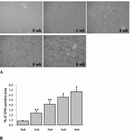
콜라겐이 앞 주의 콜라겐 %보다 통계학적으로 의미 있게 증 가하였다 (Fig. 1B). α-SMA 면역염색에서는 CCl4-투여 2주 후부터 8주까지 섬유화 진행 정도와 일치하여 형성된 섬유 사이막 내에 활성화된 간별모양세포 (hepatic stellate cells) 가 증가되어(Fig. 2A) α-SMA-양성 부위가 통계학적으로 의의 있게 증가하였다 (Fig. 2B). 또한 간섬유화 진행과 더 불어 비만세포 수적 변화를 보기 위해 toluidine blue염색 한 결과 정상대조군의 경우 문맥부위에 없거나 1개 내지 2 개 정도가 출현하였으며, 그 모양은 구형 또는 난원형으로 관찰되었다(Fig. 3A). 그러나 CCl4-투여 2주군에서는 섬유 화의 증가와 더불어 6주까지 통계학적으로 의미 있는 증가 를 보이다가 8주군에서는 통계학적으로 매우 의미 있게 감 소하였다 (Fig. 3B).
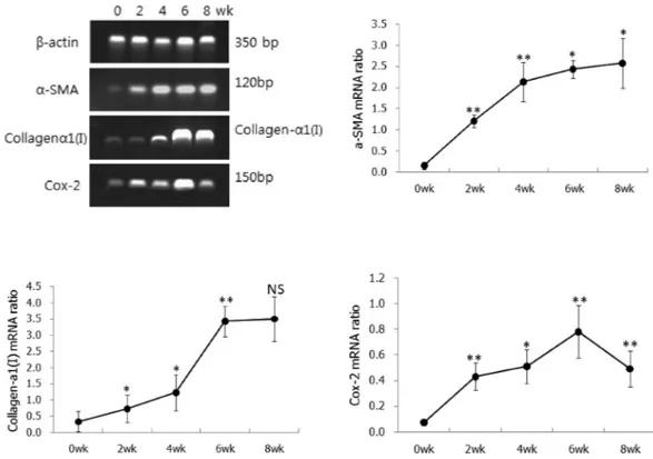
한편, 면역염색 결과를 mRNA 수준에서 확인하기 위하여 RT-PCR에 의해 α-SMA, collagen-a1(I) 그리고 COX-2 mRNA의 발현률을 조사한 결과 정상 대조군에 비해 CCl4- 투여군에서 2주군부터 통계학적으로 의미 있게 증가하기 시작하여 6주까지 섬유화 정도가 심할수록 증가하다가 8주 에서는 α-SMA mRNA 발현률은 약간 의의 있게 증가하였 으나, collagen-a1(I) mRNA 발현률은 약간 의미 있는 증가 를 보이지 않았으며, COX-2 mRNA 발현률은 매우 의의 있 는 감소를 보였다 (Fig. 4).
COX-2 면역염색에서는 COX-2 양성세포가 정상 대조 군에서는 거의 관찰되지 않았으나 CCl4-투여 2주 후부터 동굴모양혈관 내피세포와 염증세포에서 약한 양성반응을 나타냈으며, 4주 후부터 간세포에서 뚜렷한 양성반응을 보
Fig 2. Immunohistochemistry for α smooth muscle actin(α-SMA) to detect activated hepatic stellate cells in liver sections at 0, 2, 4, 6 and 8 wk after CCl4 treatment. A. At 2 and 4 wk, slight positive staining is only detected in centrilobular area. Marked staining for α-SMA is found at 6 and 8 wk, along the fibrous septa. Magnification, x40 B. Quantitative analysis by morphometry of area of staining for α-SMA. Results are expressed as mean ± SD. **p<0.01 compared with normal control and 2 wk. *p<0.05, NS(no significant) compared with 4 wk and 6 wk.
였으며, 6주후부터는 간세포의 양성반응 강도가 4주에 비 해 뚜렷이 증가하였고 특히 섬유사이막 가까이에 위치한 간 세포에서 양성반응이 더욱 뚜렷하게 관찰되었으며, 문맥부 위의 증식된 담관 상피에서도 강한 양성반응을 볼 수 있었 다. 그러나 활성화된 간별모양세포는 음성반응을 나타내었 다 (Fig. 5).
고 찰
본 연구에서 우리는 CCl4로 유도된 쥐의 간조직에서 COX-2가 과발현되고 그 발현률은 섬유화의 진행정도에 따 라 증가함을 관찰할 수 있었으며, 간섬유화의 초기 발생단
계에서 COX-2의 발현과 비만세포의 수의 증가와 밀접한 관련이 있음을 알 수 있었다.
이는 Hinz 등(2001)의 연구에서 정상 심장조직의 경우 섬 유모세포 의해 세포외기질 분비와 침착이비교적 느리고 광 범위하게 진행되는 반면에 심장에서 섬유화가 일어날 경우 섬유모세포가 수축성과 단백합성능이 활발한 근섬유모세포 로 표현형이 바뀌면서 세포외기질을 신속하게 분비하여 심 장조직 사에네 많은 양을 침착시킴으로서 심장 비대가 일어 나는 것과 같은 변화로 생각된다. 섬유화 과정에서 섬유모 세포가 근섬유모세포로의 표현형 전환하는 데는 기계적인 부하와(loading)와 함께 TGF-β1 자극 등과 같은 신호전달 인자들이 관여하는 것으로 알려져 있는데, TGF-β1 자극은 섬유모세포 내에 a-smooth muscel actin이 증가시키고 이 Fig 3. Comparison of mast cell density of liver sections at 0, 2, 4, 6 and 8 wk after CCl4 treatment. A. Mast cells are observed in periportal region, along fibrous septa and in surface fibrous capsular area. Toluidine blue staining. Magnification, x100. B.
Quantitative analysis by morphometry of mast cell number per mm2 in control and CCl4-treated rats during experimental period.
Results are expressed as mean ± SD. *p<0.05, compared with 2 wk, **p<0.01, compared with 0, 4 and 6 wk.
로 인해 수축성과 세포외기질 합성능이 활발한 근섬유모세 포가 증가하게되고 심근경색 후의 심근경색 반흔을 형성하 는데 기여한다고 알려져 있다(Border와 Noble, 1994;Hinz
등, 2001). 그러나 사이질 섬유증(interstitial fibrosis)에서 근 섬유모세포가 어떠한 경로에 의해 활성화되는지에 대해서 는 여전히 잘 알려져 있지 않고 있다.
Fig 4. Comparison of α-SMA, collagen-α1(I) and COX-2 mRNA expression ratio by RT-PCR analysis in CCl4-treated liver tissues during a period of experiment. The expression ratio is expressed as a relative value for β-actin expression, house-keeping gene. α-SMA mRNA. **p<0.01, compared with 0wk, 2wk, and 4wk. *p<0.05, compared with 4wk and 6wk. Collagen- α1(I) mRNA
*p<0.05, compared with 0wk and 2wk. **p<0.01, compared with 4wk. NS(no significant) compared with 6wk. COX-2 mRNA
**p<0.01, compared with 0, 4 and 6 wk. *p<0.05, compared with 2 wk.
Fig 5. Immunohistochemistry for cyclooxygenase-2(COX-2) in liver sections at 0 wk(control)(A) and 6 wk after CCl4 treatment(B).
Positive staining is predominantly detected in hepatic cells adjacent to fibrous septa and bile duct epithelial cells. Magnification, x200.
최근 Levick 등(2009)은 비만세포가 고혈압성 심장 내의 섬유증 발생에 중심 역할을 하며, 이들 세포가 확인되지 않 은 기전에 의한 tryptase 방출과 관련이 있다는 강력한 증거 를 제시하였고, McLarty 등(2011)도 tryptase 방출 기전의 하 나로 tryptase와 protease-activated receptor 2경로 (trypt- ase/PAR-2)가 있음을 확인하였고, 콜라겐 합성의 활성화에 trypatsse/PAR-2 신호전달 경로가 중요한 역할을 담당할 것 을 시사하였다. Masamune 등(2004)도 이미 PAR2 agonist가 쥐 췌장별모양세포( pancreatic stellate cells)에서의 콜라겐 합성을 증가시키며 이는 c-Jun N-terminal kinase의 활성 화를 경유하여 이루어진다고 보고하였다. 더욱이 Gaca 등 (2002)은 간섬유증에서 thrombin 또는 tryptase에 의해 활 성화된 PAR-2 수용체가 간별모양세포(liver stellate cells)의 증식과 콜라겐 합성을 촉진하여 간섬유증을 지속시키는데 기여하는 것으로 보고하였다. 따라서 간섬유화는 tryptase- 유도 신호전달경로를 통해 tryptase와 그 수용체인 PAR2, COX-2 그리고 peroxisome proliferator-activated receptor gamma (PPARγ) 성분들의 관여로 이루어지는 것으로 추정 하고 있다(Frungieri 등, 2002; Knight 등, 2012). Tryptase/
PAR-2 경로를 통해 비만세포로부터 방출된 tryptase는 PAR2를 활성화하여 그 결과 COX-2의 발현을 상위 조절 하고 PG 합성을 자극하며, 이것은 15d-PGJ2에 대한 수용 체로 작용하는 PPARγ를 소유한 섬유모세포와 근섬유모세 포를 활성화로 이어져 간섬유화가 일어난다는 견해와 일치 한다(Abe 등, 2000; Okuda-Ashitaka 등, 1990; Altiok 등, 1997). 뿐만 아니라 tryptase는 섬유모세포 증식과 화학주 성 그리고 아교섬유 생성에 효과를 미친다는 것은 이미 여 러 연구자들에 의해 알려져 있고 섬유모세포 증식인자도 동 정되었다(Asano-Kato 등, 2005; Sawamukai 등, 2010). 이 는 Frungieri 등(2002)이 지적한대로 구조적으로 COX-2를 매우 적게 발현하는 사람 섬유모세포가 tryptase 유도 하에 PAR-2의 활성화를 통하여 세포증식을 자극하고 COX- 2 발현을 증가시키며, tryptase와 PAR2 agonist 펩티드인 SLIGKV가 PG-2합성효소인 COX-2를 유도한다는 결과를 통해서도 COX-2와 비만세포 tryptase의 관련성을 충분히 유추할 수 있다. 또한 Frungieri 등(2002)은 COX-2 mRNA 와 그 단백질의 유도는 수분 내에 일어나는 신속한 사건으
로 그 결과 생물학적으로 활성화된 COX-2는 tryptase로 유 도된 섬유모세포로부터 PGF2a와 15d-PGJ2의 두 PGs를 증 가시킨다고 하였다. 그리고 PGs와 그 수용체는 섬유모세포 의 증식과 I형 콜라겐 합성을 조절한다는 것은 이미 여러 연구에서 제시되어 있다(Akers 등, 2000; Gruber 등, 1997;
Abe 등, 2000).
따라서 섬유화 과정에서 섬유모세포의 증식을 매개하는 tryptase 신호전달경로는 COX-2의 유도, 15d-PGJ2의 합성 그리고 PPARγ를 경유하는 작용이 관여하는 것으로 생각된 다(Kersten 등, 2000; Simonin 등, 2002). 그리고 CCl4로 유 도한 간섬유화의 경우 초기에 증가된 비만세포로부터 trypt- ase/PAR-2 경로를 통해 tryptase가 방출되어 간별모양세포 를 활성화함으로써 세포외기질을 침착을 유도하고 이 과정 에서 간세포나 담관상피세포들로부터 생성된 COX-2 역할 이 중요할 것으로 사료된다. 또한 초기에 증가하기 시작한 비만세포는 6주까지 계속적으로 증가를 보이다가 그 후 감 소하는 경향을 나타내는 것은 피부절제 후 피부 비만세포도 초기에 증가하여 섬유모세포를 활성화하여 콜라겐을 침착 을 유도한 후 감소하며(Jekal et al, 2010), 피부가 자외선에 노출되었을 때 초기에 활성화되기 시작하는 피부진피 섬유 모세포에서는 COX-2가 전자가 증가하나 오히려 증식중인 섬유모세포에서는 COX-2의 전사가 감소한다는 결과와 일 치한다(Mohammed 등, 2004).
이상의 결과를 종합해 볼 때 CCl4-유도에 의한 초기 간섬 유화과정에서의 COX-2 과발현은 초기에 증가된 비만세포 로부터 방출된 tryptase에 의한 것으로 사료된다.
참고문헌
1. Abe M, Kurosawa M, Ishikawa O, Miyachi Y. Effect of mast cell-derived mediators and mast cell-related neutral prote- ases on human dermal fibroblast proliferation and type I col- lagen production. J Allergy Clin Immunol. 2000, 106:78-84.
2. Akers IA, Parsons M, Hill MR, Hollenberg MD, Sanjar S, Lau- rent GJ, et al. Mast cell tryptase stimulates human lung fibro- blast proliferation via protease-activated receptor-2. Am J Physiol Lung Cell Mol Physiol. 2000, 278:193-201.
3. Altiok S, Xu M, Spiegelman BM. PPARgamma induces cell
cycle withdrawal: inhibition of E2F/DP DNA-binding activity via down-regulation of PP2A. Genes Dev. 1997, 11:1987- 1998.
4. Appleton l. Tomlinson A, Willoughby DA. Induction of cy- clooxygenase and nitic oxide synthase in inflmmation. Adv Pharmacol. 1996, 35:27-78.
5. Asano-Kato N, Fukagawa K, Okada N, Dogru M, Tsubota K, Fujishima H. Tryptase increases proliferative activity of human conjunctival fibroblasts through protease-activated recep- tor-2. Invest Ophthalmol Vis Sci. 2005, 46:4622-4626.
6. Border WA, Noble NA. Transforming growth factor beta in tis- sue fibrosis. N Engl J Med. 1994, 331:1286-1292.
7. Cheng AS, Chan HL, Leung WK, To KF, Go MY, Chan JY, et al. Expression of HBx and COX-2 in chronic hepatitis B, cirrhosis and hepatocellular carcinoma: implication of HBx in upregulation of COX-2. Mod Pathol. 2004, 17:1169-1179.
8. Dubois RN, Abramson SB, Crofford L, Gupta RA, Simon LS, Van De Putte LB, et al. Cyclooxygenase in biology and dis- ease. FASEB J. 1998, 12:1063-1073.
9. Enerbäck L. Mast cells in rat gastrointestinal mucosa. I. Effects of fixation. Acta Pathol Microbiol Scand. 1966, 66:289-302.
10. Friedman SL. Molecular regulation of hepatic fibrosis, an in- tegrated cellular response to tissue injury. J Biol Chem. 2000, 275:2247-2250.
11. Frungieri MB, Weidinger S, Meineke V, Kohn FM, Mayerhofer A. Proliferative action of mast cell tryptase is mediated by PAR2, COX2, prostaglandins, and PPARgamma: Possible rel- evance to human fibrotic disorders. PNAS. 2002, 99:15072- 15077.
12. Gaça MD, Zhou X, Benyon RC. Regulation of hepatic stel- late cell proliferation and collagen synthesis by proteinase- activated receptors. J Hepatol. 2002, 36:362-369.
13. Gruber BL, Kew RR, Jelaska A, Marchese MJ, Garlick J, Ren S, Schwartz LB, et al. Human mast cells activate fibroblasts:
tryptase is a fibrogenic factor stimulating collagen messenger ribonucleic acid synthesis and fibroblast chemotaxis. J Immu- nol. 1997, 158:2310-2317.
14. Hinz B, Mastrangelo D, Iselin CE, Chaponnier C, Gabbiani G. Mechanical tension controls granulation tissue contractile activity and myofibroblast differentiation. Am J Pathol. 2001, 159:1009-1020.
15. Hui AY, Leung WK, Chan HL, Chan FK, Go MY, Chan KK, et al. Effect of celecoxib on experimental liver fibrosis in rat.
Liver Int. 2006, 26:125-136.
16. Kersten S, Desvergne B, Wahli W.Roles of PPARs in health and disease. Nature. 2000, 405:421-424.
17. Knight V, Tchongue J, Lourensz D, Tipping P, Sievert W.
Protease-activated receptor 2 promotes experimental liver
fibrosis in mice and activates human hepatic stellate cells.
Hepatology. 2012, 55:879-887.
18. Kwon SH, Jeong SW, Jang JY, Lee JE, Lee SH, Kim SG, et al.
Cyclooxygenase-2 and vascular endothelial growth factor in chronic hepatitis, cirrhosis and hepatocellular carcinoma. Clin Mol Hepatol. 2012, 18:287-294.
19. Levick SP, McLarty JL, Murray DB, Freeman RM, Carver WE, Brower GL. Cardiac mast cells mediate left ventricular fibrosis in the hypertensive rat heart. Hypertension. 2009, 53:1041–
1047.
20. Levi-Schaffer F, Kupietzky A. Mast cells enhance migration and proliferation of fibroblasts into an in vitro wound. Exp Cell Res. 1990, 188:42-49.
21. López-De León A, Rojkind M. A simple micromethod for collagen and total protein determination in formalin-fixed paraffin-embedded sections. J Histochem Cytochem. 1985, 33:737-743.
22. Masamune A, Kikuta K, Suzuki N, Satoh M, Satoh K, Shi- mosegawa T. A c-Jun NH2-terminal kinase inhibitor SP600125 (anthra[1,9-cd]pyrazole-6 (2H)-one) blocks ac- tivation of pancreatic stellate cells. J Pharmacol Exp Ther.
2004, 310:520-527.
23. McLarty JL, Mele´ndez GC, Brower GL, Janicki JS, Levick SP.
Tryptase/ protease-activated receptor 2 interactions induce selective mitogen-activated protein kinase signaling and col- lagen synthesis by cardiac fibroblasts. Hypertension. 2011, 58:264–270.
24. Mohammed NA, El-Aleem SA, El-Hafiz HA, McMahon RFT.
Distribution of constitutive(COX-1) and inducible(COX-2) cyclooxygenase in postviral human liver cirrhosis: a possible role for COX-2 in the pathogenesis of liver cirrhosis. J Clin Pathol. 2004. 57:350-354.
25. Okuda-Ashitaka E, Negishi M, Sugama K, Hatanaka M, Ito S. Cyclic AMP-mediated inhibition of cell growth by pros- taglandin D2 in a fibroblastic cell line (EBTr). Eicosanoids.
1990, 3:213-218.
26. Sawamukai N, Yukawa S, Saito K, Nakayamada S, Kambayas- hi T, Tanaka Y. Mast cell-derived tryptase inhibits apoptosis of human rheumatoid synovial fibroblasts via rho-mediated signaling. Arthritis Rheum. 2010, 62:952-959.
27. Shattuck-Brandt RL, Varilek GW, Radhika A, Yang F, Wash- ington MK, DuBois RN. Cyclooxygenase 2 expression is in- creased in the stroma of colon carcinomas from IL-10(-/-) mice. Gastroenterology. 2000, 118:337-345.
28. Simonin MA, Bordji K, Boyault S, Bianchi A, Gouze E, Bécu- we P, et al. PPAR-gamma ligands modulate effects of LPS in stimulated rat synovial fibroblasts. Am J Physiol Cell Physiol.
2002, 282:125-133.
29. Smith TJ, Parikh SJ. HMC-1 mast cells activate human orbital fibroblasts in coculture: evidence for up-regulation of pros- taglandin E2 and hyaluronan synthesis. Endocrinology. 1999, 140:3518-3525.
30. Jekal SJ, Kwon PS, Kim JK. Effect of 630 nm Light Emitting Diode(LED) Irradiation on Wound Healing in Streptozotocin- Induced Diabetic Rats. J Exp Biomed Sci. 2010, 16:365-376