306 www.kjtcvs.org
KJTCVS
The Korean Journal of Thoracic and Cardiovascular SurgeryCase Report
Left Ventricular Assist Device Implantation via Dual Left
Thoracotomy in an Adult Patient with Congenitally Corrected Transposition of the Great Arteries
Shin Kim, M.D., Yang-Hyun Cho, M.D., Pyo Won Park, M.D., Young Tak Lee, M.D., Tae-Gook Jun, M.D., Wook Sung Kim, M.D., Kiick Sung, M.D., Ji-Hyuk Yang, M.D., Suryeun Chung, M.D.
Department of Thoracic and Cardiovascular Surgery, Samsung Medical Center, Sungkyunkwan University School of Medicine, Seoul, Korea
ARTICLE INFO Received July 22, 2019 Revised October 31, 2019 Accepted November 4, 2019 Corresponding author Yang-Hyun Cho Tel 82-2-3410-2213 Fax 82-2-3410-0089 E-mail [email protected] ORCID
https://orcid.org/0000-0003-1685-3641
A 59-year-old man presented for possible durable ventricular assist device (VAD) implan- tation. He had previously been diagnosed with congenitally corrected transposition of the great arteries, a ventricular septal defect, an atrial septal defect, pulmonary valve stenosis, and aortic valve regurgitation. In the previous 22 years, he had undergone palliative cardi- ac surgery 3 times. VAD implantation as a bridge to transplantation was planned. Owing to severe adhesions, mesocardia, a left ascending aorta, and moderate aortic regurgitation, we performed VAD implantation and aortic valve closure via a dual left thoracotomy and partial sternotomy.
Keywords: Congenitally corrected transposition of the great arteries, Left ventricular as- sist device, Heart failure
Copyright
© The Korean Society for Thoracic and Cardiovascular Surgery. 2020. All right reserved.
This is an Open Access article distributed under the terms of the Creative Commons Attribution Non-Commercial License (http://creativecommons.org/licenses/
by-nc/4.0) which permits unrestricted non-commercial use, distribution, and reproduction in any medium, provided the original work is properly cited.
Case report
A 59-year-old man presented for possible durable ven- tricular assist device (VAD) implantation. At 37 years of age, he was diagnosed with congenitally corrected transpo- sition of the great arteries (CCTGA) with a ventricular septal defect (VSD), an atrial septal defect (ASD), pulmo- nary valve stenosis, and aortic valve regurgitation (AR). In his first cardiac surgery at another hospital, he underwent morphologic left ventricle (mLV)-to-pulmonary artery (PA) conduit placement, ASD/VSD closure, and postoperative bleeding control. At 46 years of age, he underwent a second heart surgery at our institution consisting of mLV-to-PA conduit change, tricuspid valve replacement with a me- chanical valve, residual VSD closure, aortic valve repair, left PA angioplasty, and postoperative bleeding control. At 55 years of age, he underwent heart surgery a third time (mLV-to-PA conduit change with mechanical valved con- duit). After the third operation, he took heart failure medi- cations for systemic ventricle (morphologic right ventricle [mRV]) failure. He was admitted to the hospital 3 times within the 6 months prior to the VAD consultation. Heart
failure led to renal insufficiency (estimated glomerular fil- tration rate: 34.0 mL/min/1.73 m
2) and liver cirrhosis (Child-Pugh class B). During his most recent admission, he developed cardiogenic shock and was admitted to the in- tensive care unit multiple times. Echocardiography re- vealed severe mRV dysfunction (ejection fraction, 20%) and moderate aortic regurgitation. We referred him for cardiac transplantation; however, his condition deteriorat- ed rapidly. Owing to the aortic valve regurgitation and previous cardiac pathologies, venoarterial extracorporeal membrane oxygenation was not possible. As he had type O blood, his wait time for transplantation was expected to be relatively long. Thus, we planned to implant a HeartWare VAD (HVAD; HeartWare International Inc., Framingham, MA, USA) as a bridge to transplantation.
We decided to perform a dual left thoracotomy instead of the standard sternotomy. Through a left anterolateral thoracotomy via the fifth intercostal space using a 15-cm incision, the apex of the mRV was confirmed and the sur- rounding tissue was dissected to allow for implantation.
Owing to the left aortic arch, an anterolateral thoracotomy via the second intercostal space (10-cm incision) was per-
https://doi.org/10.5090/kjtcs.19.053 pISSN: 2233-601X eISSN: 2093-6516
Korean J Thorac Cardiovasc Surg. 2020;53(5):306-309 https://doi.org/10.5090/kjtcs.19.053
pISSN: 2233-601X eISSN: 2093-6516
Korean J Thorac Cardiovasc Surg. 2020;53(5):306-309
307
Shin Kim, et al. LVAD Implantation in an Adult Patient with CCTGA
www.kjtcvs.org
KJTCVS
formed for aortic valve closure. An unplanned partial ster- notomy was performed to improve visualization of the an- terior and medial sides of the ascending aorta because the sternum and aorta were tightly adhered. Despite the upper sternotomy, the mLV-to-PA conduit and ascending aorta could not be completely divided because of severe adhe- sions. Thus, the right femoral artery and vein were cannu- lated for cardiopulmonary bypass. Aortic cross-clamping was performed through a thoracotomy, and an aortotomy was created. The cross-clamping was insufficient and blood leakage occurred. A 12F urinary balloon catheter was inserted in the gap to block the blood leakage from the medial side of the aortic cross-clamp. A single dose of cold- blood cardioplegia was delivered via a direct cardioplegic infusion catheter. The aortic valve was closed using a mod- ified Park stitch and commissuroplasty [1]. After the aortic
cross-clamping was released, we sought a suitable site for the inflow cannula by manual palpation and insertion of a 16F Foley catheter under transesophageal echocardiogra- phy guidance. The inflow cannula axis in this patient was from the anterior to the posterior and from left to right on the horizontal plane (Fig. 1A, B). The sewing ring was placed using multiple pledged horizontal mattress sutures.
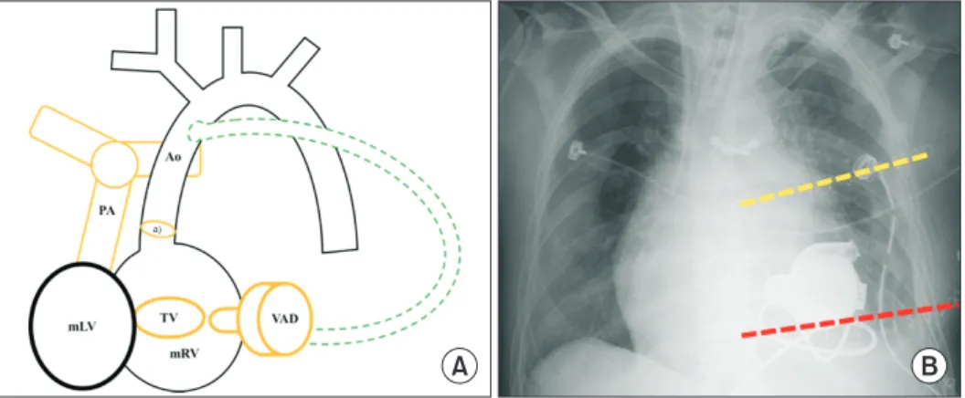
After adequate length of the outflow graft was ensured, it was trimmed for aortic anastomosis. The outflow graft was installed on the left lateral side of the ascending aorta us- ing partial aortic clamping (Fig. 2A, B). The driveline was placed using the single tunnel technique. Cardiopulmo- nary bypass was smoothly transitioned to the HVAD. The HVAD speed was set at 2,400 rpm, flow at 3.4 L/min, and power at 2.7 W. Intraoperative transesophageal echocardi- ography confirmed good inflow position. The incisions
Fig. 1. (A) Diagram of the patient’s heart. The green dotted line is the conventional implantation axis; insufficient space for the device was expected. The blue dotted line is the alternative axis applied in this case. (B) The green dotted line shows the conventional implan- tation axis. A chest computed tomography scan shows the longitudinal axis of the heart lying in the mid-sagittal plane (i.e., mesocardia).
The TV was replaced with a mechanical valve in this patient. If the VAD had been inserted along the green dotted line, the space would have been expected to be very narrow (yellow dashed arrow). VAD, ventricular assist device; mLV, morphologic left ventricle; mRV, morphologic right ventricle; MV, mitral valve; TV, tricuspid valve.
A B
Fig. 2. (A) Diagram of the patient’s postoperative status. The green dotted line is the outflow tract, which was inserted on the left side of the ascending aorta. (B) Chest radiograph taken immediately after surgery. Thoracotomy incisions for aortic valve repair and outflow anastomosis (yellow dashed line) and inflow of the pump insertion (red dashed line) are shown. VAD, ventricular assist device; mLV, morphologic left ventricle; mRV, morphologic right ventricle; TV, tricuspid valve; mLV-to-PA conduit, morphologic left ventricle-to-pul- monary artery conduit.
a)Repaired aortic valve.
A B
a)