증례7 증례5
증례8 증례6
증례9
1연세대학교 의과대학 강남세브란스병원 뇌종양센터 신경외과학교실, 2용인세브란스병원 신경외과학교실, 3홍익대학교 조각과
강민규1, 장창기2, 박현호1, 장동수3, 이규성1, 홍창기1
1Department of Neurosurgery, Gangnam Severance Hospital, Brain Tumor Center, Yonsei University College of Medicine, Seoul, Korea
2Department of Neurosurgery, Yongin Severance Hospital, Yonsei University College of Medicine, Yongin, Korea
3Department of Sculpture, Hongik University, Seoul, Korea
Min-Gyu Kang1, Chang Ki Jang2, Hun Ho Park1, Dong-Su Jang3, Kyu-Sung Lee1, Chang-Ki Hong1
계량된 톨킨슨 션트: 제4 뇌실 주변 종양에 대한 적용
J Korean Skull Base Society 15권 2호 : 90~97, 2020
종설1 원저1 원저2
증례1 원저3
증례2 증례3 증례4
Background : The Torkildsen shunt is a procedure used in the 1940s and 1950s to bypass the cerebrospinal fluid flow from the lateral ventricle to the cisterna magna, before the introduction of extracranial shunts. The aim of this study was to re-evaluate and modify the Torkildsen shunt to prevent obstructive hydrocephalus in patients with 4th ventricle tumors.
Methods : Fifteen patients underwent surgery for benign or malignant 4th ventricle tumors via the midline suboccipital approach from August 2010 to August 2016. Among them, eight received a modified Torkildsen shunt to prevent obstructive hydrocephalus. Permanent ventricular catheter is placed in the 4th ventricle from the obex to the cerebral aqueduct, bridging the 3th ventricle and the cervical subarachnoid space at the foramen magnum.
Results : Among the eight patients who underwent tumor removal with the modified Torkildsen shunt, three had hydrocephalus preoperatively, but none had hydrocephalus postoperatively. Four patients presented preoperative hydrocephalus on control group. The rate of hydrocephalus resolution after tumor removal without ventricle shunting procedure was 25%. Three patients required external ventricular drainage before tumor removal; one patient needed ventriculo-peritoneal shunt owing to progression of hydrocephalus two-month later after the operation. No shunt-related complications or malfunction were observed over a mean follow-up period of 35 months.
Modified Torkildsen shunt: Application of the historical procedure for 4th ventricle tumors
논문 접수일 : 2020년 6월 9일 논문 완료일 : 2020년 8월 1일 주소 : Department of Neurosurgery,
Gangnam Severance Hospital, Brain Tumor Center, Yonsei University College of Medicine, 211 Eonju-ro Gangnam-gu, Seoul 06273, Korea Tel : +82-2-2019-3390
Fax : +82-2-2019-2160 E-mail : [email protected]
Chang-Ki Hong
교신저자
▒ INTRODUCTION
The Torkildsen shunt was first developed in 1937 by Arne Torkildsen, a Norwegian pediatric neurosurgeon.[1,2] It is a ventriculocisternal shunt used to divert cerebrospinal fluid (CSF) flow directly from the lateral ventricle to the cisterna magna. Back in the days, the up-to-date technology of a ventriculo-peritoneal (VP) shunt was not available. So, it was a difficult problem for a pediatric patient with non- communicating or obstructive hydrocephalus at the 3rd or 4th ventricle or the aqueduct of Sylvius.
Arne Torkildsen suggested a ventriculocisternostomy, i.e., puncturing the lateral ventricle with a rubber catheter via the Frazier’s point and tunneling the catheter underneath the scalp towards the cisterna magna exposed after a midline suboccipital craniotomy with C1 laminectomy.[1] The Torkildsen shunt gained widespread use in the 1940s and 1950s before the introduction of extracranial shunts.[3-6]
The Torkildsen shunt restricts the CSF within the nervous system, and decreases the risk of its spread to the blood or peritoneal cavity in cases of cancer.[7] After the advent of the VP shunt, the Torkildsen shunt has become obsolete. However, there still have been reports regarding ventriculo-cisternal shunt.[8-10] In this study, we modified and simplified this procedure to suit modern day requirements.
To resolve and prevent obstructive hydrocephalus after removal of 4th ventricle tumors, we applied the modified
Torkildsen shunt in selected patients and compared its efficiency with that in patients who did not receive the shunt after removal of 4th ventricle tumors.
▒ METHODS
1. Ethical approval
The study was approved by the Instituitional Research Ethics Committee of the Gangnam Severance Hospital (3- 2017-0194, approved on 03.09.2017).
2. Informed consent
Informed consent was obtained from all individual participants included in the study.
3. Patient selection and application of the modified Torkildsen shunt
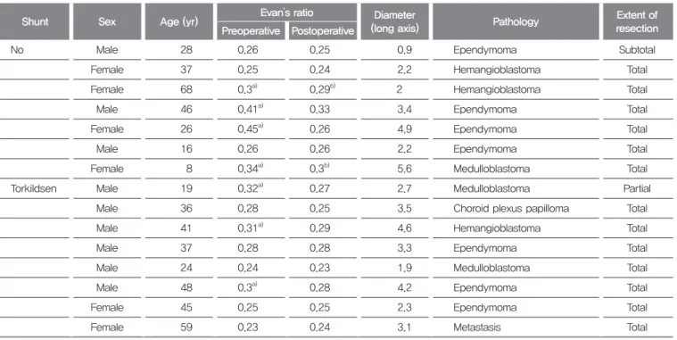
From August 2010 to August 2016, 15 patients were operated upon for benign or malignant 4th ventricle tumors. Hydrocephalus is defined using the Evan’s ratio, i.e., the ratio of the frontal horn to the maximal biparietal diameter. An Evan’s ratio > 0.3 accompanied by symptoms like headache suggests hydrocephalus. Preoperative and postoperative magnetic resonance imaging (MRI) and computed tomography (CT) scans are used for determining the Evan’s ratio (Table 1). In our patient population, seven patients had obstructive hydrocephalus before tumor removal, and the modified Torkildsen shunt was performed in three of these patients. The remaining eight patients had Conclusions : The modified Torkildsen shunt can be effective option in resolving postoperative hydrocephalus in 4th ventricle tumors. It is a simple and efficient procedure without the need for an endoscopic procedure or ventriculo-peritoneal shunt if we choose careful patient selection.
Fourth ventricle tumor, Obstructive hydrocephalus, Torkildsen shunt, Ventriculoperitoneal shunt
Key Words
a normal ventricle before tumor removal, and the modified Torkildsen shunt was performed in five of these patients.
Thus, overall, eight patients underwent the modified Torkildsen shunt to prevent obstructive hydrocephalus. The intraoperative indications we set for modified Torkildsen shunt are followed: (1) Tumor involving at least three planes of 4th ventricle; (2) Tumor invading the aqueduct of Sylvius; (3) Considerable amount of intraoperative bleeding during the resection of the tumor; (4) High risk of postoperative cerebral edema owing to substantial retraction or manipulation of adjacent structures.
4. Surgical procedure
Instead of puncturing the lateral ventricle and tunneling towards the cisterna magna, as is done in the case of the Torkildsen shunt, we inserted the catheter intradurally to connect the aqueduct of Sylvius with the cisterna magna.
A C1 laminectomy is not necessary for our procedure.
Fig. 1 shows the posterior view of the three-dimensional reconstruction of a skull undergoing the Torkildsen
shunt,[11] and the difference in the new modified Torkildsen shunt.
Preoperative MRI is a mandatory component of our procedure. The distance from the obex to the cerebral aqueduct on an MRI sagittal view and any vascular abnormality must be checked for before surgery.
All operations were performed with the patient in the prone position. Tumor resection via midline suboccipital craniotomy without C1 laminectomy was performed by two experienced surgeons using the same procedure. After tumor removal, the modified Torkildsen shunt was applied before dura closing. First, the distance from the obex to the cerebral aqueduct was measured on an MRI sagittal scan.
Then, a proximal catheter designed for the VP shunt was inserted and advanced towards the cerebral aqueduct. The distal end of the catheter was then cut according to the length measured on the MRI scan and advanced towards the obex. Finally, the distal end of the catheter was anchored to the dura with a silk tie (Fig. 2). The catheter thus connected the cerebral aqueduct with the obex.
Table 1. Summary of patient
Shunt Sex Age (yr) Evan’s ratio Diameter
(long axis) Pathology Extent of
resection Preoperative Postoperative
No Male 28 0.26 0.25 0.9 Ependymoma Subtotal
Female 37 0.25 0.24 2.2 Hemangioblastoma Total
Female 68 0.3a) 0.29b) 2 Hemangioblastoma Total
Male 46 0.41a) 0.33 3.4 Ependymoma Total
Female 26 0.45a) 0.26 4.9 Ependymoma Total
Male 16 0.26 0.26 2.2 Ependymoma Total
Female 8 0.34a) 0.3b) 5.6 Medulloblastoma Total
Torkildsen Male 19 0.32a) 0.27 2.7 Medulloblastoma Partial
Male 36 0.28 0.25 3.5 Choroid plexus papilloma Total
Male 41 0.31a) 0.29 4.6 Hemangioblastoma Total
Male 37 0.28 0.28 3.3 Ependymoma Total
Male 24 0.24 0.23 1.9 Medulloblastoma Total
Male 48 0.3a) 0.28 4.2 Ependymoma Total
Female 45 0.25 0.25 2.3 Ependymoma Total
Female 59 0.23 0.24 3.1 Metastasis Total
a)It means hydrocephalus.
b)It means ventriculomegaly.
▒ RESULTS
The mean follow-up duration after tumor resection was 35 months (2-95 months). The mean age of the patients who received the modified Torkildsen shunt was 38.6 years, while that of patients who did not receive the shunt was 32.7 years. There were no statistical differences between the two groups with respect to the age, sex,
tumor size, preoperative hydrocephalus, histological findings, and the extent of resection (Table 2).
Among the eight patients who underwent tumor removal with the modified Torkildsen shunt, three had hydrocephalus preoperatively, but none had hydrocephalus postoperatively.
Four patients presented preoperative hydrocephalus on control group. The rate of hydrocephalus resolution after
Fig. 1
A B Typical (A) and modified (B) Torkildsen
shunt three-dimensional (3D) modeling.
(A) 3D reconstructed computed tomography after the typical Torkildsen shunt procedure. Partial C1 laminectomy was performed to insert the cisternal end of the catheter. (B) 3D reconstructed computed tomography after the modified Torkildsen shunt. The catheter was placed intradurally to connect the aqueduct of Sylvius with the cisterna magna. A C1 laminectomy is not necessary in our procedure.
Fig. 2 A
B C D E
Surgical procedure of the modified Torkildsen shunt.
(A) Measure the distance from the obex to the cerebral aqueduct on a magnetic resonance imaging (MRI) sagittal view. (B) After tumor resection via midline suboccipital craniotomy, insert a catheter designed for the ventriculoperitoneal shunt towards the cerebral aqueduct. (C) Cut the distal end of the catheter according to the length measured on MRI. (D) Insert the distal end towards the obex.
(E) Anchor the distal end to the dura with a silk tie.
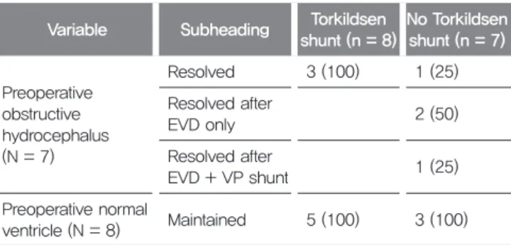
tumor removal without ventricle shunting procedure was 25%. Three patients required external ventricular drainage (EVD) before tumor removal and one patient achieved resolution of hydrocephalus; the other two patients still presented ventriculomegaly and one patient needed VP shunt two months later after the operation due to the progression of hydrocephalus. A normal ventricle could be maintained in all patients regardless of whether or not they received the Torkildsen shunt (Table 3). No shunt- related failure including catheter migration or malfunction was observed. No cases of infection were observed either.
1. Case presentation
A 36-year-old male had a mild headache. He had a brain tumor occupying the entire 4th ventricle with mild hydrocephalus. He underwent removal of brain tumor through mid suboccipital craniotomy and the telovelar approach. The tumor seemed to have originated from the
choroid plexus of the 4th ventricle. Gross total removal of the tumor was performed. There was no injury to the floor of the 4th ventricle. Then, the modified Torkildsen shunt was positioned between the cerebral aqueduct and obex without additional craniotomy (Fig. 3). No complications were observed, and the headache resolved. At the 1-year follow-up, MRI image showed mild, nearly resolved obstructive hydrocephalus.
▒ DISCUSSION
Preoperative hydrocephalus has been reported in approximately 80% of patients with posterior fossa tumor.
[12] The best case scenario would be for the obstructive hydrocephalus to resolve after tumor resection alone.
However, persistent progressive hydrocephalus after removal of tumor has been reported in about 25%-30%
of the cases.[12-17] Hence, preoperative management including EVD and endoscopic 3rd ventriculostomy (ETV) is performed, and postoperative management including EVD, ETV, VP shunt is also performed for managing hydrocephalus.
In our study, the modified Torkildsen shunt led to resolution of hydrocephalus. Among patients who did not receive the shunt, the hydrocephalus persisted in three patients-two needed a temporary EVD which were maintained until the initial postoperative period and removed on the following week, while the other patient deteriorate owing to progression of hydrocephalus and received VP shunt
Table 3. Surgical outcomes
Variable Subheading Torkildsen shunt (n = 8)
No Torkildsen shunt (n = 7)
Preoperative obstructive hydrocephalus (N = 7)
Resolved 3 (100) 1 (25)
Resolved after
EVD only 2 (50)
Resolved after
EVD + VP shunt 1 (25)
Preoperative normal
ventricle (N = 8) Maintained 5 (100) 3 (100) Values are presented as number (%).
EVD: external ventricular drainage, VP: ventriculo-peritoneal.
Table 2. Patient demographics
Variable Torkildsen shunt
No Torkildsen shunt P-value
Patients 8 7 -
Mean age (yr) 38.6 32.7 0.51
Sex 0.20
Male 6 3 -
Female 2 4 -
Mean tumor size (cm) 3.2 (1.9-4.6)
3.02
(0.9-5.6) 0.80
Preoperative hydrocephalus 3 4 0.44
Histology 0.31
Benign 5 6 -
Hemangioblastoma 1 2 -
Choroid plexus
papilloma 1 0 -
Ependymoma 3 4 -
Malignant 3 1 -
Medulloblastoma 2 1 -
metastasis 1 0 -
Gross total resection 7 (87.5) 6 (85.7) 0.91 Mean follow-up duration (mo) 35 (2-95) - Values are presented as number only. mean only, mean (range), number (%).
two month later after the operation (Table 3).
Before the advent of the VP shunt, the Torkildsen shunt was the most useful treatment for obstructive hydrocephalus.[4,5,18] The typical Torkildsen shunt involves a connection between the lateral ventricle and cisterna magna. So there is no need for CSF to go through aqueduct of Sylvius and forth ventricle. The patients who have an obstructive lesion in the 3th and 4th ventricle are good candidates for the Torkildsen shunt.
Surgical results of the Torkildsen shunt before the 1960s were not good.[5] Late complications occurred one and a quarter months to four years after the operation, leading to death in 7 out of 16 cases.[19] In the 1950s and 1960s, various surgeons reported results of the Torkildsen shunt: operative mortality rate ranged from 22% to 40%, and initial rate of arrest of hydrocephalus ranged from 58% to 77%.[3,4,18,20] However, it is worth noting that surgery at that time was performed without accurate MRI or CT scans or surgical tools. Similar to the Torkildsen shunt, other shunt methods including cardiac, peritoneal, and pleural shunts yielded poor results at that time: the mortality rate ranged from 10%-20%, and rate of arrest of hydrocephalus ranged from 50%-70%.[5] It was difficult to demonstrate the cause of mortality because in the 1930s and 1940s, radiological evaluation was very limited.
[11] So, Torkildsen shunt which needed C1 laminectomy and foramen magnum opening was very risky, and was gradually replaced by the VP shunt, which needs only a burr hole.
Upward herniation following CSF shunt before tumor removal have been reported.[21,22] Sainte-rose et al.
[15] reported a 1.5% incidence of upward herniation after preoperative ETV. Upward herniation can lead to rapid progression of mental degradation and eventually death.
It is caused by ascending cerebellar vermis above the tentorium, compressing the midbrain and also possibly occluding the posterior circulating system, including the posterior cerebral artery and superior cerebellar artery. EVD or other supratentorial shunting systems
make decompressing mainly supratentorial pressure, so infratentorial structure can go upward and make upward herniation. However, the modified Torkildsen shunt is a connection between the supratentorial and sub- infratentorial subarachnoid space passing by the infra- tentorial structure. Mismatching between the supra- and infra-tentorial pressure is minimized. Also, if the tumor recurs, CSF flow can be compromised and hydrocephalus is aggravated. In case of recurrence, the modified Torkildsen shunt can maintain normal CSF in the circulating system.
The modified Torkildsen shunt has many advantages compared to other shunting systems. First, the typical Torkildsen shunt involves C1 laminectomy and parietooccipital burr hole for ventricular puncture, but the modified Torkildsen shunt does not need C1 laminectomy and no additional craniotomy and incision, only using the craniotomy site for tumor removal. The time required for the shunting is very short, only simply finding obex and placing the tube.
Second, shunt obstruction seems uncommon because of its short ventricular catheter compared with other somatic shunts or typical Torkildsen shunt. Shunt obstruction is the most common cause of shunt malfunction.[23] Proximal catheter is the most common site, and the reported rate of distal catheter obstruction is 12%-34%. The shunt can be obstructed only in two ways: first, inside of the system by debridement, and second, outside of the system by compression or kinking. The modified Torkildsen shunt catheter is extremely short and involves less chance of obstruction due to debridement. It does not require any valve and simply connects two physiologic spaces.
Third, shunt malfunction is a very common complication, and the common cause of this is the migration of the distal tip of the shunt due to body growth in pediatric patients.
[24] The modified Torkildsen shunt, unlike cranio-somatic shunts, such as the VP shunt, is unaffected by the growth of the child.
However, the modified Torkildsen shunt does have some disadvantages too. First, in case of hydrocephalus
progression, shunt malfunction is easily checked for in case of the ventriculo-somatic shunt that has a valve by sensing the pressure of the valve. This is not possible in case of the modified torkildsen shunt. Second, the shunt catheter is placed in very important structures, the vessel and cranial nerve. In case of catheter malfunction or infection, the catheter may need to be removed, but this might be complicated by adhesion of the catheter to the surrounding structure. Removal of the catheter could create severe problems. Lastly, the shunt can get displaced causing shunt malfunction, because it is anchored only with the help of a dura suture, and the craniocervical junction is an open movable space. There are several reports showing similar cases where Torkildsen shunt catheter migration cause cervical myelopathy.[25-27]
Our study has some limitations. First, in severe hydrocephalus patients, EVD should be inserted before tumor removal. In that cases, we maintained EVD after surgery. So, it is courious that preoperative hydrocephalus was resolved only by tumor removal itself or EVD making some time to recover normal circulating system. Second, propagation or seeding of malignant tumor cells and intraoperative bleeding can trigger communicating hydrocephalus. Modified Torkildsen shunt is not enough and VP shunt may be needed in this circumstances.
Third, small sample size may weaken the safety and statistical power of this study. Lastly, long-term follow- up for monitoring possible late complication is required.
Despite this limitations, one stage catheter indwelling without further incision or craniotomy and the physiologic Fig. 3
A B
E
C
F D
Computed tomography scan and magnetic resonance imaging of case presentation.
(A, B) A 36-year-old male diagnosed as choroid plexus papilloma of the 4th ventricle with obstructive hydrocephalus. (C, D) Gross total removal was achieved and modified Torkildsen shunt was performed. (E, F) As can be seen on the images before and 1 year after surgery, mild obstructive hydrocephalus resolved with modified Torkildsen shunt and remained stable.
nature connecting supratentorial area with infratentorial subarachnoid space can be attractive alternatives of CSF diversion when we choose careful patient selection.
In conclusion, the modified Torkildsen shunt can be an effective method to resolve hydrocephalus in 4th ventricle tumors. The procedure is simple and efficient without the need for an endoscopic procedure or VP shunt. We suggest that the procedure may still prove to be valuable in the era of modern neurosurgery if we choose careful patient selection.
CONFLICT OF INTEREST
No potential conflict of interest relevant to this article was reported.
REFERENCES
1. Eide PK, Lundar T. Arne Torkildsen and the ventriculocisternal shunt: The first clinically successful shunt for hydrocephalus. J Neurosurg 2016;124:1421-8.
2. Torkildsen A, El-Toraei I. A new operative technique for the treatment of hydrocephalus externus; preliminary report. J Egypt Med Assoc 1953;36:836-46.
3. Fincher EF, Strewler GJ, Swanson HS. The Torkildsen procedure; a report of 19 cases. J Neurosurg 1948;5:213-29.
4. Paine KW, McKissock W. Aqueduct stenosis; clinical aspects, and results of treatment by ventriculocisternostomy (Torkildsen's operation). J Neurosurg 1955;12:127-45.
5. Scarff JE. Treatment of hydrocephalus: An historical and critical review of methods and results. J Neurol Neurosurg Psychiatry 1963;26:1-26.
6. Weisenberg SH, TerMaath SC, Seaver CE, Killeffer JA. Ventricular catheter development: Past, present, and future. J Neurosurg 2016;125:1504-12.
7. Fox JL, Al-Mefty O. Percutaneous ventriculocisternal shunt. Technical note.
Surg Neurol 1985;24:184-6.
8. Zhang Z, Wang H, Cheng H, Fan Y, Hang C, Sun K, et al. Management of hydrocephalus secondary to pineal region tumors. Clin Neurol Neurosurg 2013;115:1809-13.
9. Florian I , Aldea CC. Ventriculocisternal shunt redivivus: Third ventricle- cisterna magna intradural shunt: A technical note and case report. World Neurosurg 2018;116:56-9.
10. Panagopoulos D, Themistocleous M, Mitsios A, Sfakianos G. Torkildsen shunt as a salvage procedure for an infant with post-hemorrhagic hydrocephalus.
Int J Surg Case Rep 2019;54:1-6.
11. Morota N, Ihara S, Araki T. Torkildsen shunt: Re-evaluation of the historical
procedure. Childs Nerv Syst 2010;26:1705-10.
12. Schijman E, Peter JC, Rekate HL, Sgouros S, Wong TT. Management of hydrocephalus in posterior fossa tumors: How, what, when? Childs Nerv Syst 2004;20:192-4.
13. Hellwig D, Grotenhuis JA, Tirakotai W, Riegel T, Schulte DM, Bauer BL, et al.
Endoscopic third ventriculostomy for obstructive hydrocephalus. Neurosurg Rev 2005;28:1-34; discussion 35-8.
14. Santos de Oliveira R, Barros Jucá CE, Valera ET, Machado HR. Hydrocephalus in posterior fossa tumors in children. Are there factors that determine a need for permanent cerebrospinal fluid diversion? Childs Nerv Syst 2008;24:1397-403.
15. Sainte-Rose C, Cinalli G, Roux FE, Maixner R, Chumas PD, Mansour M, et al. Management of hydrocephalus in pediatric patients with posterior fossa tumors: The role of endoscopic third ventriculostomy. J Neurosurg 2001;95:791-7.
16. Morelli D, Pirotte B, Lubansu A, Detemmerman D, Aeby A, Fricx C, et al.
Persistent hydrocephalus after early surgical management of posterior fossa tumors in children: Is routine preoperative endoscopic third ventriculostomy justified? J Neurosurg 2005;103(3 Suppl):247-52.
17. Pitskhelauri DI, Konovalov AN, Kornienko VN, Serova NK, Arutiunov NV, Kopachev DN. Intraoperative direct third ventriculostomy and aqueductal stenting in deep-seated midline brain tumor surgery. Neurosurgery 2009;64:256-66; discussion 266-7.
18. Herlin L. Ventriculocisternostomy according to Torkildsen; a report of twenty- two cases. J Neurosurg 1950;7:403-11.
19. Torkildsen A. Ventriculocisternostomy. Trans Am Neurol Assoc 1948;73(73 Annual Meet.):34-6.
20. Torkildsen A. A follow-up study 14 to 20 years after ventriculocisternostomy.
Acta Psychiatr Scand 1960;35:113-21.
21. El-Gaidi MA, El-Nasr AH, Eissa EM. Infratentorial complications following preresection CSF diversion in children with posterior fossa tumors. J Neurosurg Pediatr 2015;15:4-11.
22. Epstein F, Murali R. Pediatric posterior fossa tumors: Hazards of the
"preoperative" shunt. Neurosurgery 1978;3:348-50.
23. Cozzens JW, Chandler JP. Increased risk of distal ventriculoperitoneal shunt obstruction associated with slit valves or distal slits in the peritoneal catheter. J Neurosurg 1997;87:682-6.
24. Madikians A, Conway EE Jr. Cerebrospinal fluid shunt problems in pediatric patients. Pediatr Ann 1997;26:613-20.
25. Pascual-Leone A, Dhuna A, Castillo R, Ala T. Displaced Torkildsen's shunt:
An unusual cause of cervical myelopathy. J Neurol Neurosurg Psychiatry 1991;54:654.
26. Schulder M, Maniker AH, Lee HJ. Cervical myelopathy due to migration of Torkildsen's shunt: Case report. Surg Neurol 1999;51:27-30.
27. Crotty GF, McKee KE, Klufas R, Cosgrove GR, Schmahmann JD. Progressive cervical myelopathy due to intramedullary migration of forgotten Torkildsen shunt. Neurology 2019;93:555-6.