Research Article Open Access
The effects of elbow joint angle and resistance point on muscle activation of the contralateral shoulder muscles while performing the ulnar thrust PNF pattern exercise
Bok-gi Yeo, PT·Dong-wook Han, PT, PhD
1†Department of Health Care Management Graduate School, Catholic University of Pusan
1
Department of Physical Therapy, Silla University
Received: August 9, 2015 / Revised: August 11, 2015 / Accepted: August 18, 2015
ⓒ 2015 J Korean Soc Phys Med
| Abstract |
1)PURPOSE: This study researched the effects of the changes of elbow joint angle and of arm position in PNF pattern on muscle activation of the contralateral shoulder muscles while performing PNF pattern exercise.
METHODS: The research subjects were 16 male physical therapists who had no neuromuscular or neurological disorders. To measure the muscle activation of the contrala- teral shoulder muscles, EMG electrodes were attached to the muscle valley of the middle and posterior areas of the deltoid and triceps muscles of the arm. Muscle activation while performing the ulnar thrust PNF pattern exercise was measured with the elbow joint positioned at angles of 30°, 45°, and 60°. Resistance points were at the initial, middle, and end ranges of PNF pattern exercise.
RESULTS: Muscle activation of the middle and posterior portions of the deltoid muscle increased significantly according to the changes of elbow joint angle. In each resistance point the middle range was significantly higher
†Corresponding Author: [email protected]
This is an Open Access article distributed under the terms of the Creative Commons Attribution Non-Commercial License (http://creativecommons.org/licenses/by-nc/3.0) which permits unrestricted non-commercial use, distribution, and reproduction in any medium, provided the original work is properly cited.
than at other points. A significant difference on muscle activation was demonstrated throughout each range depending on the type of muscle. Muscle activation of the middle and posterior portions of the deltoid muscle was higher than muscle activation of the triceps.
CONCLUSION: The results of this study demonstrate that the PNF pattern exercising method used in this study is a selective exercising method focusing on the deltoid muscle over the triceps muscle. In order to increase the muscle strength to the maximum level, it is necessary to provide the maximum level of resistance in the middle range of the elbow joint.
Key Words: The ulnar thrust PNF pattern, Elbow joint angle, Muscle activation
Ⅰ. Introduction
Most of the exercises designed to focus on improvement of movement and muscle strength consist of directly progressive strength training, resistance exercise, isokinetic exercise, etc. on the affected side (Häkkinen et al, 1998;
Inaba et al, 1973; Kim et al, 2001; Kisner and Colby,
2001). On the other hand, new methods for improving
muscle strength of one side of the body by enhancing
muscle strength on the opposite side of body have recently been developed (Carroll et al, 2006). In the view of biomechanics, resistance exercises on one side of the body cause muscular contraction to secure stability of the body trunk and of the limbs on the opposite side of the body (Hellebrandt, 1951). Therefore, theoretical grounds have been suggested as to how muscular exercise on one side of the body might improve muscle activation on the opposite side (Zhou, 2000). Using such principles clinics have been implementing resistance exercises on strong muscles performing indirect isometric contraction exercises which in turn cause muscle activation on physically weak areas (Kim and Yi, 2001). Likewise, contralateral effect is the reinforcement of one side of the human body by training the opposite side of the body (Lee and Caroll, 2007). Proprioceptive Neuromuscular Facilitation (PNF) is the representative treatment method in the use of such principles. In general, PNF is considered to be a useful treatment for improving the coordination of muscles and reinforcing muscle strength by using diagonal and spiral exercise patterns (Adler et al, 2008; Knott and Voss, 1968).
Furthermore, Knott and Voss (1968) have introduced the concept of irradiation for reinforcing weaker areas by applying resistance used at the time of indirect treatment on stronger areas. Therefore, PNF has been widely using contralateral effect for reinforcing muscle strength and exercise function by promoting the activity of muscles that are not directly trained through resistance exercise. In regard to contralateral effect, Munn et al (2004) have insisted that muscular exercise on one side of the body can improve muscle strength on the opposite side. Ress et al (2007) reported that muscle strength of not-trained ankles was increased by 26% when exercising with PNF.
According to Kofotolis and Kellis (2007), muscle strength and stamina of one leg was significantly increased by applying PNF leg pattern on the opposite side. Therefore, contralateral effect has already been verified in clinical settings. It is therefore regarded as a generalized treatment
by therapists. However, most of the studies dealing with contralateral effect have not considered the joint angle of the side being exercised.
Increase of muscle strength is influenced by intensity, time, and frequency of repetition of muscular contraction, as well as by joint angle. This has been demonstrated by studies indicating how joint angles are closely related to increase of muscle strength (Bandy and Hanten, 1993; Song and Kwon, 2006). In considering the length-tension relationship, it has been demonstrated that changes in joint angle cause changes in muscle length that determine physical strength or tensile power. In this regard Song and Kwon (2006) have conducted a study dealing with the effect of joint angle on muscle strength and muscular fatigue.
This study demonstrated that muscle activation of the quadriceps muscle of the thigh was increased by 68% at 90° of knee flexion posture as compared to 30° of knee flexion posture. It was also confirmed that muscular fatigue was higher at 30° of knee flexion posture. In addition, Kang et al (2013) have confirmed that changes in elbow joint angle influenced the activation of elbow flexion muscle. Yang et al (2014) also confirmed that changes in elbow joint angle influenced both muscle strength and activation on elbow flexor and extensor. Therefore, exercise focusing on contralateral effect is needed to require resistance exercise at the proper joint angle to improve muscle strength.
Nevertheless, no studies have been performed dealing
with how muscle activation is affected depending on the
joint angle in exercising the opposite side of the body using
the PNF approach. Therefore, this study was performed
to investigate how changes in elbow joint angle and
resistance point on the exercising side can influence the
activation of shoulder muscles on the opposite side.
Ⅱ. Methods
1. Subjects
This study was conducted on 16 healthy male adults who agreed to participate in the research while working as physical therapists at rehabilitation hospital K. All the subjects had no nervous or musculoskeletal diseases. They were informed of the purpose of this study and joined voluntarily. Their average age was 27.6±2.4 years and their average height was 174.6±5.2㎝. The average weight of the subjects was 69.8±10.3㎏. This study complied with the ethical standards of the Declaration of Helsinki, and written informed consent was received from each participant.
2. Measurement tools
Activation of shoulder muscles was measured by surface integrated electromyogram (MP 150, BIOPAC Systems, Inc., USA). Electrodes used were portable silver chloride electrodes with a diameter of 3cm. Sampling rate of integrated electromyogram was 1,000 Hz, and the band-pass-filter was 20 to 500Hz. Measured integrated electromyogram signals were analyzed using Acknowledge software (ver. 3.9.1).
When measuring integrated electromyogram, hair around the electrode area was removed in order to minimize skin resistance. After eliminating dead skin cells by using sandpaper, skin was maintained in a clean condition by rubbing it with cotton soaked in alcohol. Electrodes were attached onto the middle and posterior deltoid muscle belly, and the muscle belly of the triceps. A ground electrode was attached on the ulna-styloid process (Criswell, 2010).
In order to normalize the value of integrated electromyogram, maximal voluntary isometric contraction (MVIC) of all the muscles was measured. Measurement of MVIC was conducted in a sitting posture on a chair. To achieve the proper posture for measuring muscle activation of the middle deltoid muscle, shoulder joints were fully extended by extending arms outward at 90 degrees from the vertical
with palms facing the ground, with the elbow joint in a neutral position. Subjects were asked to lift their arms up, and examiners measured MVIC while providing downward resistance on the elbow. The posture for measuring the posterior deltoid muscle was the same as described above for the middle deltoid muscle. In this posture, subjects were asked to push their arms backwards, and examiners measured MVIC by providing forward resistance from the rear. In addition, measurement of the triceps was also conducted while maintaining the same 90 degree angle of the elbow joint. Subjects were asked to stretch their elbow joints, and examiners measured MVIC by providing resistance in the opposite direction of the stretching movement (Kendall et al, 2005). All measurements were conducted for five seconds and repeated three times in each posture. Subjects were given a 30-second rest after each measurement. Integrated electromyogram was measured for five seconds, multiplied by root mean square (RMS), and the average integrated electromyogram value for the middle three seconds were used to calculate
%MVIC. The first and last seconds of all measurements were not used.
3. PNF pattern exercise
All experiments were conducted by two examiners possessing PNF certification. One examiner measured integrated electromyogram, and the other examiner proceeded with the experiment according to the program. Both examiners were fully aware of all the procedures of the experiments.
However, they were not informed of the prediction of
results or the objective of the experiments. In addition,
all the subjects were provided with a detailed explanation
about experiment procedures and methods before
experiments were begun. In order to identify contralateral
effect, subjects were asked to maintain a standing posture
aligning the center-line of the body with a vertical table
that was established in advance. The body faced to the
front, and both arms were maintained for exercise with
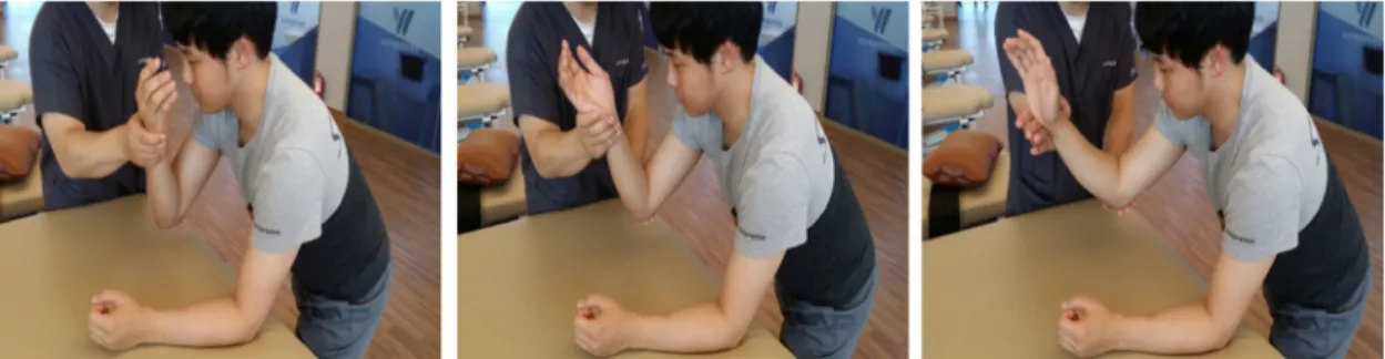
Fig. 1. Initial range(30°) Fig. 2. Middle range(45°) Fig. 3. End range(60°)
elbow flexion at 90°on the table. First of all, ulnar thrust pattern (Adler et al, 2008) was conducted on exercising portions of the patterns. While performing the ulnar thrust pattern, resistance was provided in the initial range (30°) (Fig. 1), middle range (45°) (Fig. 2), and end range (60°) (Fig. 3). Muscle activation was measured on the middle/
posterior deltoid muscle and triceps muscle on the opposite side of the body. For the collection of data, measurements were made for 5 seconds and repeated three times in each condition. Average measurements were calculated using integrated electromyogram signals for the middle three seconds only. Measurements were conducted in random order providing five-minute rest breaks in between each of the conditions.
4. Statistical analysis
The intention of this study was to identify the effect of changes in joint angle on exercising areas that provided resistance while developing PNF pattern exercises on the activation of shoulder muscles. Therefore, repeated one-way ANOVA was conducted on changes in muscle activation resulting from changes in joints. In addition, repeated one-way ANOVA was also conducted to identify whether there was a difference between muscles in each joint angle. As a result of analysis, in the event there was a difference from each type of muscle and angle, Bonferroni was conducted in order to examine differences of muscle activation between each angle and each resistance point.
SPSSWIN (ver.21.0) program was used as statistical program, and the significance level of α for verifying the statistical significance was .05.
Ⅲ. Results
1. Muscle activation of each contralateral shoulder muscle according to the changes of elbow joint angle and to each resistance point of elbow joint in the ulnar thrust PNF pattern exercise
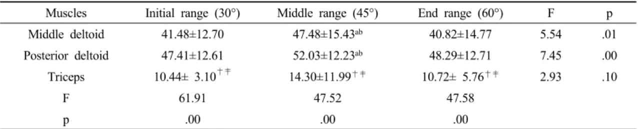
The results of identifying whether there was a difference on the activation of shoulder muscles on the opposite side according to the angle of the elbow joint on exercising parts are shown in <Table 1>. There was a difference on the muscle activation of the middle portion of the contralateral deltoid muscle according to the joint angle (p<.05). As a result of Bonferroni, muscle activation in the middle range (45°) turned out to be significantly higher than that of initial (30°) and end (60°) ranges. There was also a difference in the muscle activation of the posterior portion of the contralateral deltoid muscle depending on joint angle (p<.05). As a result of Bonferroni, muscle activation in the middle range (45°) turned out to be significantly higher than that of initial (30°) and end (60°) ranges. On the other hand, there was no difference in the triceps of arm according to joint angle.
The result of identifying the difference of muscle
Table 1. Muscle activation of each contralateral shoulder muscle according to the changes of elbow joint angle and to each resistance point of elbow joint in the ulnar thrust PNF pattern exercise (Unit: %)
Muscles Initial range (30°) Middle range (45°) End range (60°) F p
Middle deltoid 41.48±12.70 47.48±15.43
ab40.82±14.77 5.54 .01
Posterior deltoid 47.41±12.61 52.03±12.23
ab48.29±12.71 7.45 .00
Triceps 10.44± 3.10
†‡14.30±11.99
†‡10.72± 5.76
†‡2.93 .10
F 61.91 47.52 47.58
p .00 .00 .00
a: Middle range > Initial range, b: Middle range > End range
†: Middle deltoid > Triceps, ‡: Posterior deltoid > Triceps