Slipped capital femoral epiphysis (SCFE) is one of the most common disorders that affect the hips of adolescents.
In the United States, this condition affects adolescents with an incidence of 10 per 100,000.1,2) Frequently, SCFE initial-
ly presents with pain in the hip, thigh, or knee and abnor- mal gait; when the pain is referred to the knee, there may be a delay in the diagnosis.1,2) Diagnosis and management should be timely since in many long-term studies, the as- sociation between SCFE deformity and the development of early osteoarthrosis of the hip has been demonstrated.3)
The goal of treatment for SCFE is to prevent the progression of the deformity through stabilization of the proximal epiphyses.4-6) Current advances in the surgical treatment of severe and unstable SCFE are aimed at pre- venting the progression or correcting the proximal defor- mity by protecting the blood supply of the femoral head.5)
Long-Term Results of Slipped Capital Femoral Epiphysis Treated with the Modified Dunn
Procedure in a Colombian Cohort
Carlos Sarassa, MD*
,†, Daniela Carmona, MD
†,‡, Daniel Vanegas Isaza, MD
†,‡, Camilo Restrepo Rodríguez, MD
†,‡, Ana Milena Herrera Torres, MD
§*Department of Children’s Orthopedics, Clínica del Campestre, Hospital Infantil Santa Ana, Fundación Clínica Noel, CORA Group, Medellín,
†Department of Orthopedics and Traumatology, Clínica del Campestre, Medellín,
‡Orthopedics and Traumatology Residency Program, Faculty of Medicine, Universidad Pontificia Bolivariana, Medellín,
§Department of Epidemiology and Clinical Research, Clínica del Campestre, Medellín, Colombia
Background: Slipped capital femoral epiphysis (SCFE) is a severe and catastrophic disorder that affects the hips of adolescents.
Many reports about surgical procedures to treat this condition have been published, but to our knowledge, there are no published results of treatment in Latin American patients. This study describes the clinical and radiological results of the modified Dunn pro- cedure with the surgical approach described by Ganz to treat mild to severe SCFE in a cohort of Colombian pediatric patients.
Methods: We retrospectively analyzed 21 patients (22 hips) with SCFE treated with surgical dislocation of the hip from 2005 to 2017. The same pediatric orthopedic surgeon performed all operations. Clinical outcome was assessed using the range of move- ment and Merle d’Aubigné score, while radiological measurements and assessment included the slip angle and Tönnis score.
Results: The average duration of follow-up was 29 months (range, 12–72 months). Of all cases, 17 presented with acute-on-chron- ic symptoms. Preoperatively, all 22 hips were classified as poor according to the Merle d’Aubigné score. Preoperative radiological classification showed compromise grade II or III in 20 hips. Last follow-up Merle d’Aubigné score rated 17 cases as good or excel- lent (p < 0.05). The postoperative radiological classification was grade I or II in all 22 cases, and the Tönnis score was stage II in 3 cases and stage III in 4 cases.
Conclusions: Our results suggest that the modified Dunn osteotomy performed through the Ganz technique could be safely and effectively used to treat patients with mild to severe SCFE.
Keywords: Slipped capital femoral epiphysis, Surgical dislocation, Femur head necrosis
Copyright © 2021 by The Korean Orthopaedic Association
This is an Open Access article distributed under the terms of the Creative Commons Attribution Non-Commercial License (http://creativecommons.org/licenses/by-nc/4.0) which permits unrestricted non-commercial use, distribution, and reproduction in any medium, provided the original work is properly cited.
Clinics in Orthopedic Surgery • pISSN 2005-291X eISSN 2005-4408 Received March 26, 2019; Revised December 24, 2020;
Accepted December 26, 2020
Correspondence to: Ana Milena Herrera Torres, MD
Department of Epidemiology and Clinical Research, Clínica del Campestre, Calle 17 Sur #4-46, Medellín 050016, Colombia
Tel: +57-4-4442006, Fax: +57-4-4442006 E-mail: [email protected]
Multiple surgical techniques have been described, includ- ing in situ fixation, closed reduction with fixation, reduc- tion by an anterior approach, and controlled open surgical dislocation of the hip, but no consensus has been reached on the selection of the treatment method; therefore, the treatment of this condition is still a challenge.4,5,7)
Controlled surgical dislocation, as first described by Ganz et al.,8) is an effective method for the correction of deformity with low avascular necrosis (AVN) rates.4,6,8,9) However, in Latin America, there is a lack of literature from studies aimed at determining the long-term radio- logical and functional results of this surgical technique as a treatment for SCFE. This study aimed to describe the results of controlled surgical hip dislocation and the com- plications that may arise with the application of this tech- nique in a series of Colombian patients.
METHODS
Patients
A retrospective review was performed on consecutive pe- diatric patients who underwent surgical treatment with the technique of controlled dislocation for SCFE from 2005 to 2017 in our center. The ethics committee of our institution approved the study (IRB No. 55-2017). Details regarding demographic characteristics, operative notes, and patient follow-up data were compiled from the medical records, with the prior consent and assent of the parents and pa- tients, respectively. Exclusion criteria for this procedure included patients with closed proximal femoral physis, a mild slip (defined as one with < 30° of lateral slip); clinical or radiological evidence of moderate or severe cartilage damage; and preexisting AVN.
Clinical examination revealed abnormal gait or in- ability to walk, in addition to painful mobility of the hip for acute and acute-on-chronic cases, with external rota- tion of the limb and limitation of flexion, internal rota-
tion, and abduction. Slipping symptoms were categorized according to the clinical classification of O’Brien and Fa- hey10) into acute, chronic, or acute-on-chronic. The cases were also classified as stable or unstable based on Loder’s criteria.11) Preoperative and postoperative evaluation includ- ed measurements of the range of preoperative and postop- erative movement, pain, and ambulation, evaluated through the Merle d’Aubigné score. Based on the score, patients were further classified into poor (score < 13), fair (score 13 or 14), good (score 15 to 17), and excellent (score of 18).12,13)
The slipped angle was measured before and after surgery on anteroposterior hip radiographs and frog pro- jection (Figs. 1 and 2). Radiological severity was catego- rized considering the angles and relative displacement of the epiphysis in the metaphysis according to Wilson’s clas- sification14) into grade I (< 25%), grade II (25%–50%), and grade III (> 50%).
Surgical Procedure
The same senior pediatric orthopedic surgeon (CS) per- formed all operations. The modified Dunn procedure is a surgical technique for SCFE correction previously de- scribed by Ganz et al.8)
Concisely, the patient is positioned in lateral decu- bitus to perform surgical dislocation of the hip with an os- teotomy of the greater trochanter. The incision in the skin is lateral and centered on the greater trochanter, following the axis of the femur. A slide trochanteric osteotomy is then performed. Blood supply to the femoral head is pre- served through an extended retinacular soft-tissue flap.
The gluteus minimus is mobilized through the gap with the pyriformis tendon to reach the capsule. Then, a capsu- lotomy in Z shape along the acetabular insertion is made.
After the hip has been exposed, the magnitude and direction of the slip are identified. Subsequently, a provi- sional in situ fixation is performed in unstable acute cases with two 1.5-mm smooth pins; thus, mobilization of the
Fig. 1. Acute slipped capital femoral epiphysis (SCFE). (A) A radiograph of a 13-year-old patient with acute and severe SCFE. Three-year postoperative anteroposterior (B) and frog projection (C) radiographs, showing satisfactory reduction of the femoral epiphysis with no signs of avascular necrosis.
A B C
femur and dislocation of the hip can be done without risk of damaging the epiphyseal circulation. At this time, the hip is dislocated, and the pins are removed. When the SCFE is chronic, the hip is easily dislocated without the need of protecting pins.
Full exposure of the femoral neck was first achieved through complete separation of the capital epiphysis from the femoral neck (Fig. 3), which allows visualization of callus formation on the posteroinferior aspect of the neck, which is then entirely removed with a curette. The remain- ing physis of the femoral head is excised, while manually keeping the femoral head in a stable position.
The femoral head is reduced onto the neck and then stabilized by inserting a threaded nail through the fovea capitis in an anterograde manner (Fig. 4). To further sta- bilize the femoral head, under fluoroscopic guidance, a second threaded nail is placed from distal to proximal in a
retrograde manner towards the physis (Fig. 5). Before any manipulation, a 2-mm drill hole is made on the epiphysis to visualize bleeding that indicates the viability of the hip after reduction of the epiphysis (Fig. 6).
The condition of the acetabular labrum and the ar- ticular cartilage is evaluated in all hips. The hip is reduced, then a capsulorrhaphy is performed, and the greater tro- chanter is repositioned and fixed with three 3.5-mm corti- cal screws.
Evaluation and Postoperative Follow-up
The patients were monitored periodically. Hip radiographs were used to measure the slip percentage and the Tönnis score.15) Measurements of the Merle d’Aubigné score were Fig. 2. Right acute-on-chronic and left chronic slipped capital femoral epiphysis (SCFE). (A) A radiograph showing a bilateral compromise in a 16-year- old patient with severe acute-on-chronic SCFE of the right hip and mild chronic SCFE of the left hip. The patient had a preexisting condition of falciform anemia. In the right hip, the Dunn modified procedure was performed, while in situ fixation was done to the left hip. Six-year postoperative anteroposterior (B) and frog projection (C) radiographs, showing satisfactory reduction with no impingement at the right hip and mild cam of the left hip.
No signs of avascular necrosis was present in any of the hips.
A B C
Fig. 3. Open exposure. A photograph showing the anterior capsulotomy and the longitudinally opened retinaculum that exposes the femoral head and neck.
Fig. 4. Fixation through fovea. A photograph showing fixation of the femoral epiphysis with antegrade passage of a 3.5-mm fully threaded wire.
taken during follow-up. The number of patients to whom prophylactic fixation of the contralateral hip was per- formed was also recorded.
Statistical Analysis
The values of the radiographic measurements and the scores of the different scales were expressed as means and standard deviations. The changes between the preopera- tive values and those of the last follow-up were determined with the Student t-test. Values of p < 0.05 were considered significant.
RESULTS
In total, 22 hips (10 right and 12 left) were retrospectively reviewed in 21 patients (5 women and 16 men). The aver- age follow-up was 28.9 months (range, 12–72 months).
The age of the patients ranged from 10 to 18 years, with an average of 13.5 years at the time of surgery. Figs. 1 and 2 present the cases of 2 patients: 13 and 16 years of age with follow-up of 3 years and 6 years, respectively.
All patients presented with pain as an initial symp- tom, accompanied by abnormal gait. Concerning the pre- sentation of symptoms, there were 2 patients with acute SCFE, 3 patients with chronic SCFE, and 17 acute-on- chronic cases. The average time during which the symp- toms were present before the consultation was 5 weeks.
Four patients required hospitalization for soft traction use before surgery.
The preoperative clinical classification according to the Merle d’Aubigné score was poor in all 22 hips (median, 10; range, 5–12). The preoperative radiological classifica- tion showed compromise grade II and III in 8 hips and 12
hips, respectively (Table 1). In the immediate postopera- tive evaluation, only 1 patient presented with a complica- tion in the form of dehiscence of the surgical wound. No acute postoperative cases of instability were detected in the immediate radiological evaluation.
At the last follow-up, 12 patients presented with no pain; 3 presented with mild pain with regular activity; 2 presented with mild pain with activity that improved by resting; and 4 showed limited movement due to pain. In the last evaluation, the clinical classification according to the Merle d’Aubigné score was good and excellent in 11 hips and 6 hips, respectively, with a median of 16 for total cases. The postoperative radiological classification was grade I and II in 17 cases and 5 cases, respectively (Table 1).
Postoperative Tönnis score showed stage II in 3 cases and stage III in 1 case (Table 1). As complications, 2 patients had re-slipping, 2 AVN, and 3 chondrolysis. Pro- phylactic fixation of the contralateral hip was performed in 5 patients.
DISCUSSION
Surgical hip dislocation (modified Dunn procedure) in pediatric patients with SCFE in this Colombian cohort showed very satisfactory results regarding functional recovery and radiological stabilization with a low rate of complications. Mid- and long-term results indicate that this surgical technique allows patients to recover the range of movement and ambulation with no or little pain. Fur- thermore, we showed that timely intervention with the Fig. 5. Retrograde fixation. A photograph showing retrograde fixation
from the lateral cortex to the subchondral femoral epiphysis with a 3.5- mm fully threaded wire.
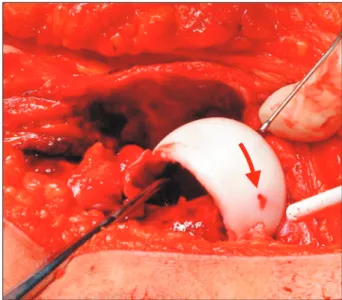
Fig. 6. Perfusion verification. A photograph showing good and permanent perfusion of the epiphysis during the whole procedure, verified through a 2-mm perforation. Arrow: point at the perforation.
Table 1. Patients Demographics, Preoperative and Postoperative Clinical Classifications and Scores CaseSexAge (yr)
PreoperativePostoperative Fahey and O'Brien classificationLoder classifcationWilson's classificationMerle d'Aubigné scoreWilson's classificationMerle d'Aubigné scoreTönnis scoreComplication 1Male13Acute-on-chronicUnstableGrade III > 50%11Grade I < 25%10Stage IIChondrolysis 2Male18Acute-on-chronicUnstableGrade II 25%–50%9Grade II 25%–50%16Stage 0 3Male14Acute-on-chronicUnstableGrade III > 50%11Grade I < 25%10Stage IIChondrolysis/AVN 4Male13Chronic StableGrade II 25%–50%11Grade I < 25%17Stage 0 5Male15AcuteUnstableGrade II 25%–50%7Grade II 25%–50%15Stage 0 6Male15Acute-on-chronicUnstableGrade II 25%–50%7Grade I < 25%18Stage 0 7Male16Chronic StableGrade III > 50%12Grade I < 25%16Stage 0 8Male16Acute-on-chronicStableGrade I < 25%10Grade I < 25%17Stage 0 9Male15Chronic StableGrade III > 50%10Grade I < 25%13Stage IIChondrolysis 10Male11AcuteUnstableGrade III > 50%11Grade I < 25%17Stage 0 11Female10Acute-on-chronicStableGrade II 25%–50%11Grade II 25%–50%15Stage 0 12Male17Acute-on-chronicStableGrade III > 50%9Grade I < 25%18Stage 0 13Male17Acute-on-chronicUnstableGrade II 25%–50%5Grade I < 25%10Stage IIIAVN 14Female12Acute-on-chronicStableGrade III > 50%9Grade I < 25%15Stage 0 15Female10Acute-on-chronicStableGrade III > 50%7Grade I < 25%15Stage 0 16Male15Acute-on-chronicStableGrade III > 50%10Grade II 25%–50%15Stage IMild re-slipping 17Female11Acute-on-chronicStableGrade I < 25%10Grade I < 25%17Stage 0 18Male13Acute-on-chronicStableGrade III > 50%10Grade I < 25%18Stage 0 19Male11Acute-on-chronicStableGrade III > 50%7Grade I < 25%18Stage 0 20Male12Acute-on-chronicUnstableGrade III > 50%10Grade I < 25%18Stage 0 21Male13Acute-on-chronicStableGrade II 25%–50%9Grade II 25%–50%14Stage 0Mild re-slipping 22Female11Acute-on-chronicStableGrade II 25%–50%10Grade I < 25%18Stage 0 AVN: avascular necrosis.
modified Dunn procedure could lower the incidence of catastrophic complications such as AVN and chondrolysis.
To the best of our knowledge, this is the first report of a series of patients from Latin America. Our country may have similar epidemiology of SCFE compared with other countries of the region. Thus, this significant cohort is a representative sample of SCFE patients with results that may apply to the Latin American population. The results of this study contribute to the existing literature, suggesting that the modified Dunn procedure is a safe and effective procedure for the treatment of moderate and se- vere SCFE.
The size of our sample is comparable to other re- ported series such as the one from Slongo et al.6) in 2010 (n = 23), Massè et al.16) in 2012 (n = 20), Sankar et al.17) in 2013 (n = 27), Madan et al.18) in 2013 (n = 28), Ziebarth et al.19) in 2017 (n = 43), and Elmarghany et al.20) in 2017 (n = 30). SCFE treatment aims to prevent early hip osteoarthritis caused by femoroacetabular impingement with an anterior cam effect. Anatomical restoration of the femoral head and acetabulum close to normality avoids the early signs of os- teoarthritis.2,3,5)
The modified Dunn procedure using Ganz et al.8) surgical hip dislocation is ideal for providing physeal stability, allowing anatomical restoration of the proximal femur, to preserve the blood supply to the epiphysis and to avoid residual deformity with minimal shortening. It also has the advantage of allowing intra-articular exploration to identify any additional chondral pathology and remov- ing the metaphyseal callus while minimizing the risk of
AVN.8,21) Even though this surgical technique is recom-
mended for acute and acute-on-chronic SCFE, it should be used carefully in patients with chronic SCFE and only by experienced surgeons.
There is still significant controversy about the treat- ment of unstable or severe SCFE with the modified Dunn procedure due to the high variability in the long-term results described in several publications.5,7,22) Most of the variability has been accounted for by the experience of the different surgical teams and the severity of the condition of each of the reported series. In the current study, we pre- sented intermediate and long-term results of the surgical hip dislocation performed by one experienced surgeon in patients with mild to severe SCFE. The surgical technique was executed following the procedure, as described by Ganz et al.8) and Slongo et al.,6) showing satisfactory functional re- sults in challenging cases. The majority of our patients were acute and acute-on-chronic patients with radiological mod- erate to severe SCFE, which required anatomical reduction and stabilization and thus minimized AVN.
The incidence of AVN is considered one of the in- dicators of success or failure of the modified Dunn proce- dure with overall rates ranging from 2% to 30%.4,9,17-19,23,24)
However, AVN incidences higher than 20% with the surgi- cal hip dislocation are comparable to those of closed re- duction and internal fixation for severe SCFE,4,9,22-24) while the reported incidences for mild or stable SCFE after the surgical hip dislocation range from 0% to 10%.5,7,23,24) It has been argued that the variability of the AVN rates could be due to the intervention of acute versus chronic patients.5) In our series, the 2 cases of AVN were found in patients with SCFE of acute onset.
Contralateral hip involvement in unilateral SCFE has been reported in 14%–63% of the cases.25-28) Prophy- lactic fixation of the unaffected hip has been shown to reduce the risk of a contralateral slip, with minimal risk of complications that include fracture, adverse reaction to osteosynthesis material, and chondrolysis among others.
The contralateral hip prophylactic fixation is better suited for patients of younger age at presentation, with severe slip, obesity, or metabolic condition, and those in which long-term follow-up may be difficult.27,28) In our cohort, prophylactic fixation of the unaffected hip was performed in 5 patients indicated mainly by the severity of the slip and the sociodemographic conditions, which made almost impossible the follow-up.
This study is not without limitations, which include the retrospective design, limited information for the peri- odic follow-ups, shortage of preoperative clinical scores, and lack of a control group. Nevertheless, it is worthy of notice that the surgical hip dislocation can give the same favorable outcomes in Latin American patients with mild to severe SCFE as in patients from other regions and eth- nicities in previously published reports.
In agreement with what has been previously dem- onstrated in several publications, we showed that surgical dislocation of the hip could be used successfully to treat pediatric patients with moderate to severe SCFE. The inci- dence of complications in our cohort was low despite the severity of the condition at the time of the intervention.
To our knowledge, this is the first report of the results of the modified Dunn procedure in a Colombian and Latin American cohort, which underscores the importance and novelty of this work given the comparable sample size with that of other previous reports.
CONFLICT OF INTEREST
No potential conflict of interest relevant to this article was reported.
ACKNOWLEDGEMENTS
The authors wish to thank Dr. David Suarez (Clínica del Campestre) for his contribution to data acquisition.
ORCID
Carlos Sarassa https://orcid.org/0000-0002-0912-6668 Daniela Carmona https://orcid.org/0000-0003-1370-6507 Daniel Vanegas Isaza
https://orcid.org/0000-0002-8663-3145 Camilo Restrepo Rodríguez
https://orcid.org/0000-0002-6230-2405 Ana Milena Herrera Torres
https://orcid.org/0000-0002-7382-5631
REFERENCES
1. Loder RT, Skopelja EN. The epidemiology and demograph- ics of slipped capital femoral epiphysis. ISRN Orthop.
2011;2011:486512.
2. Gholve PA, Cameron DB, Millis MB. Slipped capital femoral epiphysis update. Curr Opin Pediatr. 2009;21(1):39-45.
3. Carney BT, Weinstein SL. Natural history of untreated chronic slipped capital femoral epiphysis. Clin Orthop Relat Res. 1996;(322):43-7.
4. Abu Amara S, Cunin V, Ilharreborde B; French Society of Pediatric Orthopaedics (SOFOP). Severe slipped capital femoral epiphysis: a French multicenter study of 186 cases performed by the SoFOP. Orthop Traumatol Surg Res.
2015;101(6 Suppl):S275-9.
5. Bittersohl B, Hosalkar HS, Zilkens C, Krauspe R. Current concepts in management of slipped capital femoral epiphy- sis. Hip Int. 2015;25(2):104-14.
6. Slongo T, Kakaty D, Krause F, Ziebarth K. Treatment of slipped capital femoral epiphysis with a modified Dunn procedure. J Bone Joint Surg Am. 2010;92(18):2898-908.
7. Mahran MA, Baraka MM, Hefny HM. Slipped capital femo- ral epiphysis: a review of management in the hip impinge- ment era. SICOT J. 2017;3:35.
8. Ganz R, Gill TJ, Gautier E, Ganz K, Krugel N, Berlemann U.
Surgical dislocation of the adult hip a technique with full ac- cess to the femoral head and acetabulum without the risk of avascular necrosis. J Bone Joint Surg Br. 2001;83(8):1119-24.
9. Sucato DJ, De La Rocha A. High-grade SCFE: the role of surgical hip dislocation and reduction. J Pediatr Orthop.
2014;34 Suppl 1:S18-24.
10. O’Brien ET, Fahey JJ. Remodeling of the femoral neck after in situ pinning for slipped capital femoral epiphysis. J Bone Joint Surg Am. 1977;59(1):62-8.
11. Loder RT, Richards BS, Shapiro PS, Reznick LR, Aronson DD. Acute slipped capital femoral epiphysis: the importance of physeal stability. J Bone Joint Surg Am. 1993;75(8):1134-
40.
12. Banaszkiewicz PA. Functional results of hip arthroplasty with acrylic prosthesis. In: Banaszkiewicz P, Kader D, eds.
Classic papers in orthopaedics. London: Springer; 2014. 19- 22.
13. d’Aubigne RM, Postel M. The classic: functional results of hip arthroplasty with acrylic prosthesis: 1954. Clin Orthop Relat Res. 2009;467(1):7-27.
14. Wilson PD. The treatment of slipping of the upper femoral epiphysis with minimal dislacement. J Bone Jt Surg Am.
1938;20(2):379-99.
15. Tonnis D, Heinecke A, Nienhaus R, Thiele J. Predetermi- nation of arthrosis, pain and limitation of movement in congenital hip dysplasia (author’s transl). Z Orthop Ihre Grenzgeb. 1979;117(5):808-15.
16. Masse A, Aprato A, Grappiolo G, Turchetto L, Campacci A, Ganz R. Surgical hip dislocation for anatomic reorientation of slipped capital femoral epiphysis: preliminary results. Hip Int. 2012;22(2):137-44.
17. Sankar WN, Vanderhave KL, Matheney T, Herrera-Soto JA, Karlen JW. The modified Dunn procedure for unstable slipped capital femoral epiphysis: a multicenter perspective.
J Bone Joint Surg Am. 2013;95(7):585-91.
18. Madan SS, Cooper AP, Davies AG, Fernandes JA. The treat- ment of severe slipped capital femoral epiphysis via the Ganz surgical dislocation and anatomical reduction: a pro- spective study. Bone Joint J. 2013;95(3):424-9.
19. Ziebarth K, Milosevic M, Lerch TD, Steppacher SD, Slongo T, Siebenrock KA. High survivorship and little osteoarthritis at 10-year followup in SCFE patients treated with a modified dunn procedure. Clin Orthop Relat Res. 2017;475(4):1212- 28.
20. Elmarghany M, Abd El-Ghaffar TM, Seddik M, et al. Surgi- cal hip dislocation in treatment of slipped capital femoral epiphysis. SICOT J. 2017;3:10.
21. Leunig M, Slongo T, Ganz R. Subcapital realignment in slipped capital femoral epiphysis: surgical hip dislocation and trimming of the stable trochanter to protect the perfu- sion of the epiphysis. Instr Course Lect. 2008;57:499-507.
22. Zaltz I, Baca G, Clohisy JC. Unstable SCFE: review of treat- ment modalities and prevalence of osteonecrosis. Clin Or- thop Relat Res. 2013;471(7):2192-8.
23. Sucato DJ. Approach to the hip for SCFE: the North Ameri- can perspective. J Pediatr Orthop. 2018;38 Suppl 1:S5-12.
24. Loder RT, Dietz FR. What is the best evidence for the treat- ment of slipped capital femoral epiphysis? J Pediatr Orthop.
2012;32 Suppl 2:S158-65.
25. Bhattacharjee A, Freeman R, Roberts AP, Kiely NT. Out- come of the unaffected contralateral hip in unilateral
slipped capital femoral epiphysis: a report comparing pro- phylactic fixation with observation. J Pediatr Orthop B.
2016;25(5):454-8.
26. Koenig KM, Thomson JD, Anderson KL, Carney BT. Does skeletal maturity predict sequential contralateral involve- ment after fixation of slipped capital femoral epiphysis? J Pediatr Orthop. 2007;27:796-800.
27. Kocher MS, Bishop JA, Hresko MT, Millis MB, Kim YJ, Kasser JR. Prophylactic pinning of the contralateral hip af- ter unilateral slipped capital femoral epiphysis. J Bone Joint Surg Am. 2004;86(12):2658-65.
28. Stasikelis PJ, Sullivan CM, Phillips WA, Polard JA. Slipped capital femoral epiphysis: prediction of contralateral in- volvement. J Bone Joint Surg Am. 1996;78(8):1149-55.