ORIGINAL ARTICLE
Elevated Gastric Antrum Erosions in Portal Hypertension Patients:
Peptic Disease or Mucosal Congestion?
Fernanda Cordeiro de Azevedo Conejo
1, Mabel Tatty Medeiros Fracassi
2, Maurício Saab Assef
1, Maurício Alves Ribeiro
3, Luiz Arnaldo Szutan
3and Fabio Gonçalves Ferreira
3Endoscopy Service1, Department of Pathology2, Department of Surgery, Liver and Portal Hypertension Group3, Santa Casa de Sao Paulo School of Medical Sciences, São Paulo, SP, Brazil
Background/Aims: Portal hypertension (PH) is a syndrome characterized by chronic increase in the pressure gradient between the portal vein and inferior vena cava. Previous studies have suggested an increased frequency of antral elevated erosive gastritis in pa- tients with PH, as well as an etiologic association; however, there has not been any histological evidence of this hypothesis to date.
Our aim was to evaluate the histological features found in elevated antral erosions in patients with portal hypertension.
Methods: Sixty-nine patients were included; 28 with and 41 without PH. All patients underwent endoscopy, and areas with elevated antral erosion were biopsied.
Results: In the PH group, 24 patients had inflammatory infiltration with or without edema and vascular congestion, and 4 patients had no inflammation. In the group without PH, all patients showed inflammatory infiltration of variable intensity. There was no stat- istical significance between the two groups in the presence of Helicobacter pylori. There as a histological similarity between the two groups, if PH patients without inflammation were excluded; however, more edema and vascular congestion were observed in the PH group (p=0.002).
Conclusions: The findings show that elevated antral erosions in patients with PH have more evident edema and vascular congestion in addition to lymphocytic infiltration. (Korean J Gastroenterol 2017;69:278-282)
Key Words: Hypertension, portal; Gastritis; Gastric antral vascular ectasia; Lymphocytes; Edema
Received May 14, 2016. Revised February 14, 2017. Accepted February 15, 2017.
CC This is an open access article distributed under the terms of the Creative Commons Attribution Non-Commercial License (http://creativecommons.org/licenses/
by-nc/4.0) which permits unrestricted non-commercial use, distribution, and reproduction in any medium, provided the original work is properly cited.
Copyright © 2017. Korean Society of Gastroenterology.
Correspondence to: Fabio Gonçalves Ferreira, Department of Surgery, Santa Casa de Sao Paulo School of Medical Sciences, Rua Apinajés 1060 ap 93, Perdizes, São Paulo, SP 05017-000, Brazil. Tel: +55-11-99211-0057, Fax: +55-11-3337-8164, E-mail: drfabioferreira@uol.com.br
Financial support: None. Conflict of interest: None.
INTRODUCTION
Since the first description of macroscopic changes occur- ring in the gastric mucosa of patients with portal hyper- tension (PH), portal hypertensive gastropathy (PHG) remains a challenge for endoscopists in the past 30 years. PHG has a controversial pathophysiology and a highly variable in- cidence in the literature, accounting for 4% to 80% of PH patients.1-5 This wide variation is likely due to the challenges associated with making a correct endoscopic diagnosis, es- pecially for milder forms or those not associated with bleeding.6 The three most accepted and used classifications
(McCormack,7 New Italian Endoscopy Club8 and Baveno Consensus9) are still widely debated, but it is clear that the simplest classification is still the most adopted with the high- est consensus among endoscopists.10
In medical practice, there is a correlation between endo- scopic findings of PHG and the presence of elevated gastric antral erosions in PH patients. The histological relationship between these gastric manifestations has to date not been fully elucidated. Although the endoscopic appearance of ero- sions is similar between patients with and without PH, the present study aimed to determine whether an increase in the chronic pressure of the portal system causes these erosions
Fig. 1. Elevated antral erosions (arrows) in portal hypertensive gastropathy.
A B
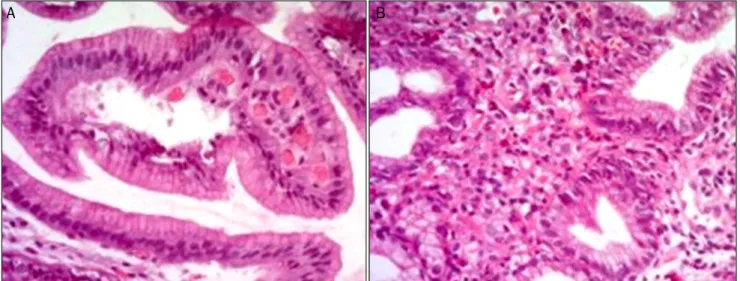
Fig. 2. Biopsies of elevated antral erosions in portal hypertensive gastropathy. (A) Edema and vascular congestion (H&E, ×200). (B) Lymphocytic in- filtration (H&E, ×400).
in PH patients or if they are a part of the large spectrum of pep- tic disease.
SUBJECTS AND METHODS
1. Patients
Consecutive patients presenting elevated antral erosions during endoscopic examination between December 2010 and June 2012, who were referred to the endoscopy unit of Santa Casa de Sao Paulo School of Medical Sciences, were enrolled in this prospective study. This study was approval by the Ethics in Human Research Committee (No. 138/10) and included in the Brazilian Registry of Clinical Trials (No.
RBR-5yr2yx-http://www.ensaiosclinicos.gov.br/rg/RBR-5yr2 yx/). We included patients of both genders, regardless of age.
We excluded the following patients: those who had previous
treatment for Helicobacter pylori (H. pylori) eradication, re- cently used antibiotics, or did not agree to participate in the study.
Overall, 69 patients admitted for dyspeptic complaints were included and divided into two groups: group I, which in- cluded 28 patients with PH and group II, which included 41 patients without PH. We apply a questionnaire to clarify the PH etiology, to ascertain whether they had used proton-pump inhibitors , non-steroidal anti-inflammatory drugs, or tobac- co, and whether they had been treated for H. pylori infection.
Patients in group I were characterized by the presence of esophageal varices in varying degrees, regardless of prior history of gastrointestinal bleeding or endoscopic treatment of varices. This group included patients with or without PHG.
In group II, patients underwent an ultrasound to identify any degree of liver disease, including homogeneous liver, normal spleen, and portal vein; study requirement for in- clusion was for the diameter to be less than 12 mm.
Endoscopic examinations were performed under sedation using midazolam (0.1 mg/kg body weight) and fentanyl (1 μg/kg body weight). Fujinon EPX 2200 videoendoscope (Fujinon Optical CO., Ltd, Tokyo, Japan) was used in conventional vid- eoendoscopy examinations to assess the esophagus, stom- ach, and duodenum. In group I patients, the presence or ab- sence of PHG was then determined. Gastric biopsies were performed on all patients for H. pylori presence using a ure- ase method; biopsies of antral elevated erosions were also performed (Fig. 1).
During the histopathological analysis, we checked for the
Table 1. Portal Hypertension Etiology of 28 Patients with Antral Elevated Erosions (Group I)
Etiology n %
Alcohol 11 39.2
Schistosomiasis 10 35.8
Hepatitis B 4 14.2
Hepatitis C 2 7.2
Alcohol and Hepatitis B 1 3.5
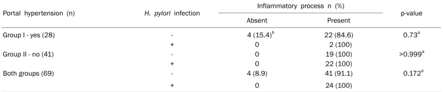
Table 2. Helicobacter pylori (H. pylori) Infection and Inflammatory Process on Both Groups
Portal hypertension (n) H. pylori infection Inflammatory process n (%)
p-value
Absent Present
Group I - yes (28) - 4 (15.4)b 22 (84.6) 0.73a
+ 0 2 (100)
Group II - no (41) - 0 19 (100) >0.999a
+ 0 22 (100)
Both groups (69) - 4 (8.9) 41 (91.1) 0.172a
+ 0 24 (100)
aStatistically non-significant, Fischer exact test; bExcluded for further analysis.
Table 3. Histopathological Analysis of 65 Patients with Elevated Antral Erosions and Lymphocytic Infiltration in Both Groups, Regarding the Presence of Portal Hypertension and PHG
Group I – portal hypertension Group II – without portal hypertension
PHG + PHG - All
Lymphocit infiltration 1a (8.3) 4a (33.3) 5b (20.8) 25b (61)
Lymphocit infiltration + edema + congestion 11a (91.7) 8a (66.7) 19b (79.2) 16b (39) Values are presented as n (%).
PHG, portal hypertensive gastropathy.
ap=0.158, statistically non-significant, Chi-squared test; bp=0.002, statistically significant, Chi-squared test.
presence or absence of inflammation in biopsies to search for factors, such as lymphocytic infiltration, edema, and vas- cular congestion (Fig. 2). One senior pathologist (MTMF) per- formed all histologic examinations.
2. Statistics
SPSS ver. 20.0 (IBM Corp., Armonk, NY, USA) was used for all statistical analyses. Differences were considered statisti- cally significant if p<0.05.
Fisher's exact test was used to assess the differences be- tween groups I and II, regarding the presence of inflammation in the gastric mucosa and to identify whether the presence of H. pylori influenced the inflammatory processes.
Chi-squared test was applied to determine whether there were significant differences between the two groups for the
presence of lymphocytic infiltration only and for the presence of lymphocytic infiltration together with edema and vascular congestion.
RESULTS
Table 1 shows the etiologies of PH in group I. H. pylori in- fection was not an important factor in justifying the presence of inflammatory process in both groups, with no significantly difference (p=0.73) when both groups were compared using Fisher's exact test (Table 2).
In group I (PH, n=28) lymphocytic infiltration alone or asso- ciated with edema and vascular congestion were present in 24 patients; however, in four patients there was no sign of lymphocytic infiltration. We excluded these four patients for further analysis. Among the remaining 24 patients with lym- phocytic infiltration, 12 belonged to the subgroup without PHG, and 12 patients belonged to the subgroup with PHG.
Histopathological evaluation revealed that in the subgroup with PHG, one individual (8.3%) presented only lymphocytic infiltration, and 11 patients (91.7%) presented lymphocytic infiltration, edema, and vascular congestion. In the subgroup without PHG, four (33.3%) presented lymphocytic infiltration alone and eight (66.7%) presented lymphocytic infiltration,
edema, and vascular congestion. We did not find any sig- nificant differences when comparing the subgroups with and without PHG between those presenting lymphocytic infiltra- tion alone and those presenting the combination of lympho- cytic infiltration, edema, and vascular congestion (Table 3).
In group II, 25 patients (61%) presented inflammation with lymphocytic infiltration alone, and 16 patients (39%) pre- sented lymphocytic infiltration with edema and vascular congestion.
Table 3 also shows a comparison between the histological features of group I (excluding the four patients without in- flammation) and group II, regarding the presence of lympho- cytic infiltration, edema, and vascular congestion.
DISCUSSION
PHG is a well-established complication of PH. Endoscopic findings range from a fine pinkish speckled pattern to a dif- fuse hemorrhage. Histological findings include tortuous and dilated vessels in the stomach submucosa and mucosal vas- cular ectasia concomitant with a few inflammatory signs.
This led McCormack et al.7 to suggest the term portal hyper- tension gastropathy, or PHG, in 1985.
In 1990, D’Amico et al.2 noted that in addition to the vas- cular changes already described, there were inflammatory changes, especially in mild PHG, whereas they observed ar- chitectural distortion with atrophy in more advanced cases of PHG.
In the literature, a few studies have evaluated the accuracy of endoscopic biopsies, mostly showing no correlation be- tween endoscopic and histological findings. Therefore, this cannot be the method that yields a definitive diagnosis of PHG. There is an increased risk of bleeding due to the coagu- lation profile and platelet dysfunction found in patients with PH.11,12 In the present study, despite the increased risk of bleeding in chronic liver disease, no significant bleeding was observed after observing the results of the biopsy.
In 2008, Assef et al.6 showed an increased frequency of elevated erosive gastritis covered with fibrin, affecting the gastric antrum of patients with PH (37.5%). Among these, 16.7% had no endoscopic diagnosis of PHG, while 50% had mild PHG and 33.3% had severe PHG. These findings moti- vated us to perform a biopsy in an attempt to elucidate how hypertensive gastropathy produces antral lesions.
In practice, we suggest that antral erosions are the primary sign of hypertensive gastropathy in the gastric mucosa be- cause even in the absence of aggressive factors to the antral mucosa, erosions are common in cirrhotic patients. In these biopsies showing erosions, we observed ectasia and edema of the mucosa, with the degree of lymphocytic infiltrate rang- ing from absent to intense.
These histological features were observed in both patients with and without PH. When comparing these groups, how- ever, it was noted that patients with PH, including both with or without PHG, showed a higher frequency of edema and vascular congestion than those without portal hypertension.
The different prevalence rates of H. pylori infection have been reported depending on the diagnostic method.13 However, we used a histopathological method and a urease test. We observed that the group without PH had a larger number of H. pylori infection compared with the group with PH; but the infection was not significantly associated with in- flammatory activity.
We expected to find obvious vascular abnormalities in the biopsies of gastric antral erosions of our samples; however, the findings were variable with respect to inflammation.
Despite the limited sample, we found that edema and vas- cular congestion were more evident in patients with PH. Both patients with and without PH exhibited inflammatory histo- logical changes in antral erosions; therefore, PH in associa- tion with PHG should not be considered the sole cause for the erosions observed in these patients.
Thus, although it was endoscopically observed that pa- tients with PH had more antral elevated erosions than pa- tients without PH, it is necessary to conduct follow-up studies with larger samples to determine the cause of hypertensive gastropathy in the gastric antrum. Perhaps this is the main limitation of this study. Meeting the criteria for inclusion in the PHG group and the risk of bleeding in antral biopsies may have reduced our sample number.
The results obtained in this present study that included 69 patients with and without PH demonstrated increased ede- ma and vascular congestion in biopsies of antral elevated erosions from PH patients.
REFERENCES
1. Sarin SK, Sreenivas DV, Lahoti D, Saraya A. Factors influencing development of portal hypertensive gastropathy in patients with
portal hypertension. Gastroenterology 1992;102:994-999.
2. D'Amico G, Montalbano L, Traina M, et al. Natural history of con- gestive gastropathy in cirrhosis. The Liver Study Group of V.
Cervello Hospital. Gastroenterology 1990;99:1558-1564.
3. Calès P, Oberti F, Bernard-Chabert B, Payen JL. Evaluation of ba- veno recommendations for grading esophageal varices. J Hepatol 2003;39:657-659.
4. Kim MY, Choi H, Baik SK, et al. Portal hypertensive gastropathy:
correlation with portal hypertension and prognosis in cirrhosis.
Dig Dis Sci 2010;55:3561-3567.
5. Choe WH. Portal hypertensive gastropathy and gastric antral vas- cular ectasia. Korean J Gastroenterol 2010;56:186-191.
6. Assef MS, Valentino W, Nakamura RK, Camunha MA, Colaiacovo R, Rossini LG. The study of magnifying endoscopy for the diag- nosis of portal hypertensive gastropathy. Gastrointest Endosc 2010;71:AB379.
7. McCormack TT, Sims J, Eyre-Brook I, et al. Gastric lesions in portal hypertension: inflammatory gastritis or congestive gastropathy?
Gut 1985;26:1226-1232.
8. Primignani M, Carpinelli L, Preatoni P, et al. Natural history of por-
tal hypertensive gastropathy in patients with liver cirrhosis. The New Italian Endoscopic Club for the study and treatment of esophageal varices (NIEC). Gastroenterology 2000;119:181-187.
9. de Franchis R, Baveno V Faculty. Revising consensus in portal hy- pertension: report of the baveno V consensus workshop on methodology of diagnosis and therapy in portal hypertension. J Hepatol 2010;53:762-768.
10. de Macedo GF, Ferreira FG, Ribeiro MA, Szutan LA, Assef MS, Rossini LG. Reliability in endoscopic diagnosis of portal hyper- tensive gastropathy. World J Gastrointest Endosc 2013;5:323-331.
11. Misra SP, Dwivedi M, Misra V, et al. Endoscopic and histologic ap- pearance of the gastric mucosa in patients with portal hypertension.
Gastrointest Endosc 1990;36:575-579.
12. Chaves DM, Sakai P, Mucenic M, Iriya K, Iriya Y, Ishioka S.
Comparative study of portal hypertensive gastropathy in schisto- somiasis and hepatic cirrhosis. Endoscopy 2002;34:199-202.
13. Testerman TL, Morris J. Beyond the stomach: an updated view of helicobacter pylori pathogenesis, diagnosis, and treatment.
World J Gastroenterol 2014;20:12781-127808.