Korean J Gastroenterol Vol. 58 No. 6, 353-356 http://dx.doi.org/10.4166/kjg.2011.58.6.353
CASE REPORT
Korean J Gastroenterol, Vol. 58 No. 6, December 2011 www.kjg.or.kr
췌장에 발생한 콜레스테롤 육아종 1예
정찬우, 김봉재, 박성오, 조아라, 하종건, 박승근, 김혜숙
1메리놀병원 내과, 해부병리과1
A Case of Cholesterol Granuloma in Pancreas
Chan Woo Jung, Bong Jae Kim, Sung Oh Park, A Ra Jo, Jong Kun Ha, Seung Keun Park and Hye Sook Kim1 Departments of Internal Medicine and Pathology1, Maryknoll Medical Center, Busan, Korea
Cholesterol granuloma is a histological term used for the description of a tissue response to a foreign body such as cholesterol crystals. Cholesterol granuloma is histologically characterized as fibrous granulation tissue containing cholesterol crystals within surrounding giant cells. Cases of cholesterol granuloma of the pancreas are very rare. We report a case of a 47-year old male who had a cholesterol granuloma of the pancreas. Abdominal CT showed 24 mm-sized cyst in the pancreas and peri-pancre- atic regional mass infiltrating to the stomach. PET-CT revealed increased 18F-FDG uptake at the cyst and peri-pancreatic mass. Thus, Whipple's operation was done. The disease was confirmed by surgical pathologic examination of the tissue. Pathologic examination of resected specimen showed numerous cholesterol crystals surrounded by multinucleated foreign body giant cells. We report on this case and give a brief review of the literature. (Korean J Gastroenterol 2011;58:353-356) Key Words: Cholesterol granuloma; Pancreas
Received September 16, 2010. Revised January 18, 2011. Accepted January 18, 2011.
CC This is an open access article distributed under the terms of the Creative Commons Attribution Non-Commercial License (http://creativecommons.org/licenses/
by-nc/3.0) which permits unrestricted non-commercial use, distribution, and reproduction in any medium, provided the original work is properly cited.
교신저자: 박승근, 600-730, 부산시 중구 대청동 4-12, 메리놀병원 내과
Correspondence to: Seung Keun Park, Department of Internal Medicine, Maryknoll Medical Center, 4-12, Daecheong-dong, Jung-gu, Busan 600-730, Korea. Tel:
+82-51-461-2338, Fax: +82-51-465-7470, E-mail: [email protected] Financial support: None. Conflict of interest: None.
서 론
콜레스테롤 육아종은 콜레스테롤 결정에 대한 이물 반응으 로 생기며 주로 만성 중이염에서 약 13%까지 동반되는 것으 로 보고된다.1,2 그 외에도 드물게 복막, 귀밑샘, 림프구, 신장, 간, 유방 등에서 발생한 예가 보고되었다.3-6 조직학적으로는 콜레스테롤 결정과 함께 거대 세포(giant cell)등이 포함된 섬 유 육아조직을 말한다.7 콜레스테롤 육아종이 췌장에 발생하 는 경우는 매우 드물며, 췌장 샘 꽈리세포 종양(solid and cy- stic acinar cell tumor)의 특징적인 소견으로 보고된 적은 있 으나 암과는 상관없이 단독으로 발생한 경우는 찾아볼 수가 없었다.8 이에 저자들이 경험한 췌장의 콜레스테롤 육아종 1 예를 문헌 고찰과 함께 보고하는 바이다.
증 례
47세 남자가 1주 전부터 시작된 상복부 통증 및 2 kg의 체중 감소를 주소로 개인 의원을 방문하여 초음파 검사를 받 았고, 췌장 체부에 덩어리가 보인다 하여 본원 소화기 내과로 의뢰되어 내원하였다. 과거력에서는 약 5년 전 당뇨를 진단받 고 치료 중이었고 그 외 특이 병력은 없었다. 주 4-5회 회당 소주 1병 반의 음주력과 30년간 하루 1갑의 흡연력이 있었고, 가족력은 누나가 당뇨로 치료 중이었다. 신체 검진에서 복부 는 부드럽고 평탄하였으며 덩어리는 촉지되지 않았다. 내원 시 혈압은 125/80 mmHg, 체온은 36.7oC, 맥박은 88회/분, 호흡수는 18회/분이었다. 말초 혈액검사에서 백혈구 4,100/
mm3, 혈색소 13.7 g/dL, 혈소판 224,000/mm3이었고 CRP 6.72 mg/L이었다. 혈청 생화학검사에서 AST 12 IU/L, ALT 10 IU/L, ALP 86 IU/L, GGT 27 IU/L, 총 단백 8.0 g/dL,
354
정찬우 등. 췌장에 발생한 콜레스테롤 육아종 1예The Korean Journal of Gastroenterology
Fig. 1. (A) Non-enhanced abdominal computerized tomography showed ro- und cystic mass on the pancreas body portion with multiple peripheral rim calcifications. (B) Enhanced abdomi- nal computerized tomography, 4 months later, showed diffuse thicken- ing of the wall on the pancreas and the invasion of soft tissue mass to peri-pancreatic region (black arrow).
Fig. 2. PET-CT showed increased 18F-FDG uptake in the rim of the cystic mass and peri-pancreatic mass.
FDG, fluoro-deoxy-glucose.
Fig. 3. Gross specimen showed fibrous encapsulated cyst and dark brown colored hard materials in the cyst and peri-pancreatic region.
FDG, fluoro-deoxy-glucose.
알부민 5.0 g/dL, 총 빌리루빈 0.53 mg/dL, 혈액 요소 질소 15.1 mg/dL, 크레아티닌 0.8 mg/dL이었고, 총 콜레스테롤은 148 mg/dL이었다. CEA는 1.39 ng/mL, AFP는 3.6 ng/mL 이었고, CA19-9은 -116.47 U/mL로 상승되어 있었다. B형 간염 항원, 항체는 음성이었고, C형 간염 항체도 음성이었다.
혈청 아밀라아제는 75 mg/dL, 리파아제는 138 mg/dL이었 다. 복부 CT에서 췌장에 다발성의 석회화 소견과 함께 췌관이 확장되어 있었고, 췌장의 체부에 36 mm 크기의 낭종이 관찰 되었고 부분적으로 낭종 벽의 석회화 소견이 관찰되었다(Fig.
1A). 환자의 임상 증상이 호전되었고, 복부 CT 결과 가성 낭 종으로 생각되어 일단 경과 관찰을 하기로 하고 외래에서 추 적 관찰하였다. 4개월 후 다시 복부 CT를 하였으며 이전에 비해 낭종의 크기는 24 mm로 줄어 들었으나 낭종의 벽이 전체적으로 두꺼워졌고 위벽을 침범하는 새로운 연부조직 덩 어리가 관찰되었다(Fig. 1B). PET-CT에서는 복부 CT에서 보 였던 췌장 체부의 낭종의 벽을 따라 standardized uptake value (SUV) 6.55의 18F-FDG의 섭취 증가가 관찰되었고, 위 벽을 침범하는 연부조직 덩어리에서도 SUV 7.01의 18F-FDG 의 섭취 증가가 관찰되었다(Fig. 2). 이상의 결과로 췌장 체부 의 악성 종양이 의심되어 수술을 시행하였다. 수술의 육안 소 견에서 췌장 체부에 섬유조직으로 둘러싸인 낭종이 관찰되었 고, 췌장의 일부 및 CT에서 위벽을 침범하는 것으로 보이던
연부조직 부분은 흑갈색의 물질로 채워져 있었다. 낭종의 내 부는 흑갈색의 물질 및 액체로 가득차 있었다(Fig. 3). 췌장 체부의 이 낭종성 병변은 콜레스테롤 육아종의 퇴행변성에 의 한 이차적인 변화로 인해 발생된 것으로 콜레스테롤 육아종의 일부로 생각된다. 현미경 소견에서는 림프구를 포함한 염증 세포들이 침윤되어 있었고 출혈의 흔적이 남아 있으면서 바늘 모양의 콜레스테롤 열(cleft)들이 관찰되었다. 그리고 그 주위 로 이물 대식 세포(foreign body giant cell)들의 침윤이 관찰 되어 있어 콜레스테롤 육아종으로 확진되었다(Fig. 4). 환자는 수술 후 8개월째 특별한 증상 없이 외래에서 추적 관찰 중이다.
고 찰
콜레스테롤 육아종은 조직학적으로 거대 세포 등을 포함한 섬유 육아조직 내에 콜레스테롤 결정이 있는 상태를 말한다.1 1894년에 중이의 콜레스테롤 육아종이 처음 보고되었다.9 주 로 중이에서 발생하고 복막, 귀밑샘, 림프구, 신장, 간, 유방, 안와 등에도 발생한 예가 보고되고 있다.2-6,10 그러나 췌장의 콜레스테롤 육아종이 췌장 샘 꽈리세포 종양의 특징적인 소견 으로 보고된 적은 있으나 암과는 상관없이 단독으로 발생한
Jung CW, et al. A Case of Cholesterol Granuloma in Pancreas
355
Vol. 58 No. 6, December 2011
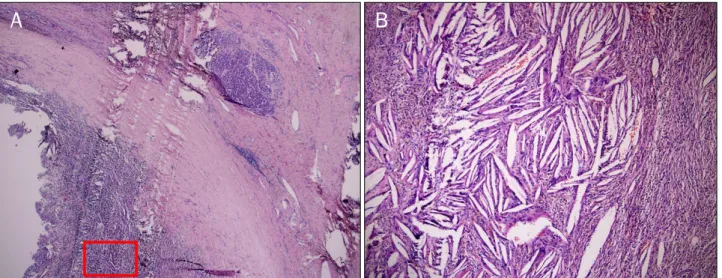
Fig. 4. Microscopic finding of resected specimen. It showed a large number of spindle shaped cholesterol crystals (cholesterol cleft) surrounded by multinucleated giant cells and hemosiderin deposits (A: H&E, ×40, B: H&E, ×400).
경우는 찾아볼 수가 없었다.8
콜레스테롤 육아종의 정확한 발생 기전은 아직까지 잘 모 르지만 만성적이고 반복적인 염증 반응이 일차적인 역할을 하 는 것으로 알려져 있다.3 췌장의 콜레스테롤 육아종은 췌장의 반복적인 염증 반응에 의한 출혈에 의해 발생하며 출혈의 불 완전한 흡수로 혈액과 그 분해 산물을 대식 세포가 탐식하여 육아종성 염증 반응을 일으켜 콜레스테롤 육아종을 형성하는 것으로 알려져 있다.11 췌장에서의 콜레스테롤 육아종은 췌장 의 다른 종괴성 질환과의 감별이 힘들며 조직학적인 진단을 통해서만 확진이 가능하다.12 이번 예에서도 CT에서 췌장 체 부에 낭성 종괴가 발견되어 췌장의 가성 낭종을 동반한 만성 췌장염으로 진단하여 추적 관찰하였으며 추적 관찰 동안 크기 는 줄었으나 주위 조직으로 침범 소견을 보여 수술을 시행하 게 되었고 조직병리검사를 통해 콜레스테롤 육아종으로 확진 을 하게 되었다. 조직병리 검사에서 이차적으로 콜레스테롤 육아종을 일으킬 수 있는 종양을 의심할 만한 소견은 전혀 관찰되지 않았다. Heaton 등7에 의하면 종괴 내부의 물질에 대한 세포학적 검사를 시행하여 이물 대식 세포와 콜레스테롤 결정 및 혈액 분해 물질을 관찰함으로써 콜레스테롤 육아종의 진단이 가능하다고 한다. 조직 검사에서 콜레스테롤 결정, 염 증 세포와 이물 대식 세포로 구성되는 섬유 육아조직이 발견 될 경우 이 질환으로 진단된다.7,11 이러한 병리조직학적 특징 으로 인해 췌장 내 종괴를 형성하는 다른 모든 질환들과 감별 이 가능하게 된다. 콜레스테롤 육아종의 치료는 수술적 절제 로서 대개 악성 종양과 감별이 안되어 절제술 후에 진단되는 경우가 많다고 한다.10
이번 증례는 CT에서 췌장의 악성 종양이 의심되어 수술을 시행하였고, 조직병리학적 검사상 콜레스테롤 육아종으로 진
단된 경우로 수술 후 현재 특이 소견이 없는 상태로 경과 관찰 중이다.
REFERENCES
1. Ochiai H, Yamakawa Y, Fukushima T, Nakano S, Wakisaka S.
Large cholesterol granuloma arising from the frontal si- nus--case report. Neurol Med Chir (Tokyo) 2001;41:283-287.
2. Chin HS, Han DY, Jang WI, Lee IH, Kim YS, Lee BD. Clinical analy- sis of cholesterol granuloma in the middle ear. Korean J Otorhinolaryngol-Head Neck Surg 2009;52:974-979.
3. al-Amer AF, Walia HS, Madda JP. Cholesterol granuloma of the peritoneum. Can J Surg 1990;33:410-413.
4. Grignon DJ, Kirk ME, Haines DS. Cholesterol granulomas in lymph nodes draining a benign ovarian neoplasm. Arch Pathol Lab Med 1985;109:1124-1126.
5. Thevendran G, Al-Akraa M, Powis S, Davies N. Cholesterol gran- uloma of the kidney mimicking a tumour. Nephrol Dial Transplant 2003;18:2449-2450.
6. Reynolds HE, Cramer HM. Cholesterol granuloma of the breast:
a mimic of carcinoma. Radiology 1994;191:249-250.
7. Heaton RB, Ross JJ, Jochum JM, Henry MR. Cytologic diagnosis of cholesterol granuloma. A case report. Acta Cytol 1993;37:
713-716.
8. Klöppel G, Morohoshi T, John HD, et al. Solid and cystic acinar cell tumour of the pancreas. A tumour in young women with fa- vourable prognosis. Virchows Arch A Pathol Anat Histol 1981;
392:171-183.
9. Manasse P. Ueber granulationsgeshwulst mit fremdkoerrie- senzellen. Virchows Arch 1894;136:245.
10. Chang JH, Lee SH, Chung WS. Three cases of orbitofrontal cho- lesterol granuloma. J Korean Ophthalmol Soc 2005;46:1228- 1234.
356
정찬우 등. 췌장에 발생한 콜레스테롤 육아종 1예The Korean Journal of Gastroenterology 11. Lee SW, Cha SH, Park DJ, Song GS, Choi CH, Lee YW. Cholesterol
granuloma of frontal bone. J Korean Neurosurg Soc 2001;30:
777-780.
12. Yoon WJ, Yoon YB, Lee KH, et al. The cystic neoplasms of the pancreas in Korea. Korean J Med 2006;70:261-267.