There is inconsistency in the data on age-related tumor in- volvement because some authors postulate that older patients have a greater propensity for AS, while others suggest that AS favors those of a younger age7. In general, all age groups can be affected, though it is prevalent among older white men and often presents as a cutaneous lesions1. The median age of those who develop AS, according to Yang et al.3, is 60 to 70 years, and the mean age, according to Fanburg-Smith et al.8, is 55 years. Sun exposure has been implicated in the etiol- ogy of this tumor2. ASs are highly aggressive and can arise in association with some recognized clinical conditions, such as chronic lymphedema and previous radiotherapy, or they can arise de novo2. AS is initially inconspicuous and is often diagnosed as a simple bruise. It expands with gradual cen- trifugal infiltration, eventually resulting in nodule formation and skin ulceration2. AS presents as an extremely aggressive lesion with propensity for local recurrence and distant metas- tasis, leading to poor prognosis3.
The overall prognosis of AS is poor, even compared with that of other head and neck sarcomas2. The five-year survival in the head and neck is 10% to 35%3,4, and approximately half of AS patients die within 15 months of diagnosis4. Early detection and treatment are essential to control this highly malignant soft tissue cancer4.
We present a case of a 35-year-old male with primary AS of the cheek initially misdiagnosed as sclerozing hemangio- ma and a review of relevant literature.
I. Introduction
Angiosarcoma (AS), also known as malignant hemangio- endothelioma, is a rare and aggressive malignant tumor of endothelial cells of vascular or lymphatic origin. These tu- mors typically have a poor prognosis, and they represent less than 1% of all malignant tumors1 and between 1% to 2% of soft tissue sarcomas2-4. About 50% to 52% of all ASs occur in the head and neck3,5 and are found predominantly on the scalp and face of elderly men2,5. One study showed that ASs represented 11.2% of all sarcomas in the the head and neck region2.
AS is extremely rare in the oral cavity and salivary glands, accounting for 1% of all ASs reported in the literature1,4. Although the tumor can occur in any location, the most com- mon sites are soft tissue, skin6, trunk, and limbs7.
The tumor type is represented equally between sexes3,7; however, Fanburg-Smith et al.8 suggest a mild male predomi- nance (ratio 1.07:1).
Benjamin Fomete
Department of Maxillofacial Surgery, Ahmadu Bello University Teaching Hospital, P.O. Box 3772, Zaria, Kaduna, Nigeria
TEL: +234-8034515494 E-mail: [email protected]
This is an open-access article distributed under the terms of the Creative Commons Attribution Non-Commercial License (http://creativecommons.org/licenses/by-nc/4.0/), which permits unrestricted non-commercial use, distribution, and reproduction in any medium, provided the original work is properly cited.
CC
Primary oral soft tissue angiosarcoma of the cheek:
a case report and literature review
Benjamin Fomete1, Modupe Samaila2, Sunday Edaigbini3, Rowlan Agbara1, Uche Albert Okeke1
1Department of Maxillofacial Surgery, 2Department of Pathology, 3Division of Cardiothoracic Surgery, Department of Surgery, Ahmadu Bello University Teaching Hospital, Zaria, Nigeria
Abstract(J Korean Assoc Oral Maxillofac Surg 2015;41:273-277)
Angiosarcoma is a rare and aggressive malignant tumor that has a poor prognosis. It represents less than 1% of all malignancies occurring in the oral cavity and salivary glands. We present a 35-year-old male with angiosarcoma of the cheek following traumatic injury and a review of the current litera- ture.
Key words: Primary, Angiosarcoma, Cheek, Oral, Soft tissue
[paper submitted 2015. 3. 16 / revised 1st 2015. 5. 11, 2nd 2015. 6. 1 / accepted 2015. 6. 10]
Copyright Ⓒ 2015 The Korean Association of Oral and Maxillofacial Surgeons. All rights reserved.
hemangioma. The mass was surgically removed via an extra- oral approach under general anesthesia. The whole specimen (Fig. 4) was sent to the histopathology laboratory of our hos- pital and was reported as AS.(Fig. 5)
He had an uneventful recovery and was transferred to the oncology team for further management on the seventh post- operative day. His management was interrupted by a health worker strike, and he was discharged home. He was seen at the clinic for follow-up three months later. No obvious or residual tumour was seen, and he declined further medical intervention, including chemotherapy and radiotherapy.
The tumor specimen was in the form of two irregular shaped tissues, one of which was covered with skin and the other ulcerated, measuring 8×7.5×3 cm and 9×9×6 cm, re-
II. Case Report
A 35-year-old male was seen at the maxillofacial surgery clinic of our hospital with a four-year history of left facial swelling of insidious growth. He reported a history of assault by armed robbers who hit him with a gun on the left cheek, where he developed swelling eight months later. Three months prior to his presentation at our center, he presented at a peripheral hospital where he underwent a surgical pro- cedure and received a histopathological report of sclerosing hemangioma.
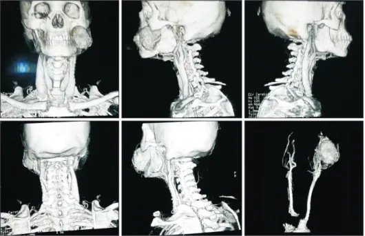
At presentation, the left cheek mass was covered with a dressing (Fig. 1), which on exposure revealed an ulcerated mass with no discharge or bleeding. The mass measured 10 cm by 7 cm and was firm to the touch. It was attached to underlying structures and protruded intraorally with indenta- tion of the overlying mucosa. A computerized tomographic angiogram of the cheek (Fig. 2) revealed a rounded, isodense and brightly enhancing mass over the body and anterior to the ramus of the left hemimandible. The enhancement was the same density as the adjacent left facial artery, which coursed through the medial aspect of the mass. A diagnosis of a left facial artery pseudo-aneurysm was made. Other tests such as full blood chemistry, electrolyte count, creatinine, and ran- dom blood glucose were within the normal range for his age and sex.
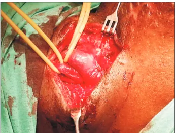
He was prepared for surgery, and on the operating table, the feeder vessel was ligated (Fig. 3) to prevent torrential bleeding from the presumed pseudoaneurysm or sclerosing
Fig. 1. Patient lesion prior to surgery.
Benjamin Fomete et al: Primary oral soft tissue angiosarcoma of the cheek: a case report and literature review. J Korean Assoc Oral Maxillofac Surg 2015
Fig. 2. Computed tomography angio- gram.
Benjamin Fomete et al: Primary oral soft tissue an- giosarcoma of the cheek: a case report and literature review. J Korean Assoc Oral Maxillofac Surg 2015
the skin and superficial soft tissue3. The etiology of AS is not fully understood2, although the sun has been implicated as a causative agent in Caucasian patients5. Chronic lymphedema and irradiation have been described as the most important eti- ological factors in the pathogenesis of AS3. Only about 10%
of ASs are associated with chronic lymphedema, even though it is the most widely accepted sign of AS in skin and soft tis- sue lesions3. Radiation-induced AS usually occurs in the ir- radiated fields many years after radiotherapy, with a median time of 8.6 years having been reported3. AS is also said to occur in pre-existing or intermediate vascular lesions. Several cases of AS arising spontaneously from hemangiomas or vas- cular malformation have been reported, but the mechanism is not clear3. Even though most authors think trauma can only alter an already existing lesion, it has been reported as an spectively, together weighing 179 g. The cut section revealed
grey, white, solid, and hemorrhagic nodular surfaces.
Histology sections showed keratinized pigmented skin overlying a fibrocollagenized dermis and a malignant spindle cell tumor growing in sheets, with anastomosing dilated vas- cular channels. The cells comprising the tumor were plump and round to spindle-shaped with hyperchromatic to vesicular nuclei, prominent nucleoli, and scanty cytoplasm. There was minimal fibrocollagenized stroma. Other areas showed hem- orrhage, fibrin deposits, and necrosis.
III. Discussion
AS is said to account for about 2% of all soft tissue sarco- mas, while oral AS is very rare3,4,9. It has a predisposition for
Fig. 3. Isolation of major vessels.
Benjamin Fomete et al: Primary oral soft tissue angiosarcoma of the cheek: a case report and literature review. J Korean Assoc Oral Maxillofac Surg 2015
Fig. 4. Excised specimen cut open.
Benjamin Fomete et al: Primary oral soft tissue angiosarcoma of the cheek: a case report and literature review. J Korean Assoc Oral Maxillofac Surg 2015
A B C
Fig. 5. A. Intraluminal buds of tumor cells (H&E staining, ×40). B. Sheets of epitheliod tumour cells (H&E staining, ×40). C. Strong positivity with highlights of neoplastic vascular channels (CD31 staining, ×40).
Benjamin Fomete et al: Primary oral soft tissue angiosarcoma of the cheek: a case report and literature review. J Korean Assoc Oral Maxillofac Surg 2015
that almost one-third of oral and salivary gland ASs are the rare epitheloid variant.
Immunohistochemical analysis is essential for differentiat- ing AS from similar lesions such as hemangioendothelioma, hemangiopericytoma, epithelioid sarcoma, Kaposi’s sar- coma6,11, perivascular epithelioid tumor (malignant Pecoma), spindle cell carcinoma, malignant melanoma, intravascular endothelial hyperplasia, epithelioid AS, and lymphangiosar- coma6.
Positive staining for factor VII and Ulex europaeus agglu- tinin has been used to diagnose vascular pathology. CD31, a membrane glycoprotein, is the best vascular marker of endo- thelial phenotypes, particularly in poorly differentiated AS11, where factor VIII and U. europaeus are negative. Positive staining for thrombomodulin and E-selectin also support the diagnosis of AS11. Tumors showing vasoformative channels and intracytoplasmic vacuoles on hematoxylin and eosin sec- tions are likely angiogenic. Positive reactions to F-VIII-RA, CD34, and CD31 endothelial markers indicate endothelial characteristics4,6. The presence of red blood cells in the vaso- formative channels suggests that the tumor is not a lymphatic tumor but a vascular tumor6.
Surgery is the main stay of treatment, and local primary tu- mors can be effectively excised. However, there is a high as- sociated rate of local recurrence and distant metastasis, which negatively impacts patient survival9. According to Köhler et al.2, the lesion must be excised with adequate three-dimen- sional surgical margins, which can be difficult to achieve in the head and neck region.
A combined modality of treatment, including postoperative radiotherapy, is recommended. The general outcome of pri- mary AS of the oral cavity is good, with a mean survival pe- riod of 7.3 to 7.6 years, compared to primary cutaneous and deep soft tissue tumors6,11. Our patient underwent surgery and declined oncological treatment. Because most patients pres- ent with systemic metastasis, chemotherapy is often involved in the treatment of AS. Complete remission of AS has been reported with the use of liposomal doxorubicin alone or in as- sociation with radiotherapy2,3.
Though there was no evidence of residual disease on fol- low-up, the risk of recurrence without further adjunct therapy cannot be overlooked. Due to its rarity, AS is associated with no significant or clearly definitive prognostic factors2. Lesion size has been used as the only significant predictor, while histology has had no impact. Additionally, the lesion site was considered a significant prognostic factor as tumors in the scalp had a worse prognosis. However, Köhler et al.2 found etiological factor3.
Fanburg-Smith et al.8 reported 29 cases with 22 primary AS lesions in the tongue (n=9), parotid gland (n=4), lip (n=4), submandibular gland (n=3), soft palate (n=1), and hard palate (n=1) and seven secondary lesions in the gingivae (n=4) and parotid gland (n=3). The tongue, parotid gland, and lip are commonly involved in adults8.
Our case occurred in the cheek. AS can arise from either a vascular or lymphatic origin, and predisposing factors to its development include irradiation, trauma, and advanced age.
Nagata et al.4 reported that 35 cases have been documented in the English literature.
Our patient reported a definite history of trauma, although the etiology and triggering factors are largely unknown in many cases.
AS has been categorized into five broad groups based on a wide range of associated clinical scenarios: lymphedema- associated AS, radiation-induced AS, post-breast cancer AS, soft tissue AS, and cutaneous AS.
AS of the oral cavity can involve oral soft tissue, minor salivary glands, or bones10. Our patient’s AS affected the oral soft tissue of the cheek. Several reports of primary AS in- volving the oral soft tissue, lip, palate, tongue, parotid gland, and submandibular glands are also documented. The clinical presentation is often a mass and/or bleeding, pain, bruis- ing, persistent hematoma, edema and ulceration, or round or ovoid nodules that appear red to bluish in color3,4,6,8.
The clinical differential diagnoses include pyogenic granu- loma, hemangioma, hemagioendothelioma, Kaposi’s sarco- ma, and ulcerative squamous cell carcinoma3,4. Our working diagnosis in this case was sclerosing hemangioma based on the histology report and the computed tomography (CT) scan the patient brought from another center.
Primary ASs are often misdiagnosed as other lesions be- cause of their rarity. Our case was initially reported as scle- rosing hemangioma, and while a CT scan supported a facial artery aneurysm, other missed diagnoses include pyogenic granuloma1. Delayed or missed diagnosis is associated with poor treatment outcome1.
According to Nagata et al.4, AS can show a wide spectrum of histological differentiation, from a well differentiated neoplasm showing anastomosing vascular channels lined by atypical endothelial cells with low mitotic activity to poorly differentiated tumors without prominent vasoformative ac- tivity, composed of solid sheets of epithelial-like or spindle cells4. Fanburg-Smith et al.8 stated that the most common morphologic type of AS is solid spindled vasoformative, but
MK, Kowalski LP. Cutaneous angiosarcoma of the head and neck:
report of 23 cases from a single institution. Otolaryngol Head Neck Surg 2008;139:519-24.
3. Yang XJ, Zheng JW, Zhou Q, Ye WM, Wang YA, Zhu HG, et al.
Angiosarcomas of the head and neck: a clinico-immunohistochem- ical study of 8 consecutive patients. Int J Oral Maxillofac Surg 2010;39:568-72.
4. Nagata M, Yoshitake Y, Nakayama H, Yoshida R, Kawahara K, Nakagawa Y, et al. Angiosarcoma of the oral cavity: a clinicopatho- logical study and a review of the literature. Int J Oral Maxillofac Surg 2014;43:917-23.
5. Lydiatt WM, Shaha AR, Shah JP. Angiosarcoma of the head and neck. Am J Surg 1994;168:451-4.
6. Terada T. Angiosarcoma of the mandibular gingiva. Int J Clin Exp Pathol 2011;4:791-3.
7. Sarachev E, Mateeva G. Angiosarcoma of the oral cavity. Int Med Assoc Bulgaria 2006;12:33-4.
8. Fanburg-Smith JC, Furlong MA, Childers EL. Oral and salivary gland angiosarcoma: a clinicopathologic study of 29 cases. Mod Pathol 2003;16:263-71.
9. Trinh NQ, Rashed I, Hutchens KA, Go A, Melian E, Tung R. Un- usual clinical presentation of cutaneous angiosarcoma masquerad- ing as eczema: a case report and review of the literature. Case Rep Dermatol Med 2013. doi: 10.1155/2013/906426.
10. Sumida T, Murase R, Fujita Y, Ishikawa A, Hamakawa H. Epulis- like gingival angiosarcoma of the mandible: a case report. Int J Clin Exp Pathol 2012;5:830-3.
11. Chadha V, Rawat DS, Prasad B. Primary angiosarcoma of the tongue: a case report. Int J Otolar Head Neck Surg 2012;1:51-2.
that grade was the most important prognostic factor.
Metastatic spread is usually hematogenous, and a common site of involvement is the lungs. Lymphatic spread is reported in less than 20% of ASs involving the head and neck region11. In our case, no lymph node involvement or distant metastases were observed at initial presentation or during the follow-up period; however, we did not carry out any lymph node sur- vey.
Primary AS of the cheek is rare, and its diagnosis and treat- ment still pose a challenge in clinical settings.
Conflict of Interest
No potential conflict of interest relevant to this article was reported.
References
1. Ramadhan A, Willén H, Thor A. Angiosarcoma of the mandible:
metastasis from a primary tumour of the right atrium of the heart.
Case Rep Clin Med 2013;2:53-7.
2. Köhler HF, Neves RI, Brechtbühl ER, Mattos Granja NV, Ikeda