ORIGINAL ARTICLE
팽대부 선근종의 임상적, 병리학적, 면역조직화학염색의 특징
최용혁, 김미진, 한정호, 윤순만, 채희복, 윤세진, 강민호1, 성노현2, 최재운3, 박선미
충북대학교 의과대학 내과학교실, 영상의학교실1, 병리학교실2, 외과학교실3
Clinical, Pathological, and Immunohistochemical Features of Adenomyoma in the Ampulla of Vater
Yong Hyeok Choi, Mi Jin Kim, Joung-Ho Han, Soon Man Yoon, Hee Bok Chae, Sei Jin Youn, Min Ho Kang1, Rohyun Sung2, Jae Woon Choi3 and Seon Mee Park
Departments of Internal Medicine, Radiology1, Pathology2, and Surgery3, Chungbuk National University College of Medicine, Cheongju, Korea
Background/Aims: Ampullary adenomyoma is a benign lesion whose malignant potential has yet to be confirmed. Despite its benign nature, adenomyoma is frequently misdiagnosed as a carcinoma or adenoma and is overtreated by extensive surgery.
This study was performed to analyze the clinical, pathological, and immunohistochemical features of adenomyomas in the ampulla of Vater.
Methods: Nine cases of adenomyoma in the ampulla of Vater, diagnosed in Chungbuk National University Hospital between 2008 and 2011, were enrolled in this study. We reviewed the clinical data on the symptoms, laboratory data, and radiologic findings of the abdominal computed tomography and endoscopic retrograde cholangiopancreatography. For pathological analysis, all the slides were reviewed by one pathologist, and immunohistochemical stainings with antibodies against cytokeratin 7 (CK7), cytokeratin 20 (CK20), α-smooth muscle actin (α-SMA), and Ki-67 antigen were performed.
Results: All the cases were CK7 positive and CK20 negative. A strong cytoplasmic expression of α-SMA was confirmed in all cases. The Ki-67 index was less than 1% in eight cases and 5% in one case. Four cases underwent endoscopic papillectomy, and one case received surgical ampullectomy during colorectal cancer surgery. Five cases that underwent endoscopic or surgical treatment remained symptom-free for three years. Four cases that were closely observed with repeated endoscopic examinations exhibited no interval changes in the papillary lesions.
Conclusions: Endoscopic biopsy and immunohistochemistry can aid in the diagnosis of ampullary adenomyomas. Endoscopic papillectomy or surgical ampullectomy is adequate for the treatment of symptomatic ampullary adenomyomas. (Korean J Gastroenterol 2013;62:352-358)
Key Words: Adenomyoma; Ampulla of Vater; Immunohistochemistry
Received August 1, 2013. Revised September 1, 2013. Accepted September 12, 2013.
CC This is an open access article distributed under the terms of the Creative Commons Attribution Non-Commercial License (http://creativecommons.org/licenses/
by-nc/3.0) which permits unrestricted non-commercial use, distribution, and reproduction in any medium, provided the original work is properly cited.
교신저자: 박선미, 361-711, 청주시 흥덕구 내수동로 52, 충북대학교 의과대학 내과학교실
Correspondence to: Seon Mee Park, Department of Internal Medicine, Chungbuk National University College of Medicine, 52 Naesudong-ro, Heungdeok-gu, Cheongju 361-711, Korea. Tel: +82-43-269-6019, Fax: +82-43-27-3252, E-mail: [email protected]
Financial support: This work was supported by the research grant of the Chungbuk National University in 2012. Conflict of interest: None.
INTRODUCTION
Adenomyoma in the Vaterian system is a benign tumor-like lesion that has been observed in different sites throughout the biliary tract. The gallbladder is the most frequent location
of this lesion, which is rarely found in the extrahepatic biliary tract or ampulla of Vater.1 Unlike adenoma, adenomyoma is a benign lesion whose malignant potential has not yet been confirmed. According to the World Health Organization (WHO) classification, adenomyoma is defined as a pro-
Table 1. Clinical Features of Nine Cases of Ampullary Adenomyoma
Case Age (yr)/sex
Clinical
symptom CT finding Endoscopic finding Endoscopic biopsy
AST/ALT (IU/L)
ALP (IU/L)
T-bil
(mg/dL) Treatment 1 70/M Abdominal
pain
Focal enhancing lesion at AOV and CBD dilation (14 mm)
15 mm sized ampullary mass with granularity
Adenomyoma 78/30 268 0.57 Endoscopic papillectomy
2 71/M Abdominal pain
Non-specific finding 12 mm sized ampullary mass
Adenomyoma 55/92 432 1.83 Endoscopic papillectomy 3 72/M Incidental 15×12 mm sized well
defined nodule at AOV and CBD dilatation (13 mm)
12 mm sized ampullary mass with villous mucosa
Dysplasia
→adenomyoma
25/11 180 0.36 Endoscopic papillectomy
4 53/M Abdominal pain
Non-specific finding 10 mm sized lobulated lesion
Chronic inflammation
→adenomyoma
25/33 300 1.46 Endoscopic papillectomy
5 75/M Incidental (colon cancer)
Focal enhancing lesion at AOV with diffuse IHD and CBD dilatation (11 mm)
Bulging and lobulated papilla
Adenomyoma 17/15 230 0.55 Surgical ampullectomy
6 75/F Incidental Mass (11 mm) at AOV, IHD and CBD dilatation (10 mm)
Bulging and lobulated papilla
Adenomyoma 23/16 251 0.36 Close
observation
7 64/F Incidental CBD dilatation (10 mm) Bulging papilla Adenomyoma 44/33 178 0.60 Close observation 8 57/F Incidental Non-specific finding Enlarged and lobulated
papilla
Atypical epithelial proliferation
→adenomyoma
36/29 174 1.02 Close observation
9 65/F Incidental Not performed Bulging and lobulated papilla
Adenomyoma 29/28 273 0.56 Close
observation M, male; F, female; AOV, ampulla of Vater; CBD, common bile duct; IHD, intrahepatic duct; T-bil, total bilirubin.
liferation of duct-like structures accompanied by hyperplasia of smooth muscle cells.2 The symptoms of adenomyoma vary depending on the location. Adenomyoma of the gallbladder is asymptomatic or results in mild upper quadrant abdominal pain. However, adenomyoma of the biliary tree can produce symptoms such as jaundice or abdominal pain due to the ob- struction of bile flow.3,4 Despite its benign nature, ad- enomyoma is responsible for biliary obstruction and is usu- ally misdiagnosed as a carcinoma or adenoma. As a result, the lesion is frequently overtreated with radical surgery.1 Proper diagnosis before surgery can prevent unnecessary radical resection and result in treatment with endoscopic papillectomy or close observation.
This study was performed to analyze the clinical, patho- logical, and immunohistochemical features of adenomyo- mas in the ampulla of Vater and to compare the clinicopatho- logic features of other cases reported in the literature.
SUBJECTS AND METHODS
Nine cases of adenomyoma in the ampulla of Vater, diag-
nosed in Chungbuk National University Hospital (Cheongju, Korea) between 2008 and 2011, were enrolled in this study.
They were identified according to the WHO diagnostic criteria of endoscopic biopsy or resected specimens by pathologic review. Four patients underwent endoscopic papillectomy, while surgical ampullectomy was performed on one patient during colorectal cancer surgery. Four patients were diag- nosed by repeated endoscopic examinations with biopsy and were followed-up for more than two years. Clinical data about any symptoms such as abdominal pain or jaundice and labo- ratory tests of AST, ALT, GGT, ALP, and total bilirubin at the time of diagnosis were obtained from the medical records. The findings from the abdominal CT and ERCP were reviewed by one radiologist. The size of the ampullary lesion, the diameter of the common bile duct (CBD), and the presence of intra- hepatic duct (IHD) dilatation were evaluated. Endoscopic findings including the size of the mass and gross features were reviewed by one endoscopy expert. For the pathological data, all the specimens were fixed in 10% formalin and em- bedded in paraffin. The 4-μm sections were stained using a hematoxylin and eosin stain. In each case, all the slides were
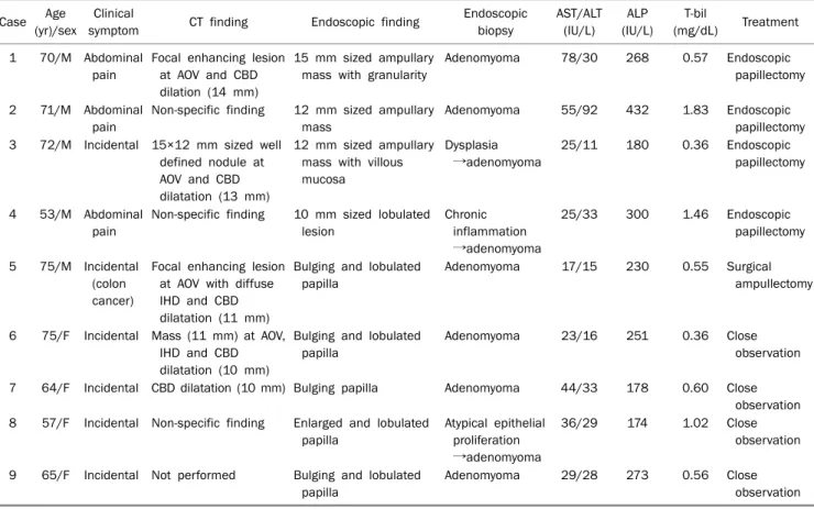
Fig. 1. Radiologic findings of adenomyomas in the ampulla of Vater. Abdominal CT (A, axial view; B, coronal view) showed a 1.4-cm mass protruding into the duodenal lumen with a dilated common bile duct (arrows). (C) Endoscopic retrograde cholangiopancreatography showed a dilated biliary tract (arrow).
reviewed by one pathologist. Representative sections were selected for immunohistochemical staining with antibodies against cytokeratin 7 (CK7; Clone OV-TL12/30; dilution, 1 : 400; Neomarker, Fremont, CA, USA), cytokeratin 20 (CK20;
Clone Ks20.8; dilution, 1 : 100; Novocastra, Newcastle Upon Tyne, UK), α-smooth muscle actin (α-SMA; asm-1; dilution, 1 : 150; Novocastra), and Ki-67 antigen (MM1; dilution, 1:
150; Novocastra). CK7, CK20, and α-SMA findings were ex- pressed as positive or negative, while Ki-67 antigen findings were expressed as a percentage of the immunohistoche- mistry.
RESULTS
1. Clinical features
The ages of the nine patients ranged from 57 to 75 years (mean, 66.9 years), and the male to female ratio was 5:4.
Six patients were asymptomatic, and lesions were found in- cidentally during esophagogastroduodenoscopy for health screening in five patients and during a colon cancer work-up in one patient. Three patients complained of mild nonspecific abdominal pain. Six patients had normal laboratory tests, and three patients exhibited mild abnormalities in liver func- tion tests (Table 1).
2. Radiologic and endoscopic features
An abdominal CT scan showed mild dilatation of the CBD (10-14 mm) in five cases, mild dilatation of the IHD in two cas- es, and the presence of a focal nodular mass (11-15 mm) in four cases (Fig. 1). Three cases showed no abnormal findings
on the abdominal CT scan. Endoscopic findings of an en- larged major papilla and villous granularities around the pap- illary orifice could be identified in all cases (Fig. 2). All the cas- es were diagnosed by endoscopic examinations including gross findings and biopsies. One case underwent extensive biopsy after sphincterotomy. Six cases were diagnosed in the first examination, while three cases were diagnosed in the second examination.
3. Pathologic features
Upon histologic examination, all the cases had similar morphology. Hyperplastic glandular lobules were located in the ampulla of Vater and were covered by a single-layer epi- thelium consisting of cuboidal and columnar epithelial cells.
There was no cellular atypia or mitotic changes in these cells.
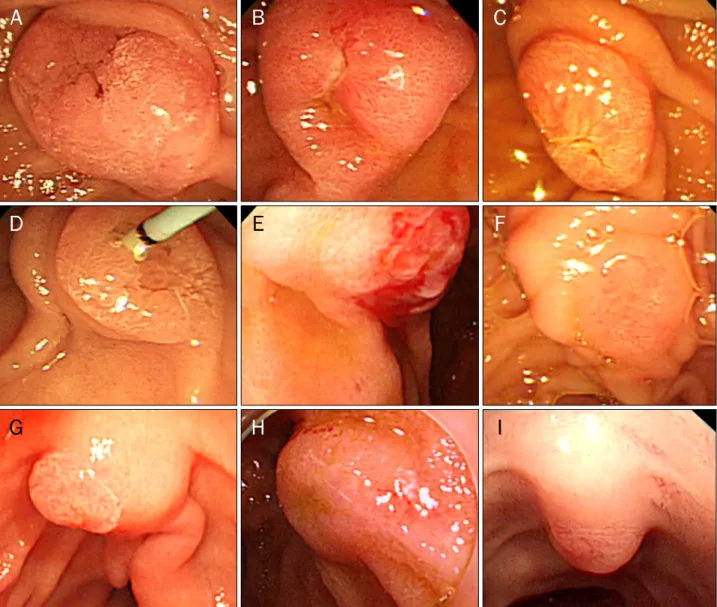
The hyperplastic glandular lobules were surrounded by hy- perplastic mesenchymal tissues that consisted of muscle fi- bers, fibroblasts, myofibroblasts, and capillaries (Fig. 3A).
The proliferative activity estimated by the Ki-67 index was de- termined to be less than 1% in eight cases and 5% in one case (Fig. 4B). The myofibroblastic phenotype of most spindle cells was confirmed by the strong cytoplasmic expression of α-SMA without desmin expression in all cases (Fig. 4C, D). All the cases were CK7 positive and CK20 negative (Fig. 4E-H).
This means that that the glandular epithelial cells of the ad- enomyoma originated from pancreatic or biliary tracts.
4. Treatments
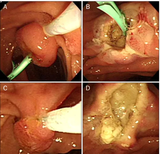
Four patients underwent endoscopic papillectomy as the usual procedure. After grasping the mass at the base with a
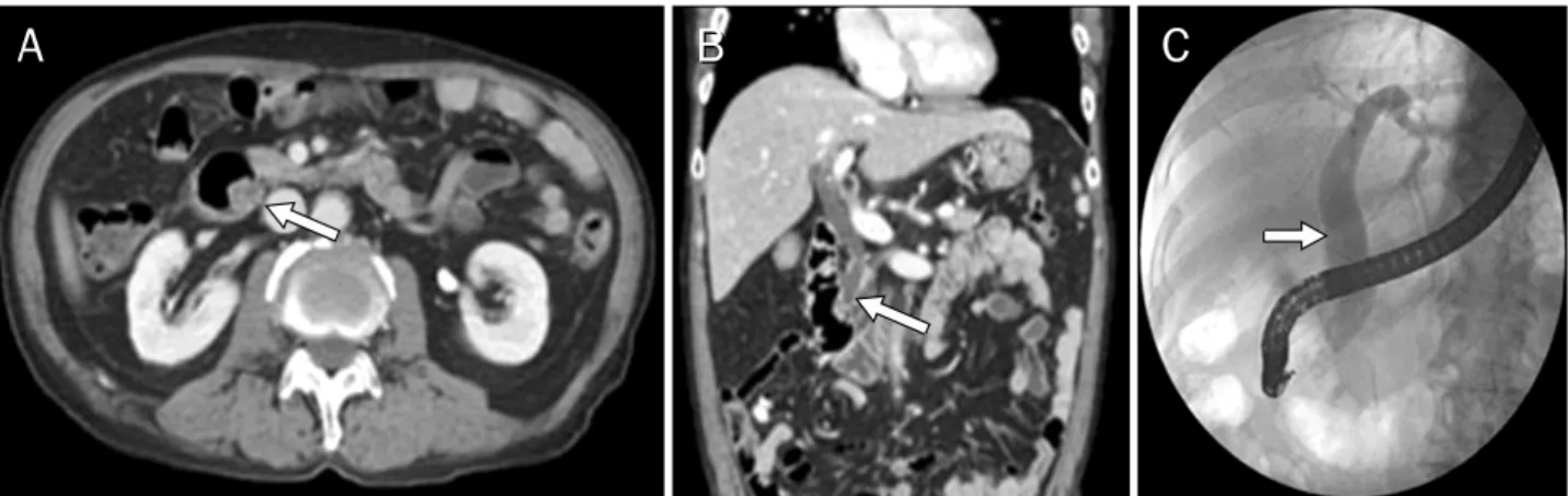
Fig. 2. Endoscopic view of ampullary adenomyomas. Enlarged major papilla and villous and granular mucosa around the papillary orifice could be identified in all cases; each case matched from A to I.
snare, resection was performed using electrocautery (Fig. 3).
A pancreatic stent was placed before or after the papil- lectomy by the decision of an endoscopist. Surgical ampullec- tomy was completed on one patient during colorectal cancer surgery. During the procedure, no remarkable complications occurred. Five cases that underwent endoscopic or surgical resection remained symptom-free for three years. The four cases that received repeated endoscopic examinations ex- hibited no interval changes in the papillary lesions.
DISCUSSION
The histological aspect of ampullary adenomyoma is char- acterized by multiple lobules of glands, mainly located in the
muscle layers of the Vaterian system, resulting in hypertrophy of the sphincter of Oddi.1 The histogenesis of adenomyomas remains uncertain. The most widely accepted hypothesis is that these lesions may represent a form of an incomplete het- erotopic pancreas.1 Some authors explained the histo- genesis of adenomyomas as hyperplastic smooth muscle tis- sue undergoing secondary muscle proliferation because of the stimulation of misplaced epithelium, muscle mis-ar- rangement, or by an aberrant growth invading and distorting the normal muscle tissue.5 Other authors considered ad- enomyoma as a part of an involutive process of the fi- broadenomatous type due to aging.6
Adenomyoma of the ampulla of Vater is very rare. Four cas- es of adenomyoma in the ampulla of Vater have been re-
Fig. 3. Pathological and immunohistochemical features of adenomyomas in the ampulla of Vater. (A) Hyperplastic glandular lobules covered by a single layer of epithelium were surrounded by hyperplastic mesenchymal tissues composed of muscle fibers, fibroblasts, myofibroblasts, and capillaries (H&E, ×200). (B) Immunohistochemical staining with the Ki67 antibody level detected at less than 1% (×40). (C, D) α-SMA expression (arrows) in the myofibroblastic component (×40 and ×400, respectively). (E, F) Strong CK7 expression (arrows) in the epithelial lining of the glandular structures (×40 and ×400, respectively). (G, H) No CK20 expression (arrows) in the epithelial lining of the glandular structures (×40 and ×400, respectively).
Fig. 4. Endoscopic papillectomy for an ampullary adenomyoma. (A) Endos- copic papillectomy was performed after pancreatic stent insertion. (B) Endoscopic view of the stent placed in the biliary duct after papillectomy. (C) Endoscopic papillectomy was per- formed without prior pancreatic stent insertion. (D) Endoscopic view of the major papilla after papillectomy.
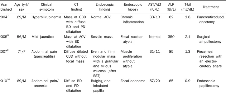
Table 2. Clinical Features of Four Cases of Ampullary Adenomyoma Reported in the Literature Year
published
Age (yr)/
sex
Clinical symptom
CT finding
Endoscopic finding
Endoscopic biopsy
AST/ALT (IU/L)
ALP (IU/L)
T-bil
(mg/dL) Treatment 20047 69/M Hyperbilirubinemia Mass at CBD
with diffuse BD and PD dilatation
Normal AOV Chronic inflammation
33/13 62 1.8 Pancreaticoduod enectomy
20058 56/M Mild jaundice Mass at AOV with BD dilatation
Sessile mass Focal nuclear atypia
Normal 350 2.1 Surgical ampullectomy
20079 74/F Abdominal pain (pancreatitis)
Diffuse dilated CBD without focal mass
Even and firm nodular mass with a granular and villous mucosa (after EST)
Muscle proliferation without atypia
31/11 85 1.3 Piecemeal
resection with an electro- cautery snare
201010 69/M Abdominal pain/
anorexia
Diffuse BD and PD dilatation
Bulging and lobulated papilla
Focal adenoma 57/20 85 0.9 Endoscopic papillectomy
M, male; F, female; CBD, common bile duct; BD, bile duct; PD, pancreatic duct; AOV, ampulla of Vater; EST, endoscopic sphincterotomy;
T-bil, total bilirubin.
ported in Korea (Table 2).7-10 Most cases presented with ab- dominal pain, jaundice, pancreatitis, or dilatation of the bile duct that was found through an abdominal CT scan. Endos- copic treatment was performed in two cases,9,10 while surgi- cal resection was performed in two other cases.7,8 One case, which presented with acute pancreatitis, was treated by large particle biopsy using an electrocautery snare. After treat- ment, the patient’s symptoms improved, and no complica- tions occurred during the follow-up period.9 The other case underwent papillectomy by ERCP.10 Two cases7,8 underwent surgical resection by pancreaticoduodenectomy because malignant potential could not be excluded (Table 2).
Since ampullary adenomyoma is a benign lesion, exten- sive surgery is not necessary. Most patients who have re- ceived extensive surgery such as pancreaticoduodenectomy have had their ampullary adenomyomas mistaken as malig- nant lesions before surgery. Proper preoperative diagnosis can prevent unnecessary surgery. The proper diagnosis of ampullary adenomyomas is sometimes very difficult to ach- ieve through abdominal CT or endoscopic exam. Usually the diagnostic accuracy of endoscopic biopsies for ampullary le- sions is not high. One study showed that it was 62%.11 However, our study revealed that all cases could be diag- nosed as adenomyomas through a single or repeated endo- scopic biopsies. Most cases showed an enlarged nodular papilla with villous and mucosal granularities. ERCP can be
a useful diagnostic tool for this lesion since it allows for the inspection of the periampullary region and visualization of the pancreatobiliary tracts. Moreover, a proper biopsy speci- men can be taken from the lesion through a lateral scope rather than a direct scope.12 Pathologic features of speci- mens obtained from proper biopsy sites are useful for diag- nosis when there are findings of glandular epithelial cells and smooth muscles proliferation without mitosis or atypical changes.
There is no established treatment for adenomyoma of the ampulla of Vater. Endoscopic sphincterotomy or endoscopic papillectomy can be helpful in restoring adequate biliary drainage.3 When surgical treatment is decided upon, intra- operative frozen sections could result in limited resection in- stead of extensive surgery.13,14 In this study, we performed endoscopic treatment or close observation in all cases ex- cept one. One patient underwent surgical ampullectomy dur- ing colorectal cancer surgery, since malignancy could not be completely excluded despite the preoperative diagnosis of benign disease.
Special immunohistochemical staining can show the char- acteristics of adenomyoma compared to neoplastic lesions in the ampulla of Vater. One study reported on the function of mucin in differentiating between adenomyoma and malig- nant lesions.15 Another study investigated the expression patterns of CK7, CK20, α-SMA, and Ki-67 in ampullary
adenomyomas.16 CK7 and CK20 are expressed differently according to the tissue origin - CK7 is expressed from the pan- creato-biliary tract, while CK20 is expressed from the gastro- intestinal tract. These markers have been used for the identi- fication of the site of origin. α-SMA recognizes the alpha- smooth muscle isoform of actin and exhibits no cross re- action with actin from fibroblasts, striated muscle, or myocardium. α-SMA stains smooth muscle cells in vessel walls, gut walls, and the myometrium.17 The Ki-67 protein is a cellular marker for proliferation and is present during all ac- tive phases of the cell cycle, but is absent from resting cells.18 A low expression level of Ki-67 means that the adenomyoma has no malignant potential. Our nine cases showed compat- ible patterns of adenomyoma that consisted of CK7 positive epithelium, α-SMA positive smooth muscle hyperplasia, and a low Ki-67 index.1,8 These results indicate that immunohis- tochemistry performed on the biopsy tissue of the ampulla of Vater before treatment is very useful in confirming ampullary adenomyoma.
In conclusion, awareness of ampullary adenomyomas is very important in clinical situations because of their clinical and endoscopic similarities to ampullary tumors like ad- enomas and carcinomas. The method of treatment based on the diagnosis can affect the patient’s quality of life. The im- munohistochemical results of ampullary lesions can help in the diagnosis of adenomyoma. Immunohistochemical cri- teria like CK7 positivity and CK20 negativity and low pro- liferative activity in the epithelial cells should be considered as indicating a diagnosis of adenomyoma of the ampulla of Vater. Endoscopic biopsy and immunohistochemistry of am- pulla of Vater lesions can help in the diagnosis of ampullary adenomyoma. Endoscopic papillectomy or surgical ampul- lectomy is sufficient for symptomatic ampullary adeno- myomas. For asymptomatic ampullary adenomyomas, close observation is recommended. These strategies can avoid un- necessary surgeries such as laparotomies or pancreati- coduodenectomy.
REFERENCES
1. Handra-Luca A, Terris B, Couvelard A, Bonte H, Flejou JF.
Adenomyoma and adenomyomatous hyperplasia of the Vaterian system: clinical, pathological, and new immunohistochemical
features of 13 cases. Mod Pathol 2003;16:530-536.
2. Albores-Saavedra JH, Sobin LH, Gibson JB. Histological typing of tumours of the gallbladder and extrahepatic bile ducts. Berlin:
Springer-Verlag, 1991.
3. Hammarström LE, Holmin T, Stenram U. Adenomyoma of the ampulla of Vater: an uncommon cause of bile duct obstruction.
Surg Laparosc Endosc 1997;7:388-393.
4. Narita T, Yokoyama M. Adenomyomatous hyperplasia of the pap- illa of Vater: A sequela of chronic papillitis? Ann Diagn Pathol 1999;3:174-177.
5. Clarke BE. Myoepithelial hamartoma of the gastrointestinal tract. Arch Pathol 1940;30:143-152.
6. Fernandez-Cruz L, Pera C. A histological study of the sphincter of Oddi. In: Delmont J, ed. The sphincter of Oddi. Proceedings of the 3rd Gastroenterology Symposium. Nice: Gastroenterology Symposium, 1976:13-20.
7. Kim JW, Jang JY, Han SS, Choi MK, Kim SH, Park YH. Adenomy- oma of the Vaterian ampulla. Korean J Hepatobiliary Pancreat Surg 2004;8:258-261.
8. Jang KT, Heo JS, Choi SH, et al. Adenomyoma of ampulla of Vater or the common bile duct: a report of three cases. Korean J Pathol 2005;39:59-62.
9. Kwon TH, Park do H, Shim KY, et al. Ampullary adenomyoma pre- senting as acute recurrent pancreatitis. World J Gastroenterol 2007;13:2892-2894.
10. Lee BU, Bang JS, Yang SH, et al. A case of ampullary adenomy- oma associated with dilatations of pancreatic and biliary ducts.
Korean J Gastrointest Endosc 2010;40:391-395.
11. Menzel J, Poremba C, Dietl KH, Böcker W, Domschke W. Tumors of the papilla of Vater--inadequate diagnostic impact of endo- scopic forceps biopsies taken prior to and following sphinc- terotomy. Ann Oncol 1999;10:1227-1231.
12. Bedirli A, Patiroglu TE, Sozuer EM, Sakrak O. Periampullary ad- enomyoma: report of two cases. Surg Today 2002;32:1016- 1018.
13. Kayahara M, Ohta T, Kitagawa H, Miwa K, Urabe T, Murata T.
Adenomyomatosis of the papilla of Vater: a case illustrating diag- nostic difficulties. Dig Surg 2001;18:139-142.
14. Läuffer JM, Baer HU, Maurer CA, et al. Adenomyoma of the distal common bile duct mimicking cholangiocarcinoma. Dig Dis Sci 1998;43:1200-1204.
15. Higashi M, Goto M, Saitou M, et al. Immunohistochemical study of mucin expression in periampullary adenomyoma. J Hepato- biliary Pancreat Sci 2010;17:275-283.
16. Duval JV, Savas L, Banner BF. Expression of cytokeratins 7 and 20 in carcinomas of the extrahepatic biliary tract, pancreas, and gallbladder. Arch Pathol Lab Med 2000;124:1196-1200.
17. Sheehan M, O'Briain DS. False-positive immunoreactivity with muscle-specific actins in non-Hodgkin's lymphomas. Arch Pathol Lab Med 1995;119:225-228.
18. Scholzen T, Gerdes J. The Ki-67 protein: from the known and the unknown. J Cell Physiol 2000;182:311-322.