59
Alterations of the Apoptosis Genes and Their Products in Non-small Cell Lung Cancer Tissues
Apoptosis is a principal type of cell death, and it has a profound effect on the development of cancer. It is also well known that anti-cancer agents induce apoptosis, and defects in the apoptosis pathways reduce the treatment sensitivity. Of the many pathways that induce apoptosis, the mechanisms of the intrinsic and extrinsic apoptosis pathways are well established. Non-small cell lung cancer (NSCLC) is a leading cause of cancer death worldwide, yet the exact molecular mechanisms of its development remain unclear. Apoptosis deregulations may underlie the development and pathogenesis of NSCLC.
This review discusses the general mechanisms of apoptosis, the constituents of the apoptosis machinery and the alterations of the apoptosis-related genes in NSCLC. (J Lung Cancer 2008;7(2):59 64)
Key Words: Non-small cell lung cancer, Apoptosis, Mutation, Expression
Nam Jin Yoo, M.D. and Sug Hyung Lee, M.D.
Department of Pathology, College of Medicine, The Catholic University of Korea, Seoul, Korea
Received: August 23, 2008 Accepted: October 13, 2008 Address for correspondence Sug Hyung Lee, M.D.
Department of Pathology, College of Medicine, The Catholic University of Korea, 505, Banpo-dong, Seocho-gu, Seoul 137-701, Korea
Tel: 82-2-590-1188 Fax: 82-2-537-6586
E-mail: [email protected] This study was supported by a grant of the National Cancer Control R&D Program 2008, Ministry for Health, Welfare and Family Affairs (0820080).
서 론
Apoptosis는 세포사멸(cell death)의 주된 발생기전으로 조 직의 항상성, 세포분화 및 발생에 중요한 역할을 하며, 세포 사멸 조절 이상은 퇴행성 질환, 종양, 에이즈 등 여러 질병 을 유발한다(1,2). Hanahan과 Weinberg는 암 발생 및 진행에 필수 불가결한 6가지의 표현형을 기술하였으며, 그들 중 하 나가 암세포의 apoptosis 회피현상이라 정의하였다(1). 이는 apoptosis 회피현상이 암의 발생 및 진행에서 보조적인 기전 이 아닌 핵심기전임을 제시하는 것이다. 암세포의 성장은 암세포의 증식과 세포사멸의 불균형에 의하여 생기며, 세 포사멸 결손은 암세포의 수명연장을 초래하고 유전자의 이 상을 축적하게 하여 전이를 포함한 암의 진행을 용이하게 한다. 암에 대한 항암제 치료 및 방사선 치료 역시 암세포의 세포사멸을 유발시키는 치료방법이므로(2), 세포사멸 연구 는 암의 발병기전을 밝히는 것뿐 아니라, 치료의 기전을 규 명하고, 치료의 내성기전을 밝히는데 중요하다.
세포사멸을 유발하는 경로는 많지만, 내인성(intrinsic) 경
로와 외인성(extrinsic) 경로로 나누는 것이 가장 흔한 분류 법이다(2). 외인성 경로는 Fas, tumor necrosis factor-related apoptosis-inducing ligand (TRAIL) receptor 같은 death recep- tor에 의해서 유발되고, 내인성 경로는 성장인자의 소실, 저 산소증, 방사선 조사, 항암제 등에 의해서 유발된다(2). 내인 성 경로는 미토콘드리아를 중심으로 일어나며 bcl-2 family 에 의해서 주로 조절된다. Caspase는 단백질분해효소로 cystein protease에 속하며 apoptosis의 시작 및 진행에 중요한 역할을 수행한다(2). Apoptosis를 유발하고 신호전달에 관여 하는 단백질은 많지만 실제로 세포를 사멸로 유도하는 단 백질을 apoptosis machinery로 명명하며 이를 이루는 주요 구성원이 death receptor, bcl-2 family, caspase 등이다(2).
비소세포폐암은 여러가지 세포사멸 자극에 대해 저항적 이라는 것이 잘 알려져 있다(3). 이제까지 비소세포폐암에 서 밝혀진 대표적인 세포사멸 기전의 이상은 Fas, TRAIL receptor, caspase-3의 돌연변이 등과 Fas, bcl-2 등의 이상발 현이다(4∼8). 하지만, apoptosis 관련 유전자의 돌연변이 및 발현연구가 일부 유전자에 제한적으로 이루어져 있고, 이
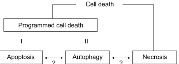
Fig. 1. Current classification of cell death. Cell death can be categorized into programmed cell death and necrosis. The programmed cell death is further classified into type I (apoptosis) and type II (autophagy) programmed cell death.
The interconnection of apoptosis, autophagy and necrosis remains unclear.
들에 대한 임상적 의미는 불확실하다. 본 리뷰는 세포사멸 의 분류, apoptosis와 암의 관계, death receptors, 비소세포폐 암의 death receptors 이상, bcl-2 계열 유전자, 비소세포폐암 의 bcl-2 계열 유전자 이상, caspases, 비소세포폐암의 caspase 이상에 관하여 기존의 논문정보를 요약하고 apoptosis 이상 과 비소세포폐암의 특징을 연계하고자 한다.
세포사멸(Cell Death) 분류
세포사멸에 관한 정의 및 분류는 다양하며 기존의 분류 에 추가로 다른 종류의 세포사멸이 계속 밝혀지고 있다. 하 지만, 세포사멸을 apoptosis, autophagy, necrosis로 분류하는 것이 현재 가장 일반적인 분류이다. Apoptosis를 program- med cell death type I, autophagy를 programmed cell death type II로 분류하는 것도 일반적이다(9)(Fig. 1). Programmed cell death는 종종 apoptosis의 동의어로 사용되기도 한다. Apop- tosis가 형태 및 생화학적 특성에 근거한 정의인 반면 programmed cell death는 ‘유전적으로 정해진’ 세포사멸이라 는 모호한 정의이며 ‘예정이 안된’ 병리학적 자극에 대한 세포사멸이라는 necrosis의 반대 개념으로 사용된다(9). 하 지만, 발생이나 노화에 나타나는 세포사멸 같이 program- med cell death의 적용이 명확한 경우도 있지만, 항암제 투여 시에 나타나는 세포사멸 같이 ‘유전적으로 정해진’ 것인지 혹은 ‘예정이 안된’ 것인지가 불명확한 경우가 많아 적용이 어려운 현실이다(9).
Apoptosis는 Kerr 등(10)에 의해 명명된 특정한 형태의 세 포사멸로 세포가 둥글어지고, 위족(pseudopod)은 위축되고, 세포의 부피가 줄어들고, 크로마틴이 농축되고, 핵이 분쇄 되는 형태를 보인다. 생화학적으로 DNA ladder, caspase 활 성화 등이 apoptosis의 특징이지만, 이런 현상이 관찰되지 않는 apoptosis도 많이 보고되므로 이들이 apoptosis의 정의
에 필수적인 것은 아니다(9).
Apoptosis와 암
종양 생성과정이 다단계로 일어나며, 각 단계가 정상세 포를 transformation시키는 유전학적 이상에 의해 나타난다 (1). 종양의 genome은 다양한 부위에서 이상이 발생하고 그 이상은 점 돌연변이로부터 유전자의 전좌에 이르기까지 다 양하게 나타난다. 중요한 유전자에 나타나는 돌연변이는 암의 다양한 능력을 부여하는데 이는 성장자극 없이도 세 포성장을 유도하는 능력, 세포성장 억제 신호에 둔감하게 반응하는 능력, 무한히 증식하는 능력, 혈관생성을 유도하 는 능력, 침윤하고 전이하는 능력, 세포사멸을 회피하는 능 력 등이다(1).
bcl-2가 세포생존을 촉진하는 종양유전자로 확인된 후 (11), apoptosis를 길항하는 과정이 암을 유발하는 과정에서 필요하다는 것이 널리 알려져 있다. 암세포의 클론 확장과 암 종괴의 성장은 암세포의 증식과 세포사멸의 불균형에 의한 결과라는 것이 시험관내 실험, 동물실험, 암 조직의 분 석에서 다양하게 입증된 바 있다(1,2). 종양유전자 c-Myc과 발현 쥐에서 나타나던 증식과 apoptosis가 bcl-2의 동시 발현 에 의해 apoptosis가 억제되고 종양의 발생이 가속화한다는 것이 입증된 바 있다(12). 종양세포는 충분한 산소와 영양 분이 제한된 경우가 많고 이에 의한 apoptosis 자극도 많지 만, 종양세포 중 apoptosis 회피능력을 획득한 세포가 선택 되어 증식할 수 있는 능력을 부여 받는다. 암세포가 전이하 기 위해서는 혈류에서 살아남고 다른 조직에 침윤해야 한 다. 정상 상피세포는 suspension 상태가 지속되거나 적절한 생존 자극이 주어지지 않는 타 장기에서의 생존은 제한적 이지만, 암세포는 이러한 apoptosis 자극 상황에서도 생존하 고 전이할 수 있는 apoptosis 회피 능력이 있다(1). 따라서, apoptosis 회피 현상은 암의 발생부터 전이에 이르는 전 과 정에 밀접한 영향을 미치는 것을 알 수 있다.
1) Death receptors
가장 잘 알려진 death receptor는 tumor necrosis factor (TNF) receptors, Fas (CD95/Apo-1) and TNF-related apoptosis- inducing ligand (TRAIL) receptors이며, 이들의 death ligand는 각각 TNF, Fas ligand, TRAIL이다(2,13). 세포 표면에 존재하 는 수용체인 death receptor들은 특이적인 death ligand와 결 합하여 apoptosis 신호를 세포 내로 전달한다. 즉, death ligand와 trimer 상태로 결합한 death receptor는 adaptor (FADD 또는 TRADD)와 결합하고 순차적으로 caspase-8 혹
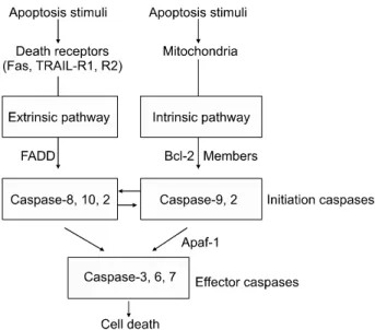
Fig. 2. Two apoptosis pathways. Apoptosis pathways are largely categorized in two pathways: the intrinsic and extrinsic pathways. The extrinsic apoptosis is initiated with activation of death receptors, while the intrinsic pathway is initiated from mitochondria. Initiator caspases in each pathway relay the apoptosis signal to effector caspases that eventually kill the cells.
은 10과 결합 후 extrinsic 혹은 intrinsic apoptosis pathway를 따라 세포사멸을 유도한다(Fig. 2).
2) 비소세포폐암의 death receptor 이상
Death receptor는 다양한 종류의 암에 잘 발현이 되므로 발현자체에 따라 해당 암이 apoptosis 자극에 민감한 지를 예측할 수 없고, 이 사실은 신호경로 상에 apoptosis를 억제 하는 기전이 있다는 증거를 제시한다(14). Death receptor의 돌연변이는 가장 잘 알려진 이 경로의 억제기전으로, T-급 성 림프구성 백혈병, 다발성 골수종, 림프종, 비소세포폐암, 방광암, 흑색종 등 다양한 암에서 규명된 바 있다(4,15∼
19).
편평상피암 30예, 샘암 24예, 세기관지페포암 11예 등 총 65예의 비소세포폐암을 분석한 연구에서 Fas 유전자는 돌 연변이가 7.7%에서 나타났다(4). 이 연구에서 모든 돌연변 이는 missense mutation이었으며, 4개는 death domain, 1개는 transmembrane domain을 코딩하는 부위에 나타났다. 이때, Fas 단백질은 발현이 잘 되는 것으로 나타나 돌연변이 Fas 가 세포사멸억제 기능을 나타낼 가능성이 높을 것으로 예 상되었다(4). Fas 경로 유전자의 돌연변이가 폐암의 전이와 연관성 역시 연구된 바도 있다. Shin 등(20)은 림프절 전이 가 있는 43예, 전이가 없는 37예의 비소세포폐암을 분석한 결과, Fas 경로 유전자의 돌연변이가 대부분 전이성 폐암에
서 나타나며, 전이성 폐암에서도 원발병소가 아닌 림프절 전이 병소에서 의미있게 돌연변이가 증가하는 것을 관찰하 였다. 이 연구에서는 돌연변이가 Fas 유전자에 국한되지 않 고 FADD와 caspase-10에서도 나타남이 확인되었다. 또한, transfection system을 통해 돌연변이 Fas, FADD, caspase-10 을 세포에 주입한 결과 돌연변이가 대부분 apoptosis를 억제 하는 것을 관찰하였다(20).
돌연변이 이외에 Fas 단백질의 발현 여부 및 발현 패턴 역시 apoptosis를 억제할 수 있는 기전이다. Nambu 등(7)은 42예의 폐샘암을 대상으로 Fas 발현을 조사하여 48%의 폐 샘암만이 Fas를 발현하고, 발현하는 경우도 대부분 정상 위 치인 세포표면에 존재하지 않고 세포질 내에 존재함을 밝 혔다. 세포질 내에 존재하는 Fas는 apoptosis 유발능력이 현 저하게 감소함이 확인되어 이 기전이 폐암의 apoptosis 회피 에 중요함을 나타냈다(7).
TRAIL receptor는 TRAIL-R1 (DR4), TRAIL-R2 (DR4), DcR1, DcR2, osteoprogeterin 등이 알려져 있으며, TRAIL- R1, TRAIL-R2가 apoptosis를 유발하는데 중요한 반면 나머 지는 apoptosis 기능을 억제하거나 apoptosis 기능의 연관성 이 잘 안 알려진 상태이다(2,13). TRAIL-R1 및 TRAIL-R2의 돌연변이는 비소세포폐암 및 유방암에서 규명된 바 있다 (5,21). Lee 등(5)은 104예의 비소세포폐암을 분석하여 11예 (10.6%)에서 TRAIL-R2 돌연변이를 발굴하였다. 이 중 9개 가 missense mutation, 1개가 silent mutation, 1개는 splice-site mutation이었는데, 특이하게 모든 missense mutation이 death domain을 코딩하는 exon 9에서 나타났다. Death domain은 세포질에 존재하는 부위로 adaptor에 apoptosis signal을 전달 하는 중요한 부위이다(2). 이는 Fas 돌연변이와 동일한 결과 로 death receptor 돌연변이가 death domain이라는 apoptosis 신호전달에 중요한 부위에 밀집해서 일어난다는 사실을 보 여준 결과였다(17∼19).
bcl-2 계열 유전자
Apoptosis의 내인성 경로는 bcl-2 family에 의해서 주로 조 절되는데(Fig. 2), bcl-2 family는 크게 apoptosis 유발성과 apoptosis 길항성으로 나뉜다(2). 이들 bcl-2 family 구성원은 서로 결합하여 homodimer 및 heterodimer로서 복잡한 조절 체계를 구성하며, bcl-2 family 단백질들의 상대적 비율은 apoptosis의 반응 정도를 결정하는 중요한 인자이다(2). 포유 동물의 bcl-2 family 단백질은 약 30종류가 밝혀져 있고, 이 들 모두에는 한 종류 이상의 bcl-2 homology (BH) domain이 있다(2). BH domain 중 BH3 domain은 apoptosis를 유발하는
데 필요한 domain이다. Apoptosis 유발성 bcl-2 family 단백 질에는 PUMA, BAD, Hrk, bcl-G, Noxa, BIM, bcl-rambo, BAX, BIK, BAX, BID, BMF 등이 있으며, 이들은 모두에는 BH3 domain이 있고 대부분의 경우 apoptosis 유발을 BH3 domain의 존재에 의존한다(1). Apoptosis 길항성 bcl-2 family 는 bcl-2, bcl-XL, Mcl-1, bfl-1, bcl-W, Boo 등이 있다(1). 내인 성 apoptosis의 자극은 이들 길항성, 유발성 bcl-2 family 단 백질의 균형을 깨고 bax, bak 단백질을 mitochondria에서 활 성화 시킨 후, mitochondria에 존재하는 cytochrome c 등의 단백질을 세포질로 유출시킨다(1). 유출된 cytochrome c는 apaf-1, caspase-9와 결합하여 커다란 단백질 복합체를 형성 하고 caspase-3, caspase-7 등의 caspase를 활성화하여 중요 단백질을 분해하고 세포를 죽음에 이르게 한다(Fig. 2).
1) 비소세포폐암의 bcl-2 계열 유전자 이상
Apoptosis 유발성 bcl-2 계열 유전자의 불활성화 돌연변이 가 apoptosis를 억제하여 암 발생을 촉진시킬 수 있다는 가 설 하에 많은 돌연변이 연구가 이루어졌다. 위암 및 대장암 의 bak 돌연변이, 림프종의 bik 돌연변이, 위암 및 폐암의 bax 돌연변이, 대장암의 bad 돌연변이 등이 보고되었지만, 매우 낮은 빈도로 나타나며 일부 암에 국한된 돌연변이만 보고 된 바 있다(22-25). 이는 death receptor의 돌연변이와는 달리 apoptosis 유발성 bcl-2 계열 유전자 돌연변이가 암 발 생에 비교적 중요한 역할을 하지 않는다는 것을 제시하는 연구결과이다. 비소세포폐암에서는 apoptosis 유발성 bcl-2 계열 유전자 돌연변이 연구가 Noxa, bad, BNIP3, PUMA 등 에 대해 이루어졌지만 발견이 되지 않았다(26∼29).
비소세포폐암에서 bcl-2 계열 유전자의 돌연변이의 역할 이 중요하지 않다고 밝혀진 반면 이들 유전자의 발현은 임 상적으로 중요한 의미를 나타낸다. bcl-2의 발현은 10∼50%
정도의 비소세포폐암에서 나타나고 환자의 생존기간 연장 과 연관된 마커임이 여러 연구를 통해서 확인되었다 (8,30,31). 역시, bax의 발현도 비소세포폐암 환자의 증가된 생존과 연관성이 관찰 되었다(32). 이들 결과는 apoptosis 유 발성 혹은 길항성 인자의 발현이 항상 암세포의 생존을 감 소 혹은 연장하여 환자의 생존을 증가 혹은 감소시킨다는 논리와 직접적으로 연계되지는 않음을 보여준다. 이는 환 자의 예후가 많은 단백질, 이들의 다양한 기능, 이들의 상호 관계, 암세포의 미세환경 등 다양한 요소에 의해 결정되는 것을 제시한다.
2) Caspases
Caspase는 단백질분해효소로 cystein protease로 기질의
aspartic acid 부위를 절단하여, apoptosis의 시작 및 진행에 중요한 역할을 수행한다(2). 포유동물의 caspase 단백질은 14종류가 밝혀져 있고, 사람에서는 caspase1-10, 14의 11종 류가 밝혀졌다(1). 이들의 구조는 prodomain, large protease subunit, small protease subunit으로 구성되어 있다. Caspase는 기시 caspase (initiation caspase)와 수행 caspase (effector caspase)로 나눌 수 있으며, 기시 caspase는 caspase-2, 8, 9, 및 10, 수행 caspase는 caspase-3, 6, 및 7로 나뉜다(2).
3) 비소세포폐암의 caspase 이상
Caspase는 apoptosis를 유발할 수 있는 능력이 있어 putative tumor suppressor gene으로 분류될 수 있으며, caspase 기능의 불활성화는 암세포의 apoptosis를 방해하여 암 발생에 기여 할 수 있다. Caspase의 불활성화를 유발할 수 있는 돌연변이 는 다양한 암에서 caspase-3, 5, 6, 7, 8, 9, 10, 14가 분석되었 고, 이 중 caspase-3, 7, 8, 10의 돌연변이는 높은 빈도로 발견 되었다(6,33∼40). Caspase-3의 돌연변이는 1.2∼4.1%의 빈 도로 대장암, 폐암, 위암, 간암, 다발성골수종, 림프종에서 나타났다(6). Caspase-5는 coding sequence에 A(10) mononu- cleotide repeat이 있으며, microsatellite mutator phenotype (MMP)의 위암, 대장암, 자궁내막암에서 frameshift mutation 되고 불활성화되는 것이 알려져 있다(33). Caspase-6은 대장 암 및 위암의 2%에서 돌연변이가 나타나며(34), caspase-9은 대부분의 고형암에서 돌연변이가 발견되지 않았다(38).
caspase-7은 대장암, 식도암, 두경부암의 2∼3%에서 돌연변 이가 발견되었고 발견된 돌연변이가 대부분 기능적으로 wild-type caspase-7의 apoptosis를 불활성화하는 것이 증명되 었다(35). Caspase-8의 돌연변이는 위암, 대장암에서 각각 10%, 5%의 높은 빈도로 관찰되었는데, 특징적으로 돌연변 이의 종류가 missense보다는 frameshift mutation이 많은 것 이 특징이었다(36,37). 또한, 기능적으로 caspase-8 돌연변이 들이 wild-type caspase-8의 apoptosis를 불활성화하는 것이 증명되었다(36,37). Caspase-10의 돌연변이는 비소세포폐암 및 림프종에서 각각 5%, 14%의 높은 빈도로 관찰된 바 있 다(4,39). Caspase-14의 돌연변이는 대부분의 고형암에서는 발견되지 않았으며 대장암의 1%에서만 관찰되었다(40).
비소세포폐암에서 caspase의 돌연변이를 정리하면 caspase- 3, 6, 7, 8, 9, 10, 14가 분석되었고, 이 중 caspase-3에서 2.2%, caspase-10에서 5%의 돌연변이가 발굴된 것을 알 수 있다 (4,6,34,35,37,38,40). 저자의 미발표 실험에서 caspase-1, 2, 4, 5 역시 비소세포폐암에서 돌연변이가 발견되지 않아 비소 세포폐암에서의 caspase 돌연변이에 의한 불활성화는 흔하 지 않다는 것을 알 수 있다.
Caspase의 발현은 종양의 종류와 caspase의 종류에 따라 다양하게 나타나는데, 위암에서 caspase-3, 8, 9, 10의 발현은 정상점막세포에 비해 증가하고(41), caspase-2, 6, 7은 감소 하는 것이 보고된 바 있다(42). 대장암에서 caspase-7의 발현 감소도 알려져 있다(43). 비소세포폐암에서는 caspase-3, 6, 8, 9의 발현이 정상조직에 비해 증가한 것으로 나타났다 (44). 소세포폐암에서 caspase-8은 프로모터의 과메칠화에 의해서 발현이 감소되는 것으로 나타나서(45), 비소세포폐 암과 대조적이다. 종양세포의 과도한 caspase 발현이 암 발 생에 어떤 영향을 주는지는 아직 정확한 기전이 밝혀지지 않았으나, 암에서 나타나는 증가된 apoptosis index를 설명 하는 기전일 가능성이 있다.
고안 및 결론
이상의 기술을 통해서 많은 종류의 암에서 apoptosis machinery를 이루는 주요 단백질인 death receptor, bcl-2 family, caspase와 그들을 코딩하는 유전자들이 돌연변이 되 고 이상발현 되는 것을 알 수 있었다. 비소세포폐암도 예외 없이 이들 유전자의 돌연변이와 발현 이상이 관찰되었다.
하지만, 이들 이상이 어떻게 조절되며 암 발생에 구체적으 로 어떻게 영향을 주는지에 관한 기능연구는 미흡한 실정 이다. 또한, death receptor, bcl-2 family, caspase 이외에 FADD, RAIDD, TRADD 등의 adaptor, inhibitor of apoptosis protein (IAP)인 XIAP, c-IAP1, c-IAP2, survivin, smac/Diablo, AIF, HtrA2, cytochrome c 등의 mitochondrial factor들이 종합 적으로 어떤 이상을 가지고 있는지가 비소세포폐암에서 규 명되어야 할 것이다(1,2). 세포사멸은 apoptosis 이외에 necrosis, autophagy 등 다양하며 더 많은 종류의 세포사멸이 속속 밝혀지고 있고, 이들은 서로 독립적인 것이 아니라 유 기적 연결관계를 가지고 연속적으로 작용하거나, 길항적 혹은 자극적으로 작용하므로 이들에 대한 종합적 분석 또 한 필요하다. 최근 apoptosis machinery를 자극시켜 암세포 치료에 적용하고자 하는 많은 시도가 이루어지고 있다. Putt 등(46)은 대장암에서 과도하게 발현된 caspase-3을 small molecule drug으로 활성화시켜서 암세포를 효과적으로 살해 할 수 있는 방법을 선보였으며, Oltersdorf 등(47)은 apoptosis 길항성 bcl-2 family를 억제할 수 있는 small molecule drug이 고형암 치료에 효과가 있음을 제시한 바 있다. 이들 연구는 암에 대한 apoptosis 연구가 암 발병을 규명하는 것뿐 아니 라 이를 통해 환자의 암 치료에도 적용할 수 있는 중개연구 로서의 중요성이 있음을 보여주는 것이다. 향후, 비소세포 폐암에 대한 apoptosis 연구는 비소세포폐암의 발병기전 규
명 및 치료법 개발에 중요한 역할을 할 것으로 예상한다.
REFERENCES
1. Hanahan D, Weinberg RA. The hallmarks of cancer. Cell 2000;100:57-70.
2. Reed JC. Mechanisms of apoptosis. Am J Pathol 2000;157:
1415-1430.
3. Fennell DA. Caspase regulation in non-small cell lung cancer and its potential for therapeutic exploitation. Clin Cancer Res 2005;11:2097-2105.
4. Lee SH, Shin MS, Park WS, et al. Alterations of Fas (Apo-1/
CD95) gene in non-small cell lung cancer. Oncogene 1999;18:
3754-3760.
5. Lee SH, Shin MS, Kim HS, et al. Alterations of the DR5/
TRAIL receptor 2 gene in non-small cell lung cancers. Cancer Res 1999;59:5683-5686.
6. Soung YH, Lee JW, Kim SY, et al. Somatic mutations of CASP3 gene in human cancers. Hum Genet 2004;115:112-115.
7. Nambu Y, Hughes SJ, Rehemtulla A, Hamstra D, Orringer MB, Beer DG. Lack of cell surface Fas/APO-1 expression in pulmonary adenocarcinomas. J Clin Invest 1998;101:1102- 1110.
8. Pezzella F, Turley H, Kuzu I, et al. bcl-2 protein in non- small-cell lung carcinoma. N Engl J Med 1993;329:690-694.
9. Kroemer G, El-deiry WS, Golstein P, et al. Classification of cell death: recommendations of the Nomenclature Committee on Cell Death. Cell Death Differ 2005;12 Suppl 2:1463-1467.
10. Kerr JF, Wyllie AH, Currie AR. Apoptosis: a basic biological phenomenon with wide-ranging implications in tissue kinetics.
Br J Cancer 1972;26:239-257.
11. McDonnell TJ, Deane N, Platt FM, et al. bcl-2-immunoglo- bulin transgenic mice demonstrate extended B cell survival and follicular lymphoproliferation. Cell 1989;57:79-88.
12. Strasser A, Harris AW, Bath ML, Cory S. Novel primitive lymphoid tumours induced in transgenic mice by cooperation between myc and bcl-2. Nature 1990;348:331-333.
13. Nagata S. Apoptosis by death factor. Cell 1997;88:355-365.
14. Leithäuser F, Dhein J, Mechtersheimer G, et al. Constitutive and induced expression of APO-1, a new member of the nerve growth factor/tumor necrosis factor receptor superfamily, in normal and neoplastic cells. Lab Invest 1993;69:415-429.
15. Beltinger C, Kurz E, Böhler T, Schrappe M, Ludwig WD, Debatin KM. CD95 (APO-1/Fas) mutations in childhood T-lineage acute lymphoblastic leukemia. Blood 1998;91:3943- 3951.
16. Landowski TH, Qu N, Buyuksal I, Painter JS, Dalton WS.
Mutations in the Fas antigen in patients with multiple myeloma. Blood 1997;90:4266-4270.
17. Grønbæk K, Straten PT, Ralfkiaer E, et al. Somatic Fas mutations in non-Hodgkin's lymphoma: association with extranodal disease and autoimmunity. Blood 1998;92:3018- 3024.
18. Lee SH, Shin MS, Park WS, et al. Alterations of Fas
(APO-1/CD95) gene in transitional cell carcinomas of urinary bladder. Cancer Res 1999;59:3068-3072.
19. Shin MS, Park WS, Kim SY, et al. Alterations of Fas (Apo-1/CD95) gene in cutaneous malignant melanoma. Am J Pathol 1999;154:1785-1791.
20. Shin MS, Kim HS, Lee SH, et al. Alterations of Fas-pathway genes associated with nodal metastasis in non-small cell lung cancer. Oncogene 2002;21:4129-4136.
21. Shin MS, Kim HS, Lee SH, et al. Mutations of tumor necrosis factor-related apoptosis-inducing ligand receptor 1 (TRAIL- R1) and receptor 2 (TRAIL-R2) genes in metastatic breast cancers. Cancer Res 2001;61:4942-4946.
22. Kondo S, Shinomura Y, Miyazaki Y, et al. Mutations of the bak gene in human gastric and colorectal cancers. Cancer Res 2000;60:4328-4330.
23. Arena V, Martini M, Luongo M, Capelli A, Larocca LM.
Mutations of the BIK gene in human peripheral B-cell lymphomas. Genes Chromosomes Cancer 2003;38:91-96.
24. Rampino N, Yamamoto H, Ionov Y, et al. Somatic frameshift mutations in the BAX gene in colon cancers of the micro- satellite mutator phenotype. Science 1997;275:967-969.
25. Lee JW, Soung YH, Kim SY, et al. Inactivating mutations of proapoptotic Bad gene in human colon cancers. Carcinogenesis 2004;25:1371-1376.
26. Lee SH, Soung YH, Lee JW, et al. Mutational analysis of Noxa gene in human cancers. APMIS 2003;111:599-604.
27. Lee JW, Soung YH, Nam SW, Lee JY, Yoo NJ, Lee SH.
Mutational analysis of pro-apoptotic BAD gene in non-small cell lung cancer. J Lung Cancer 2006;5:35-38.
28. Lee SH, Lee SH. Mutational analysis of pro-apoptotic BNIP3 gene in non-small cell lung cancers. J Lung Cancer 2007;6:74- 77.
29. Yoo NJ, Lee JW, Lee SH, Lee SH. Mutational analysis of PUMA gene in non-small cell lung cancers. J Lung Cancer 2006;5:92-95.
30. Moldvay J, Scheid P, Wild P, et al. Predictive survival markers in patients with surgically resected non-small cell lung carcinoma. Clin Cancer Res 2000;6:1125-1134.
31. Fokkema E, Timens W, de Vries EG, et al. Expression and prognostic implications of apoptosis-related proteins in locally unresectable non-small cell lung cancers. Lung Cancer 2006;
52:241-247.
32. Gessner C, Liebers U, Kuhn H, et al. BAX and p16INK4A are independent positive prognostic markers for advanced tumour stage of nonsmall cell lung cancer. Eur Respir J 2002;
19:134-140.
33. Schwartz S Jr, Yamamoto H, Navarro M, Maestro M,
Reventos J, Perucho M. Frameshift mutations at mono- nucleotide repeats in caspase-5 and other target genes in endometrial and gastrointestinal cancer of the microsatellite mutator phenotype. Cancer Res 1999;59:2995-3002.
34. Lee JW, Kim MR, Soung YH, et al. Mutational analysis of the CASP6 gene in colorectal and gastric carcinomas. APMIS 2006;114:646-650.
35. Soung YH, Lee JW, Kim HS, et al. Inactivating mutations of CASPASE-7 gene in human cancers. Oncogene 2003;22:8048- 8052.
36. Kim HS, Lee JW, Soung YH, et al. Inactivating mutations of caspase-8 gene in colorectal carcinomas. Gastroenterology 2003;125:708-715.
37. Soung YH, Lee JW, Kim SY, et al. CASPASE-8 gene is inactivated by somatic mutations in gastric carcinomas. Cancer Res 2005;65:815-821.
38. Soung YH, Lee JW, Kim SY, et al. Mutational analysis of proapoptotic caspase-9 gene in common human carcinomas.
APMIS 2006;114:292-297.
39. Shin MS, Kim HS, Kang CS, et al. Inactivating mutations of CASP10 gene in non-Hodgkin lymphomas. Blood 2002;99:
4094-4099.
40. Yoo NJ, Soung YH, Lee SH, Jeong EG, Lee SH. Mutational analysis of caspase-14 gene in common carcinomas. Pathology 2007;39:330-333.
41. Yoo NJ, Kim HS, Kim SY, et al. Stomach cancer highly expresses both initiator and effector caspases: an immuno- histochemical study. APMIS 2002;110:825-832.
42. Yoo NJ, Lee JW, Kim YJ, et al. Loss of caspase-2, -6 and -7 expression in gastric cancers. APMIS 2004;112:330-335.
43. Palmerini F, Devilard E, Jarry A, Birg F, Xerri L. Caspase 7 downregulation as an immunohistochemical marker of colonic carcinoma. Hum Pathol 2001;32:461-467.
44. Krepela E, Procházka J, Fiala P, Zatloukal P, Selinger P.
Expression of apoptosome pathway-related transcripts in non-small cell lung cancer. J Cancer Res Clin Oncol 2006;132:
57-68.
45. Hopkins-Donaldson S, Ziegler A, Kurtz S, et al. Silencing of death receptor and caspase-8 expression in small cell lung carcinoma cell lines and tumors by DNA methylation. Cell Death Differ 2003;10:356-364.
46. Putt KS, Chen GW, Pearson JM, et al. Small-molecule activa- tion of procaspase-3 to caspase-3 as a personalized anticancer strategy. Nat Chem Biol 2006;2:543-550.
47. Oltersdorf T, Elmore SW, Shoemaker AR, et al. An inhibitor of Bcl-2 family proteins induces regression of solid tumours.
Nature 2005;435:677-681.