Open Access
Clinical Characteristics and Outcomes of Non-small Cell Lung Cancer Patients with HER2 Alterations in Korea
Original Article
Purpose
Human epidermal growth factor receptor 2 (HER2) alterations are found in approximately 1%-3% of non-small cell lung cancers (NSCLCs). We evaluated the clinical features and out- comes of NSCLC harboring HER2 alteration detected by next-generation sequencing (NGS) in Korea.
Materials and Methods
A total of 1,108 patients who were diagnosed with NSCLC between December 2015 and December 2017 were screened and analyzed by NGS. Medical records were reviewed ret- rospectively to analyze the clinical characteristics and outcomes from various treatments.
Results
HER2 alterations were identified in 36 NSCLC patients. Of the patients, 22 (61.1%) had an exon 20 in-frame insertion mutation, 15 (41.7%) had HER2 amplification, and one had both. The median patient age was 58 years, 55.6% were male, and 50.0% were never- smokers. Adenocarcinoma was predominant (88.9%). The most common metastatic site was bone (58.3%), and 66.7% of patients were stage IV at initial diagnosis. Six patients (16.7%) had a coexistent sensitizing epidermal growth factor receptor (EGFR) mutation, and two patients (5.6%) had anaplastic lymphoma kinase (ALK) rearrangement. With a median 14 months of follow-up, the median progression-free survival of first-line treatment was 6 months (95% confidence interval, 4.172 to 7.828), and median overall survival was not reached. The proportions of adenocarcinoma, never-smokers, and metastasis to the liver were higher in the exon 20 in-frame insertion mutation group, whereas coexistence of EGFR mutation was more frequently found in the HER2 amplification group.
Conclusion
HER2-altered NSCLC showed distinct clinical features. Moreover, different characteristics were identified between the HER2 in-frame insertion mutation group and the HER2 ampli- fication group.
Key words
Non-small cell lung cancer, HER2 mutation, HER2 amplification
Kangkook Lee, MD1 Hyun Ae Jung, MD2 Jong-Mu Sun, MD, PhD2 Se-Hoon Lee, MD, PhD2 Jin Seok Ahn, MD, PhD2 Keunchil Park, MD, PhD2 Myung-Ju Ahn, MD, PhD2
1Division of Hematology-Oncology, Department of Medicine, Dongsan Medical Center, Keimyung University School of Medicine, Daegu, 2Division of Hematology-Oncology, Department of Medicine, Samsung Medical Center, Sungkyunkwan University School of Medicine, Seoul, Korea
+ + + + + + + + + + + + + + + + + + + + + + + + + + + + + + + + + + + + + + + + + + + + + + + + + + + + + + + + + + + + + + + + + + + + + + + + + + + + + + + + + + + + + + + + + + + + + + + + + + + + + + + + + + + + + + + + + + + + + + + + + + + + + + + + + + + + + + + + + + + + + + + + + + + + + + + + + + + + + + + + + + + + + + + + + + + + + + + + + + + + + + + + + + + + + + + + + + + + + + + + + + + + + + + + + + + + + + + + + + + + + + + + + + + + + + + + + + + + + + + + + + + + + + + + + + + + + + + + + + + + + + + + + + + + + + + + + + + + + + + +
Correspondence: Myung-Ju Ahn, MD, PhD Division of Hematology-Oncology, Department of Medicine, Samsung Medical Center, Sungkyunkwan University School of Medicine, 81 Irwon-ro, Gangnam-gu, Seoul 06351, Korea Tel: 82-2-3410-3452
Fax: 82-2-3410-1754
E-mail: [email protected]
Received April 8, 2019 Accepted July 25, 2019 Published Online July 26, 2019
Introduction
The identification of oncogenic driver mutations has trig- gered the development of new treatments for non-small cell lung cancer (NSCLC). Targeted therapies have shown signif- icant improvement of clinical outcomes in NSCLC patients with epidermal growth factor receptor (EGFR) mutation, ana- plastic lymphoma kinase (ALK), or c-ros oncogene1 (ROS1)
gene rearrangement.
With the advent of precision medicine using next-genera- tion sequencing (NGS), other various genetic alterations can be detected. Human epidermal growth factor receptor 2 (HER2), also known as NEU, EGFR2, or ERBB2, is a member of the EGFR family of receptor tyrosine kinases. HER2 alter- ation has been demonstrated to play important roles in the pathogenesis of certain human cancers, including breast can- cer and gastric cancer, by activating downstream signaling
via the phosphatidylinositide 3-kinase (PI3K)–protein kinase B (AKT) and mitogen-activated protein kinase kinase (MEK)–
extracellular signal-regulated kinase pathways.
HER2 alterations have emerged as novel oncogenic drivers and therapeutic targets in lung cancers [1]. The incidence of HER2 mutation in lung adenocarcinoma is 1%-5%, and fre- quency of HER2 amplification is 2%-5% [2]. Previous studies have identified distinct clinical features of NSCLCs with HER2 alteration. Mazieres et al. [3] reported that HER2- altered NSCLC shows a high proportion of adenocarcinoma (100%), female patients (69.2%), and never-smokers (52.3%).
Li et al. [4] also showed that HER2 alteration occurs predom- inantly in never-smokers (100%) and female patients (87.5%).
In contrast, Arcila et al. [5] reported that there were no asso- ciations between HER2-altered NSCLC and sex, race, or stage at diagnosis. Given the small number of patients analyzed in previous studies, the clinical features and outcomes have not been fully evaluated in advanced NSCLC harboring HER2 alterations [6].
The aims of this study are to evaluate the characteristics and clinical outcomes of NSCLC patients harboring HER2 alteration detected by NGS.
Materials and Methods
1. Patients
A total of 1,108 patients who were initially diagnosed with metastatic NSCLC was screened between December 2015 and December 2017 at Samsung Medical Center. All tissue samples were analyzed by NGS. For patients with HER2 alterations, patient characteristics, histology, initial stage at diagnosis, sites of metastasis, and response to first-line sys- temic treatment for metastatic NSCLC were retrospectively analyzed.
2. Identification of HER2 alteration
HER2 gene alterations were identified by NGS. The Onco- mine Focus Assay panel (Thermo Fisher Scientific, San Fran- cisco, CA) or the CancerSCAN panel (LabGenomics, Seong- nam, Korea) was used for analyzing HER2 alterations, inclu- ding HER2 amplification and exon 20 in-frame insertion mutation.
CancerSCAN is a targeted sequencing platform, designed at Samsung Medical Center. This customized platform inclu- ded target genes curated from the literature of requested by the researchers and clinicians and the selected genes covered variants associated with the targeted cancer therapies appro-
ved by the Korean Ministry of Food and Drug Safety and U.S. Food and Drug Administration. In this study, Cancer- SCAN version 2 was applied, encompassing 381 cancer-rela- ted genes. The genes contained in the panel are listed in S1 Table. The panel has been validated with data demonstrating 95% sensitivity and positive predictive value [7].
We also used Oncomine Focus Assay (OFA) panel, which simultaneously detects hundreds of variants across 52 genes relevant to solid tumors [8]. The biomarkers included were selected based on information in the Ion Torrent Oncomine Knowledgebase, one of the world’s largest collections of curated oncology data. OFA has shown a precise, reprodu- cible, sensitive, and accurate NGS assay for the detection of somatic genetic variants with nearly 99% of accuracy and specificity, which is commercially available [8].
Copy number variations (CNVs) of genes were detected using an in-house copy number caller. To identify somatic CNVs, we calculated the mean read depth at each exon, nor- malized by the coverage of the target regions in that sample.
This normalized read depth was further standardized by dividing by the expected coverage for a normal individual (the expected coverage at each exon was taken to be the median of the read depth at that exon across a set of normal individuals). These steps account for the variability in cap- ture efficiency and GC content at different exons. To infer the correct copy number, the amplitude of the copy numbers were then adjusted based on the estimated purity. Copy numbers greater than four were categorized as amplifica- tions.
3. Statistical analysis
Medical records were reviewed to obtain clinical informa- tion. Descriptive statistics were used to analyze the demo- graphics, histology, incidence of various sites of metastasis, EGFR mutation, and ALK rearrangement. Statistical signifi- cance was set at p < 0.05. All analyses were conducted using SPSS ver. 24.0 (IBM Corp., Armonk, NY). Either the chi-squa- red test or Fisher exact test was used, as appropriate, to ana- lyze statistical significance among patient characteristics.
Progression-free survival (PFS) was calculated from the time of first systemic treatment to radiographic progression or death. Overall survival (OS) was also calculated from the time of first systemic treatment to the date of death. PFS and OS were analyzed using the Kaplan-Meier estimation method.
4. Ethical statement
Institutional Review Board (IRB) approval was obtained from Samsung Medical Center (SMC, Seoul, Korea, SMC 2019-03-107-001). The IRB approved waiver of informed con- sent.
Results
A total of 1,108 patients diagnosed with NSCLC were screened in this study. Tissue samples from all patients were analyzed by NGS. The NGS results showed that 36 patients (3%) had HER2 alteration. The median age of HER2-altered
patients was 58 years (range, 35 to 79 years), 55.6% of the patients were male, and 50.0% were never-smokers. Adeno- carcinoma was the predominant cancer type (88.9%), and there were four cases (11.1%) of squamous cell carcinoma.
The most common metastatic site was bone (58.3%), followed by pleura (50%) and brain (36.1%). At initial diagnosis, 66.7%
of patients were stage IV. Six patients (16.7%) had a coexis- Table 1. Characteristics of the patients harboring HER2 alteration
Values are presented as number (%). HER2, human epidermal growth factor receptor 2; ECOG PS, Eastern Cooperative Oncology Group performance status; LN, lymph node; LMS, leptomeningeal seeding; ADC, adenocarcinoma; SQC, squa- mous cell carcinoma; EGFR, epidermal growth factor receptor; ALK, anaplastic lymphoma kinase.
Characteristic HER2 altered Exon20 insertion HER2 amplification
p-value
(n=36) (n=22) (n=15)
Age at diagnosis (yr) 58 ( 60.2 ( 56.4 (
Sex 0.093
Men 20 (55.6) 10 (45.5) 11 (73.3)
Women 16 (44.4) 12 (54.5) 4 (26.7)
ECOG PS 0.819
0 4 (11.1) 3 (13.6) 1 (6.7)
1 30 (83.3) 18 (81.8) 13 (86.7)
2 2 (5.6) 1 (4.5) 1 (6.7)
Tobacco 0.084
Never 18 (50.0) 14 (63.6) 4 (26.7)
Former 11 (30.6) 5 (22.7) 6 (40.0)
Current 7 (19.4) 3 (13.6) 5 (33.3)
Stage at diagnosis 0.753
I 5 (13.9) 2 (9.1) 3 (20.0)
II 3 (8.3) 2 (9.1) 1 (6.7)
III 4 (11.1) 2 (9.1) 2 (13.3)
IV 24 (66.7) 16 (72.7) 9 (60.0)
Metastasis sites
Bone 21 (58.3) 14 (63.6) 8 (53.3) 0.531
Pleura 18 (50.0) 12 (54.5) 6 (40.0) 0.385
Brain 13 (36.1) 8 (36.4) 5 (33.3) 0.850
Contralateral metastasis 11 (30.6) 6 (27.3) 5 (33.3) 0.728
Liver 7 (19.4) 7 (31.8) 0 ( 0.028
Distant LN 7 (19.4) 4 (18.2) 3 (20.0) 0.808
Adrenal 4 (11.1) 4 (18.2) 0 ( 0.131
LMS 3 (8.3) 2 (9.1) 1 (6.7) 1.000
Thyroid 1 (2.8) 1 (4.5) 0 ( 1.000
Histology 0.021
ADC 32 (88.9) 22 (100) 11 (73.3)
SQC 4 (11.1) 0 ( 4 (26.7)
EGFR(+) 0.002
Wild type 30 (83.3) 22 (100) 9 (60.0)
Mutation 6 (16.7) 0 ( 6 (40.0)
ALK(+) 0.505
Wild type 34 (94.4) 20 (90.9) 15 (100)
Mutation 2 (5.6) 2 (9.1) 0 (
Table 2. Amino acid changes of HER2 in-frame insertion mutation group
HER2, human epidermal growth factor receptor 2.
Patient No. Amino acid change Amino acid change Total (n=22)
1 E770_A771insAYVM E770_A771insAYVM 12
2 E770_A771insAYVM E770delinsEAYVM 4
3 E770_A771insAYVM G776delinsVC 3
4 E770_A771insAYVM V777delinsVGSP 2
5 E770_A771insAYVM A771_Y772insYVMA 1
6 E770_A771insAYVM - -
7 G776delinsVC - -
8 E770_A771insAYVM - -
9 E770_A771insAYVM - -
10 A771_Y772insYVMA - -
11 G776delinsVC - -
12 E770_A771insAYVM - -
13 E770_A771insAYVM - -
14 E770_A771insAYVM - -
15 E770delinsEAYVM - -
16 G776delinsVC - -
17 V777delinsVGSP - -
18 E770delinsEAYVM - -
19 E770delinsEAYVM - -
20 V777delinsVGSP - -
21 E770delinsEAYVM - -
36 E770_A771insAYVM - -
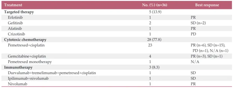
Table 3. Best response to the first-line treatment of HER2 altered non-small cell lung cancer
HER2, human epidermal growth factor receptor 2; PR, partial response; SD, stable disease; PD, progressive disease; N/A, not acquired.
Treatment No. (%) (n=36) Best response
Targeted therapy 5 (13.9)
Erlotinib 1 ( PR
Gefitinib 2 ( SD (n=2)
Afatinib 1 ( PR
Crizotinib 1 ( PD
Cytotoxic chemotherapy 28 (77.8)
Pemetrexed+cisplatin 23 PR (n=6), SD (n=15),
PD (n=1), N/A (n=1)
Gemcitabine+cisplatin 4 ( PR (n=3), SD (n=1)
Pemetrexed monotherapy 1 ( N/A
Immunotherapy 3 (8.3)
Durvalumab+tremelimumab+pemetrexed+cisplatin 1 ( SD
Ipilimumab+nivolumab 1 ( SD
Nivolumab 1 ( PR
tent sensitizing EGFR mutation, and two patients (5.6%) had ALK rearrangement (Table 1). The EGFR mutations consisted of three exon 21 L858R mutations, one exon 19 deletion, and two compound mutations (exon 18 E709K+G719A [n=1] and exon 18 G179C+exon 20 S768I [n=1]). Five of the six EGFR mutations (83.3%) were detected by NGS. The other EGFR and two ALK rearrangements were found by peptide nucleic acid–mediated polymerase chain reaction (PCR) clamping method and fluorescent in situ hybridization (FISH), respec- tively.
Among the 36 HER2-altered NSCLC patients, 22 patients (61.1%) had exon 20 in-frame insertion mutation, 15 patients (41.7%) had HER2 gene amplification, and one patient had both. The most frequently inserted amino acid sequence was E770_A771insAYVM (55%), followed by E770delinsEAYVM, G776delinsVC, and V777delinsVGSP (Table 2). Demograph- ics, including Eastern Cooperative Oncology Group perform- ance status, stage at diagnosis, and the ALK rearrangement incidence were similar between the exon 20 in-frame inser-
tion mutation group and the HER2 amplification group.
However, patients with adenocarcinoma were significantly more predominant in the exon 20 in-frame insertion muta- tion group than the HER2 amplification group (100% vs.
73.3%, respectively, p=0.021). The proportion of never-smok- ers and frequency of metastasis to the liver were also higher in the exon 20 in-frame insertion mutation group (63.6% vs.
26.7%, p=0.027; 31.8% vs. 0%, p=0.025, respectively). In con- trast, EGFR mutation was more frequent in the HER2 ampli- fication group (40% vs. 0%, p=0.002), and the proportion of male patients tended to be higher in the HER2 amplification group (73.3% vs. 45.5%, p=0.093), although the difference was not statistically significant (Table 1).
As the initial treatment, 28 patients (77.8%) were treated with cytotoxic chemotherapy (pemetrexed/cisplatin [n=23], gemcitabine/cisplatin [n=4], pemetrexed monotherapy [n=1]), and the median PFS of these patients was six months (95% confidence interval [CI], 3.969 to 8.031). Of the six patients with coexisting sensitizing EGFR mutations, four Fig. 1. Progression-free survival (PFS) and overall survival (OS) of human epidermal growth factor receptor 2 (HER2)-altered non-small cell lung cancer patients.
Probability of PFS
1.00
0 0.25 0.50
0
Time (mo) 10
5 15 20 25
0.75
36 24 8 5 5 3
All No. at risk
p=0.479 p=0.827
Probability of OS
1.0
0 0.7 0.8
0
Time (mo) 14
7 21 28 35
0.9
36 26 16 10 7 3
All No. at risk
Probability of PFS
1.00
0 0.25 0.50
0 5 10Time (mo)15 20 25 0.75
22 15
16 9
6 3
3 3 3 3
2 2 Exon 20 insertion
mutation group HER2 amplification group No. at risk
Exon 20 insertion mutation group HER2 amplification group
Probability of OS
1.0
0 0.6 0.7
0 7 14Time (mo)21 28 35 0.8
0.9
22 15
14 13
9 8
4 4 5 6
1 2 Exon 20 insertion
mutation group HER2 amplification group No. at risk
Exon 20 insertion mutation group HER2 amplification group
patients were treated with EGFR tyrosine kinase inhibitors (TKIs) as first-line treatment (erlotinib [n=1], gefitinib [n=2], or afatinib [n=1]). Two patients showed PFS of 5 months and 3 months and achieved the best response of stable disease.
Another two patients showed PFS of more than 21 months with partial response. The other two patients with EGFR mutations were treated with EGFR-TKI (gefitinib [n=2]) as second-line treatment, and the PFS were 33 and 15 months, respectively. One of the two patients with ALK rearrange- ment was initially treated with crizotinib, and the PFS was one month.
Of the 28 patients without coexisting sensitizing EGFR mutation or ALK rearrangement, three (two with exon 20 insertion mutation, one with both HER2 alterations) were able to participate in a clinical trial and were treated with immunotherapy (durvalumab/tremelimumab/pemetrexed/
cisplatin [n=1], ipilimumab/nivolumab [n=1], nivolumab [n=1]) (Table 3). Two of these patients showed PFS of 6 months and 7 months, and one patient showed sustained treatment response for 29 months until the data cut-off date.
Overall, with 14 months of median follow-up (95% CI, 8.039
to 19.961), the median PFS of first-line treatment was six months (95% CI, 4.172 to 7.828), and the median OS had not been reached. There was no statistically significant difference between the exon 20 in-frame insertion mutation group and HER2 amplification group in terms of PFS or OS (PFS, 7 months vs. 5 months, respectively; p=0.479) (Fig. 1).
Discussion
In our study, HER2-altered NSCLC was associated with a higher rate of adenocarcinoma (88.9%) and never-smokers (50%). This result is consistent with previous studies per- formed in Europe and Asia [3-5]. By contrast, there was no relationship between HER2 alteration and sex, unlike the pre- vious studies. Given that previous studies included smaller numbers of patients and were based on results from FISH or PCR, we could emphasize closer associations of adenocarci- noma histology and smoking history with the incidence of Fig. 2. Swimmers plot of progression-free survival (PFS) of first-line treatment. PR, partial response; SD, stable disease;
N/A, not acquired.
Patients No.
PFS 36
32 74 2018 1412 1 1522 1033
19
2116 342 3025 175 24
0 5 10 15 20 25 30
8 36 26 2913 3128
23 119
3527 PR Best response
PR SDSD SDPR PRPR SD SDPR SDPR
SD
SDSD SDPR SDSD SDPD N/APD
SDPR SD SDSD PRPR
SD N/ASD
SDPR
Chemotherapy Target agent Immunotherapy
HER2-altered NSCLC based on our NGS results. Early stud- ies described HER2 mutations as mutually exclusive with other oncogenic driver mutations, such as EGFR mutations [6]. However, recent reports suggested that concomitant EGFR mutation or ALK rearrangement is more common [9], and we found a relatively high rate of coexistence in this study (22.2%). Most of coexistent EGFR mutations (83.3%) were detected by NGS solely, not PCR. NGS seems to have better sensitivity in detecting EGFR mutations, which can be a key target for treatment. On the other hand, one EGFR mutation and two ALK rearrangements were found in PCR and FISH, respectively, not NGS. It means NGS is not a per- fect test, and we need a better NGS panel to detect broader mutations, including rare ones.
HER2 alteration consists of HER2 amplification and HER2 mutation, which have been revealed as in-frame insertion mutation. Li et al. [10] showed that HER2 mutations were not associated with HER2 amplification and suggested a distinct disease entity and therapeutic target for each alteration. Of the 36 HER2-altered patients in our study, 35 patients had either HER2 mutation or HER2 amplification. One patient harbored both gene alterations. We found distinct clinical features between the HER2 mutation and HER2 amplifica- tion groups. The proportions of never-smokers, adenocarci- noma, and metastasis to the liver were more frequent in the exon 20 in-frame insertion mutation group, while the HER2 amplification group showed a high proportion of EGFR mutation. Although these discrepancies should be confirmed with a bigger study, their different clinical features imply dif- ferent pathogenesis and treatment strategies in NSCLC.
Most of the HER2-altered patients (77.8%) were treated with cytotoxic chemotherapy as first-line treatment due to the unavailability of targeted agents. The median PFS of the patients was 6 months, which was similar to previous studies [11]. Other patients who were treated with EGFR-TKI, ALK inhibitor, or immunotherapy showed diverse responses (Fig. 2). There was no significant difference between the HER2 mutation group and the HER2 amplification group in terms of clinical outcomes.
Many clinical trials have attempted to find the best treat- ment for HER2-altered NSCLC. Although most of the studies used HER2 TKI or HER2-targeted monoclonal antibody (trastuzumab), which yielded disappointing results, some recent studies have produced encouraging results. A phase- II basket trial showed a therapeutic benefit of adotra- stuzumab emtansine (T-DM1) for NSCLC patients with HER2 mutations, who achieved a median 5-month PFS with acceptable toxicity [12]. Although the study size was small, based on this result, National Comprehensive Cancer Net- work guideline suggests T-DM1 as an available targeted agent for HER2-mutant NSCLC. In addition, afatinib and dacomitinib, which are pan-HER inhibitors and have been
approved as first-line treatments for metastatic EGFR-mutant NSCLC, have shown promising results in phase-II studies [13,14]. Ogoshi [15] also showed that neratinib inhibited the growth of HER2-altered NSCLC cell lines. Other therapies for HER2 alteration in lung cancer are still being introduced [16,17], and in clinical trials (NCT03505710, NCT02535507).
In our study, two patients showed an outstanding thera- peutic response. One patient (No. 22 in Fig. 2) harboring HER2 amplification with L858R mutation was treated with afatinib and showed a partial response after initial therapy.
The therapeutic effect was sustained until the data cut-off date, for 21 months. Even considering the EGFR mutation, the PFS was much longer than the results of previous LUX- lung trials [18,19]. Previously, Fang et al. [20] showed a diver- gent therapeutic benefit from afatinib according to mutation type of HER2-altered NSCLC. Patients with G776delinsVC and G778_P780dup derive the greatest benefit from afatinib, while patients with A775_G776insYVMA, co-mutations in the TP53 and PI3K/AKT/mammalian target of rapamycin (mTOR) pathway show primary resistance to afatinib [20].
One preclinical study reported the efficacy of afatinib for NSCLC with HER2 amplification [21]. Although further research is needed, based on our experience, we suggest that afatinib seems to have a sustained therapeutic effect for NSCLC patients with HER2 amplification.
Another patient (No. 36 in Fig. 2) was treated with nivo- lumab and showed the most significant PFS of more than 29 months. The patient did not have any EGFR mutations, ALK rearrangement, or programmed death 1/programmed death- ligand 1 overexpression. Interestingly, he had both HER2 amplification and exon 20 insertion mutation, and the inserted amino acid was E770_A771insAYVM. One previous case report showed that a HER2 mutant patient with exon 20 insertion (M774_A775insAYVM) exhibited a great response to nivolumab [22]. However, the coexistence of HER2 muta- tion and amplification has been reported with low incidence in several studies [3,23,24], and HER2 amplification was as- sessed by different methods in each study, which could have a big impact on results and interpretation [10]. Therefore, the exact roles of the coexistence and certain mutation types of HER2 require further study.
This study has one important limitation. Given the high incidence of EGFR mutations in Korea, a reflex test for EGFR and ALK alterations is routine practice, while NGS is per- formed mostly for EGFR/ALK-negative patients. Therefore, HER2-alteration incidence might have been overestimated in this study. Nevertheless, this is the biggest study to evaluate the clinical features of HER2-altered NSCLC in an Asian pop- ulation, as detected by NGS.
In conclusion, NSCLC patients with HER2 alterations have distinct clinical features, and NSCLC patients with HER2 in-frame insertion mutation and HER2 amplification exhib-
ited unique characteristics. Given the limited number of available effective agents to target HER2-altered NSCLC, fur- ther investigation with novel agents is needed.
Electronic Supplementary Material
Supplementary materials are available at Cancer Research and Treatment website (https://www.e-crt.org).
Conflicts of Interest
Conflict of interest relevant to this article was not reported.
1. Shigematsu H, Takahashi T, Nomura M, Majmudar K, Suzuki M, Lee H, et al. Somatic mutations of the HER2 kinase domain in lung adenocarcinomas. Cancer Res. 2005;65:1642-6.
2. Yu HA, Planchard D, Lovly CM. Sequencing therapy for gene- tically defined subgroups of non-small cell lung cancer. Am Soc Clin Oncol Educ Book. 2018;38:726-39.
3. Mazieres J, Peters S, Lepage B, Cortot AB, Barlesi F, Beau- Faller M, et al. Lung cancer that harbors an HER2 mutation:
epidemiologic characteristics and therapeutic perspectives. J Clin Oncol. 2013;31:1997-2003.
4. Li C, Sun Y, Fang R, Han X, Luo X, Wang R, et al. Lung ade- nocarcinomas with HER2-activating mutations are associated with distinct clinical features and HER2/EGFR copy number gains. J Thorac Oncol. 2012;7:85-9.
5. Arcila ME, Chaft JE, Nafa K, Roy-Chowdhuri S, Lau C, Zaidin- ski M, et al. Prevalence, clinicopathologic associations, and molecular spectrum of ERBB2 (HER2) tyrosine kinase muta- tions in lung adenocarcinomas. Clin Cancer Res. 2012;18:
4910-8.
6. Pillai RN, Behera M, Berry LD, Rossi MR, Kris MG, Johnson BE, et al. HER2 mutations in lung adenocarcinomas: a report from the Lung Cancer Mutation Consortium. Cancer. 2017;
123:4099-105.
7. Shin HT, Choi YL, Yun JW, Kim NKD, Kim SY, Jeon HJ, et al.
Prevalence and detection of low-allele-fraction variants in clin- ical cancer samples. Nat Commun. 2017;8:1377.
8. Williams HL, Walsh K, Diamond A, Oniscu A, Deans ZC. Val- idation of the Oncomine(™) focus panel for next-generation sequencing of clinical tumour samples. Virchows Arch. 2018;
473:489-503.
9. Mazieres J, Barlesi F, Filleron T, Besse B, Monnet I, Beau-Faller M, et al. Lung cancer patients with HER2 mutations treated with chemotherapy and HER2-targeted drugs: results from the European EUHER2 cohort. Ann Oncol. 2016;27:281-6.
10. Li BT, Ross DS, Aisner DL, Chaft JE, Hsu M, Kako SL, et al.
HER2 amplification and HER2 mutation are distinct molecular targets in lung cancers. J Thorac Oncol. 2016;11:414-9.
11. Scagliotti GV, Parikh P, von Pawel J, Biesma B, Vansteenkiste J, Manegold C, et al. Phase III study comparing cisplatin plus gemcitabine with cisplatin plus pemetrexed in chemotherapy- naive patients with advanced-stage non-small-cell lung can-
cer. J Clin Oncol. 2008;26:3543-51.
12. Li BT, Shen R, Buonocore D, Olah ZT, Ni A, Ginsberg MS, et al. Ado-trastuzumab emtansine for patients with HER2- mutant lung cancers: results from a phase II basket trial. J Clin Oncol. 2018;36:2532-7.
13. Lai WC, Lebas L, Milia J, Barnes TA, Gautschi O, Peters S, et al. Afatinib in patients with metastatic HER2-mutant lung can- cers: an international multicenter study. J Clin Oncol. 2017;
35(15 Suppl):9071.
14. Garrido-Castro AC, Felip E. HER2 driven non-small cell lung cancer (NSCLC): potential therapeutic approaches. Transl Lung Cancer Res. 2013;2:122-7.
15. Ogoshi Y. P3.13-35 Antitumor effect of neratinib targeting HER2-altered lung cancer. J Thorac Oncol. 2018;13(10 Suppl):
S990.
16. Chouitar J, Vincent S, Brake R, Li S. P2.13-32 TAK-788 is a novel and potent tyrosine kinase inhibitor with selective acti- vity against EGFR/HER2. J Thorac Oncol. 2018;13(10 Suppl):
S811.
17. Liu S, Li S, Hai J, Wang X, Chen T, Quinn MM, et al. Targeting HER2 aberrations in non-small cell lung cancer with osimer- tinib. Clin Cancer Res. 2018;24:2594-604.
18. Sequist LV, Yang JC, Yamamoto N, O'Byrne K, Hirsh V, Mok T, et al. Phase III study of afatinib or cisplatin plus pemetrexed in patients with metastatic lung adenocarcinoma with EGFR mutations. J Clin Oncol. 2013;31:3327-34.
19. Wu YL, Zhou C, Hu CP, Feng JF, Lu S, Lu YH, et al. LUX-Lung 6: a randomized, open-label, phase III study of afatinib (A) ver- sus gemcitabine/cisplatin (GC) as first-line treatment for Asian patients (pts) with EGFR mutation-positive (EGFR M+) advanced adenocarcinoma of the lung. J Clin Oncol. 2013;
31(15 Suppl):8016.
20. Fang W, Zhao S, Liang Y, Yang Y, Yang L, Dong X, et al. P088 Distribution of different HER2 mutations and their impact on clinical responses to afatinib in HER2-mutant lung cancer. J Thorac Oncol. 2018;13(12 Suppl):S1080.
21. Suzawa K, Toyooka S, Sakaguchi M, Morita M, Yamamoto H, Tomida S, et al. Antitumor effect of afatinib, as a human epi- dermal growth factor receptor 2-targeted therapy, in lung can- cers harboring HER2 oncogene alterations. Cancer Sci. 2016;
107:45-52.
References
22. Catania C, Passaro A, Rocco EG, Spitaleri G, Barberis M, Noberasco C, et al. Dramatic antitumor activity of nivolumab in advanced HER2-positive lung cancer. Clin Lung Cancer.
2016;17:e179-83.
23. Yoshizawa A, Sumiyoshi S, Sonobe M, Kobayashi M, Uehara T, Fujimoto M, et al. HER2 status in lung adenocarcinoma: a comparison of immunohistochemistry, fluorescence in situ
hybridization (FISH), dual-ISH, and gene mutations. Lung Cancer. 2014;85:373-8.
24. Suzuki M, Shiraishi K, Yoshida A, Shimada Y, Suzuki K, Asamura H, et al. HER2 gene mutations in non-small cell lung carcinomas: concurrence with Her2 gene amplification and Her2 protein expression and phosphorylation. Lung Cancer.
2015;87:14-22.