Received:April 14, 2016, Revised:(1st) May 21, 2016, (2nd) July 27, 2016, (3rd) August 12, 2016, Accepted:August 16, 2016
Corresponding to:So-Young Bang, Division of Rheumatology, Department of Internal Medicine, Hanyang University Guri Hospital, 153 Gyeongchun-ro, Guri 11923, Korea. E-mail:[email protected]
pISSN: 2093-940X, eISSN: 2233-4718
Copyright ⓒ 2017 by The Korean College of Rheumatology. All rights reserved.
This is a Open Access article, which permits unrestricted non-commerical use, distribution, and reproduction in any medium, provided the original work is properly cited.
Fluorine-18-Fluorodeoxyglucose Positron Emission Tomography/Computed Tomography in Diagnosis and Assessment of Takayasu Arteritis and Ulcerative Colitis
Yeon-Woo Choi1,2, Sodam Jung1,2, Tae Yang Jung1,2, Young Hwan Kim3,4, Dong Soo Han2, So-Young Bang1
1Division of Rheumatology, Department of Internal Medicine, Hanyang University College of Medicine, 2Division of Gastroenterology, Department of Internal Medicine, Hanyang University College of Medicine, 3Department of Nuclear Medicine, Hanyang University College of Medicine, 4Department of Nuclear Medicine, Kangbuk Samsung Hospital, Sungkyunkwan University School of Medicine, Seoul, Korea
Takayasu arteritis (TA) and ulcerative colitis (UC), both immune-mediated inflammatory diseases, rarely occur together. This report describes TA in a 29-year old female patient who was being treated for UC for three years. As she had left-side neck pain and headache, she was diagnosed with TA and her response to tumor necrosis factor (TNF) inhibitor was assessed by fluo- rine-18-fluorodeoxyglucose (18F-FDG) positron emission tomography (PET)/computed tomography (CT). Positive responses to the TNF inhibitor were seen by PET/CT for the TA and by endoscopy for the UC. We conclude that TNF inhibitors are effective treatments for both TA and UC. We found that PET/CT is a useful for diagnosing and assessing TA. (J Rheum Dis 2017;24:55-59) Key Words. Takayasu arteritis, Ulcerative colitis, Positron emission tomography
INTRODUCTION
Takayasu arteritis (TA) is a vasculitis involving medium and large arteries, while ulcerative colitis (UC) is an in- flammatory bowel disease that affects the colon. The causes of these two auto-immune diseases are not yet clear [1]. However, since tumor necrosis factor (TNF), a pro-inflammatory cytokine, plays a key role in the in- flammation caused by autoimmune reactions, TNF in- hibitors can be regarded as potential treatments for both diseases. For the cases of UC or TA treatment, TNF in- hibitors in particular infliximab have been used in pa- tients who relapse or steroid dependence occurs [1,2].
Fluorine-18-fluorodeoxyglucose (18F-FDG) positron emis- sion tomography (PET)/computed tomography (CT) has been used as a diagnostic and follow-up tool for TA [3-5].
Since it is an imaging modality that reflects metabolic sta- tus, it assesses TA activity in terms of vascular metabolic uptake.
We described a patient with TA already suffering from UC, and evaluated treatment responses by means of PET/CT.
CASE REPORT
A 29 year old female patient visited our hospital com- plaining of left neck pain, headache and claudication of the upper extremities over the previous 2 months. She had been treated for UC for 3 years by taking azathioprine (AZA) and 5-aminosalicylic acid (ASA). But she also de- veloped diarrhea and intermittent hematochezia 2 months ago.
At the time of her visit, her heart rate was 95 beats/mi- nute, body temperature was 36.9oC, and respiration rate was 20/minute. Reduced blood pressure was noted on her left arm (100/84 mmHg) compared to her right arm (124/85 mmHg). Arterial pulse was also diminished on the left brachial artery. Tenderness was observed at car-
Figure 1. (A) Neck computed tomography images showing bilateral common carotid arteries, subclavian arteries, and aortic arch (arrows) surrounded by soft tissue density with enhancement, with left side of both common carotid arteries and subclavian arteries being more severe than the other side. (B) Angiographic images representing that the vertebral artery as well as the bilateral com- mon carotid arteries and subclavian arteries were abnormally narrow as well (arrows).
otid artery of her left neck. There was no neurological abnormality.
Laboratory findings were as follow: white blood cell count, 9,900/mm3 (4,000∼10,000/mm3); hemoglobin, 7.9 g/dL (13.0∼17.0 g/dL); and platelet count, 610,000/mm3 (150,000∼400,000/mm3). Erythrocyte sedimentation rate (ESR) and C-reactive protein (CRP) were elevated to 61 mm/hour (0∼20 mm/hour) and 12 mg/dL (0∼0.5 mg/dL), respectively. Other laboratory findings were within nor- mal limits, including liver function and renal function.
Anticardiolipin immunoglobulin M (IgM)/IgG, anti-be- ta-2-glycoprotein1 IgM/IgG and lupus anticoagulant were all negative. Complement (C) 3, C4, IgG, IgM, and IgA were all normal. However, a perinuclear anti-neu- trophil cytoplasmic antibody (p-ANCA) was positive.
Contrast enhanced neck CT performed to evaluate the tenderness of the neck revealed that the bilateral com- mon carotid arteries, subclavian arteries, and aortic arch were all surrounded by soft tissue densities with en- hancement, with the left side of both common carotid ar- teries and subclavian arteries being more severely af- fected than the right sides (Figure 1A). In addition, an- giography showed that parts of the vertebral artery as well as the bilateral common carotid arteries and subclavian arteries were abnormally narrowed (Figure 1B). No coro- nary artery lesion was observed.
Based on her age, the difference between right and left brachial blood pressure, the claudication of the upper ex-
tremities and arteriogram abnormalities (lesions of the common carotid artery, thoracic aorta and abdominal aor- ta), the patient was diagnosed as TA according to the di- agnostic criteria of K. Ishikawa [6]. Since the disease had spread to all segments of the aorta including the abdomi- nal aorta as well as the aortic arch, the TA was classified specifically as type V TA [7].
PET/CT performed to evaluate the inflammatory status of TA revealed increased uptake of FDG in the thoracic and abdominal aorta as well as in both common carotid arteries and subclavian arteries, suggesting active in- flammation (Figure 2B). In addition to the increased FDG uptake, the contour of entire aorta was irregular and exit of inferior mesenteric artery was abnormally narrow, al- though that of celiac axis and superior mesenteric artery were normal. Meanwhile, there was no uptake on colon.
Sigmoidoscopy was performed to evaluate the disease ac- tivity of UC and revealed a loss of vessel wall and redness of the mucosa between rectum and sigmoid colon (Figure 2A). Since the mucosa was friable, even a light touch caused membrane bleeding, with some spontaneous bleeding as well.
In order to treat her TA and UC, both of which were highly active, high dose steroid (1 mg/kg) was adminis- tered combined with AZA and 5-ASA. However, there were no improvement in symptoms and laboratory data, including ESR and CRP. Therefore, we decided to use in- fliximab to control these two diseases together. We ad-
Figure 2. (A) Sigmoidoscopy which is done before the administration of the infliximab, representing a loss of vessel wall, redness of mucosa and spontaneous bleeding (black arrows). (B) Positron emission tomography (PET)/computed tomography (CT) before the administration of the infliximab. There is increase of uptake of fluorodeoxyglucose at both common carotid arteries, subclavian arteries and thoracic and abdominal aorta (white arrow). (C) Sigmoidoscopy after 3 months of the treatment, although the redness of mucosa was still left, the spontaneous bleeding disappeared. (D) PET/CT after 3 months of the treatment. The uptake of both com- mon carotid arteries, subclavian artery and aorta baseline have been decreased (arrow).
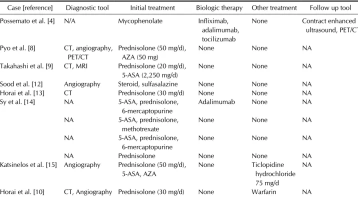
Table 1. The table of cases with TA and UC in terms of diagnostic tool, treatment and follow up
Case [reference] Diagnostic tool Initial treatment Biologic therapy Other treatment Follow up tool Possemato et al. [4] N/A Mycophenolate Infliximab,
adalimumab, tocilizumab
None Contract enhanced ultrasound, PET/CT Pyo et al. [8] CT, angiography,
PET/CT
Prednisolone (50 mg/d), AZA (50 mg)
None None NA
Takahashi et al. [9] CT, MRI Prednisolone (20 mg/d), 5-ASA (2,250 mg/d)
None None NA
Sood et al. [12] Angiography Steroid, sulfasalazine None None NA
Horai et al. [13] CT Prednisolone (30 mg/d) None None NA
Sy et al. [14] NA 5-ASA, prednisolone,
6-mercaptopurine
Adalimumab None NA
NA 5-ASA, prednisolone,
methotrexate
None None NA
NA 5-ASA, prednisolone,
6-mercaptopurine
None None NA
NA Prednisolone None None NA
Katsinelos et al. [15] Angiography Prednisolone (50 mg/d), 5-ASA, AZA
None Ticlopidine
hydrochloride 75 mg/d
NA
Horai et al. [10] CT, Angiography Prednisolone (30 mg/d) None Warfarin NA
Note that we have utilized PET/CT as an assessment and follow up tool, for the co-existence of TA and UC. TA: Takayasu arteritis, UC: ulcerative colitis, NA: not available, PET/CT: positron emission tomography/computed tomography, AZA: azathioprine, ASA:
amino salicylic acid.
ministered infliximab at a dose of 5 mg/kg, as induction therapy at 0, 2, and 6 weeks, and continued treatment ev- ery 8 weeks.
The clinical symptoms including neck pain, headache, claudication of upper extremities, hematochezia, and ab- dominal pain have been all improved after induction ther- apy of infliximab. Moreover, ESR and CRP fell from 61 mm/hour to 17 mm/hour and 12 mg/dL to 0.1 mg/dL, re- spectively after 3 months.
Follow-up PET/CT and sigmoidoscopy were performed 3 months after infliximab use. The increased FDG up- takes on both carotid arteries, subclavian arteries and the aorta seen on previous scan significantly decreased on the follow-up scan (Figure 2D). On the follow-up sigmoido- scopy, the friable, inflamed mucosa showed a significant improvement with no sign of spontaneous bleeding (Figure 2C).
In short, clinical symptoms, laboratory data, PET/CT and endoscopy are all improved. Up to the time of writing, the patient has been receiving infliximab every 8 weeks as maintenance dose, with no worsening of symptoms.
DISCUSSION
It has been well known that immunologic abnormalities are present in TA and UC [8]. In TA, natural killer-cell and T cells infiltrate the aorta, and the levels of several cyto- kines such as TNF, interleukin (IL)-6, or IL-18 increase.
These cytokines also increase in UC patients, implying that UC is also associated with autoimmune phenomena [1]. Among that cytokines, TNF is targeted by TNF inhibitor. TNF inhibitor is known as an effective treat- ment for TA [1]. Also, it, particularly infliximab, can be used for treating the active or glucocorticoid-refractory UC [2]. In this case, the TA and UC were treated together, using infliximab.
UC can occur together with other autoimmune diseases, not only TA but also lupus erythematosus, autoimmune hemolytic anemia, chronic hepatitis and so on [9]. Hence we should approach UC patients systemically, not just concentrating on one part of the body [9]. Indeed the fe- male patient described here was initially diagnosed as UC and only diagnosed as TA 3 years later on the basis of her neck pain and headache. At the time of diagnosis, the pa- tient in this case was revealed to have positive P-ANCA.
This result can be associated with UC [10,11]. High prev- alence of P-ANCA about 40 to 60% in UC has been re- ported [11]. The cases about coexistence of TA and UC
are rarely reported over the world and most of them are Japanese. In Korea, there are only a few cases, including the case reported by Pyo et al. [8]. To the patient with both TA and UC, we used PET/CT for assessing not only the disease activity but also treatment response, which is the unique point of our report. Other cases are summar- ized at the Table 1 including diagnostic, treatment and fol- low up tool [4-10,12-15].
The disease activity of TA was evaluated from 18F-FDG PET/CT, it showed good performance in the assessment of vessel inflammation.
We conclude that PET/CT can be a good tool for diag- nosing and assessing the treatment response in TA patients.
SUMMARY
Biological treatments should be considered for UC and TA patients refractory to current immunosuppressive treatments. The PET/CT could be used to assess TA and its response to treatment.
CONFLICT OF INTEREST
No potential conflict of interest relevant to this article was reported.
REFERENCES
1. Clifford A, Hoffman GS. Recent advances in the medical management of Takayasu arteritis: an update on use of bio- logic therapies. Curr Opin Rheumatol 2014;26:7-15.
2. Ford AC, Sandborn WJ, Khan KJ, Hanauer SB, Talley NJ, Moayyedi P. Efficacy of biological therapies in inflammatory bowel disease: systematic review and meta-analysis. Am J Gastroenterol 2011;106:644-59.
3. Alibaz-Oner F, Aydin SZ, Direskeneli H. Advances in the di- agnosis, assessment and outcome of Takayasu's arteritis.
Clin Rheumatol 2013;32:541-6.
4. Possemato N, Macchioni P, Germanò G, Pipitone N, Versari A, Salvarani C. Clinical images: PET-CT and contrast- en- hanced ultrasound in Takayasu's arteritis. Rheumatology (Oxford) 2014;53:447.
5. Alibaz-Oner F, Dede F, Ones T, Turoglu HT, Direskeneli H.
Patients with Takayasu's arteritis having persistent acute- phase response usually have an increased major vessel up- take by 18F-FDG-PET/CT. Mod Rheumatol 2015;25:752-5.
6. Arend WP, Michel BA, Bloch DA, Hunder GG, Calabrese LH, Edworthy SM, et al. The American College of Rheuma- tology 1990 criteria for the classification of Takayasu arteritis. Arthritis Rheum 1990;33:1129-34.
7. Mason JC. Takayasu arteritis--advances in diagnosis and management. Nat Rev Rheumatol 2010;6:406-15.
8. Pyo JY, Park JS, Song CH, Lee SW, Park YB, Lee SK.
Takayasu arteritis associated with ulcerative colitis and op- tic neuritis: first case in Korea. Korean J Intern Med 2013;
28:491-6.
9. Takahashi N, Tanabe K, Sugamori T, Sato M, Kitamura J, Sato H, et al. Association between Takayasu arteritis and ul- cerative colitis - case report and review of serological HLA analysis. Med Sci Monit 2011;17:CS81-4.
10. Horai Y, Miyamura T, Shimada K, Takahama S, Minami R, Yamamoto M, et al. A case of Takayasu's arteritis associated with human leukocyte antigen A24 and B52 following reso- lution of ulcerative colitis and subacute thyroiditis. Intern Med 2011;50:151-4.
11. Schulte-Pelkum J, Radice A, Norman GL, Lόpez Hoyos M, Lakos G, Buchner C, et al. Novel clinical and diagnostic as- pects of antineutrophil cytoplasmic antibodies. J Immunol
Res 2014;2014:185416.
12. Sood A, Midha V, Sood N, Bansal M. Coexistence of Takayasu's arteritis with ulcerative colitis. J Assoc Physi- cians India 2006;54:151-2.
13. Horai Y, Satoru O, Lapalme-Remis S, Sumiyoshi R, Naka- shima Y, Suzuki T, et al. Takayasu arteritis developing dur- ing treatment of ulcerative colitis with infliximab. Mod Rheumatol 2013;23:572-6.
14. Sy A, Khalidi N, Dehghan N, Barra L, Carette S, Cuthbertson D, et al. Vasculitis in patients with inflammatory bowel dis- eases: A study of 32 patients and systematic review of the literature. Semin Arthritis Rheum 2016;45:475-82.
15. Katsinelos P, Tsolkas P, Beltsis A, Chatzimavroudis G, Papaziogas B, Kapelidis P, et al. Takayasu's arteritis and ul- cerative colitis in a young non-Asian woman: a rare asso- ciation. Vasa 2005;34:272-4.