대한치주과학회지 : Vol. 35, No. 4, 2005
The Effect of single Low-Power CO 2 Laser irradiation on human PDL fibroblast Proliferation & Differentiation
In-Kwon Jang
1,2ㆍTae-Gyun Kim
1,2ㆍUi-Won Jung
1,2Chang-Sung Kim
1,2,3ㆍSeong-Ho Choi
1,2,3ㆍKyoo-Sung Cho
1,2,3Jung-Kiu Chai
1,2ㆍChong-Kwan Kim
1,2,31Department of Periodontology, College of Dentistry, Yonsei University,
2Reasearch Institute for Periodontal Regeneration,
3Brain Korea 21 project for Medical Science
Ⅰ. Introduction
Laser is widely used in oral surgery1,2), endodontics, periodontology3) and restorative dentistry4). Lasers can be classified as sur- gical(high power) and non-surgical(low power) according to therapeutic purposes. Parti- cularly low-power laser, has been increa- singly used for the treatment of injuries of soft and hard tissue.1)
Low-power lasers are widely used as ti- ssue stimulator, to improve wound repair and anti-inflammatory and analgesic effects.
5,6)Experimental reports have also suggested that laser would accelerate wound repair.
6~11) This effect could be explained by chan-
ge in mitotic acivity.12,13) Favourable results were achieved in examinations of hard ti- ssue, as bone fractures in mice showed a faster formation of bone tissue with a tighter mesh of trabeculae after three weeks of daily irradiation with Helium-Neon la- ser.14) And Ozawa et al.(1995) achieved a significant increase in the total area of bone nodules with a Gallium-Aluminium -Arse- nide laser(GaAlAs) in a dose-dependent man- ner.15)(10.8-108J/cm2/day) Another studies re- port that low-power laser promotes growth and differentiation of cells through many experiments.6,12,16~21) This shows that low- power laser may give positive effects in re- generation of periodontal tissue by pro-
*This study was supported by a grant of the Korea Health 21 R&D Project, Ministry of Health & Welfare, Republic of Korea(03-PJ1-PG1-CH08-0001)
*Corresponding author: Chong-Kwan Kim. Department of Periodontology, College of Dentistry, Yonsei University, Shinchon-dong, Seodaemun-gu, Seoul, 120-752, Korea
moting growth and differentiation of cells related to regeneration, especially hard tissue regeneration, and this could be applied in destroyed periodontal tissue.22~24)
The energy of wavelength of commercially available CO2laser is 10.6 ㎛, which falls in the far infrared range. This particular wavelength of energy is effectively absorbed by water. Consequently, the CO2laser would appear to be the best choice for use on hyd- rated, composite mineralized tissue such as bone.25) Also, low-power laser therapy using CO2lasers is known to activate surrounding cells and tissue. Shiozaki et al.(2005) achi- eved that low-power CO2lasers induced not only mineralization but also osteoblast dif- ferentiation.26) And Tajima et al.(2003) re- ported that low-power CO2 laser accelerates new bone formation within the marrow cavity subjacent to the laser treatment site.27) In spite of successful clinical and experimental results, the mechanisms un- derlying low-power CO2laser effects are still poorly understood.
The aim of this study is to evaluate the effect of low-power CO2laser in proliferation of cultured human PDL fibroblast and to further understand laser effect in perio- dontal regeneration. In addition, this study tried to find out the most effective degree of energy and power density in which cell pro- liferates and differentiates to osteoblastic cells after laser irradiation. Through this, we intend to make the base of clinical perio- dontal regeneration procedure by low-power
CO2laser therapy.
Ⅱ. Materials and Methods
1. PDL cell separation and culture
The human PDL fibroblast was gathered and cultured from a healthy premolar extracted for orthodontic treatment. Before extraction, plaque and calculus were re- moved with the use of a periodontal curette.
The extracted tooth was then rinsed with HBSS to remove the blood and other foreign bodies, the PDL cells were collected from the middle portion of root surface.
ɑ-MEM including 10% fetal bovine serum, 100U/ml penicillin, 100mg/ml streptomycin and 0.5mg/ml amphotericin-B was used as the culture media with an environment of 3 7℃, 100% humidity and 5% CO2. Culture medium was changed every 2 or 3 days.
2. Experiment design
The experiments were divided into 1) MTT assay, 2) ALP detection assay and 3) ALP activity assay. The control group was not applied to laser. Experimental groups were divided into 4 groups by applying different irradiating distances from cultured cell to laser tip; 2Cm(Focal spot=∅0.74mm), 3Cm(Focal spot=∅1.04mm) and different irra- diating time; 1 second, 3 seconds.
3. Application of Laser
2)
Laser-treated specimens were irradiated with a CO2 laser‡ using a focused beam of 0.74, 1.04mm diameter focal spot, wave- length of 10.6 ㎛. Laser parameters were 0.5W of power delivered at 50Hz with con- tinuous mode. The operator wore the pro- tection glasses for the risk of damaging the eyes due to the direct vision of the laser and the refraction of the beam.
4. MTT assay
After seeding PDL fibroblast at 96 well plate, the cells were cultured it in α-MEM including 5% FBS and was made sure that cells were attached to the plate. One day after seeding, laser was irradiated and MTT assay was done every 0,3, and 5 days later.
Different laser treatment conditions were applied with different distances from cul- tured cell to laser tip and irradiating time.
The distances were 2cm and 3cm with time condition of 1 second, 3 second. And the control group was not exposed to laser.
The samples were measured by using cell proliferation assay kit(CHEMICON International Inc, Temecula, CA, USA) on absorbant of 570 nm wave, and prior to measuring, the cul- tured cells were incubated by removing reagent out of the kit as maker allowed.
5. ALP detection assay
After seeding PDL fibroblast at 24 well
plate, the cells were cultured it in α-MEM including 10% FBS till it become semi- confluenced. Laser was applied and ALP was detected after 0,3, and 15 days. At this time different laser treatment condition was used. Different laser treatment conditions were applied with different distances from cultured cell to laser tip and irradiating time. The distances were 2cm and 3cm with time condition of 1 second, 3 second. And the control group was not exposed to laser.
Detection was done by using TRACP &
ALP double-stain kit(Takara Bio Inc, Seta 3-4-1, Otsu, Shiga 520-2193, japan)and samples were viewed under light microscope. Before sample detection, the cells were incubated by removing the reagent out of the kit as the manufacturer allowed. And the control group was not exposed to laser.
The samples obtained in accordance to time were recorded by photographs.
6. ALP activity assay
After seeding PDL fibroblast at 6 well plate, the cells were cultured in α-MEM including 10% FBS till it become semi- confluenced. Laser was applied and ALP was detected after 0,3,5,7, and 10 days. At this time different laser treatment condition was used. Apply laser at distance, from cultured cell to laser tip, of 2cm, 3cm and apply each different irradiation time, 1sec, 3sec. And the control group was not exposed to laser.
ALP activity of the cell was measured by the method of Lowry et al.28) with p-nitro-
‡OPELASER O3SII, Yoshida Dental MFG. CO., Japan
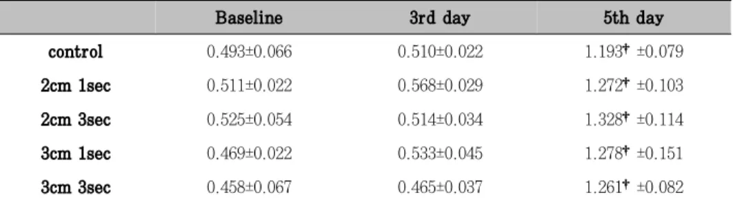
Table 1. MTT assay(group means ± SD; O.D.: optical density)
Baseline 3rd day 5th day
control 0.493±0.066 0.510±0.022 1.193†±0.079
2cm 1sec 0.511±0.022 0.568±0.029 1.272†±0.103
2cm 3sec 0.525±0.054 0.514±0.034 1.328†±0.114
3cm 1sec 0.469±0.022 0.533±0.045 1.278†±0.151
3cm 3sec 0.458±0.067 0.465±0.037 1.261†±0.082
Figure 2. MTT assay (O.D.: optical density)
*: statistically significant differ- ence compared to baseline(p< 0.05) phenyl phosphate as a substrate and was
normalized by the total protein content of the cell which was determined by Bio-Rad Protein assay kit.(BioRad 2000, Alfred Nobel Drive Hercules, CA 94547, USA) Briefly, after finishing the laser irradiation, the medium was removed and the cells were washed twice with Tris-buffered saline. The cells were detached from the culture dish with a scraper after the addition of Tris-buffered saline. ALP activity was then assayed using Alkaline Phosphatate Substrate Kit.(Wako Pure Chemical Industries, Osaka, japan)
7. Statistical Analysis
1-way ANOVA and tukey's test for mul- tiple comparison was used to determine the
statistical significance of MTT assays and ALP activity between different treatment groups at baseline and at indicated time points after laser irradiation. Repeated Measures ANOVA was used to determine the statistical significance of the MTT and ALP activity within groups in comparison with baseline.
Ⅲ. Results
1. MTT assay
On the 5th day after laser irradiation, statistically significant increase of cells were found compared to the baseline.
At the baseline, 3rd day and 5th day of laser irradiation there were no significant
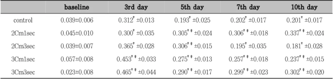
Table 2. ALP activity (group means ± SD; unit/mg)
baseline 3rd day 5th day 7th day 10th day
control 0.039±0.006 0.312†±0.013 0.193†±0.025 0.202†±0.017 0.201†±0.017 2Cm1sec 0.045±0.010 0.300†±0.035 0.305†‡±0.024 0.306†‡±0.018 0.337†‡±0.024 2Cm3sec 0.039±0.007 0.365†±0.028 0.306†‡±0.015 0.195†±0.035 0.181†±0.028 3Cm1sec 0.057±0.008 0.453†‡±0.033 0.275†‡±0.013 0.257†‡±0.018 0.237†‡±0.015 3Cm3sec 0.023±0.008 0.465†‡±0.044 0.290†‡±0.017 0.299†‡±0.023 0.302†‡±0.020
†: statistically significant difference compared to baseline(p<0.05)
‡: statistically significant difference compared to control group(p<0.05) difference of cell increase among the groups
(Table 1, Figure 1).
2. ALP detection assay
On the 3rd and 15th day after laser irradiation, ALP positive cells were found in control group.(arrow head)More ALP positive cells were found in laser irradiation groups compared to control group on the 3rd day.
When it came to the 15th day, much more ALP positive cells were found in 2cm,1sec group than other groups on the 15th day.
(Figure 3, 4, 5)
3. ALP activity
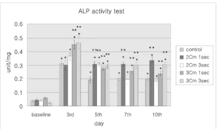
Statistically significant increase of ALP activity was seen on the 3rd, 5th, 7th, and 10th day after laser irradiation compared to the baseline(Table 2). The control and 3cm, 3sec group showed statistically significant decrease of ALP activity between the 3rd and 5th day and no significant increase till the 10th day.
On the other hand 2cm, 3sec and 3cm,
1sec groups showed statistically significant decrease of ALP activity between the 3rd and 5th day, and significant decrease of ALP activity till the 10th day.
Especially 2cm,1sec group showed increase of ALP activity not statistically significant from the 3rd day to the 10th day.
On the 3rd day after laser irradiation 3cm, 1sec and 3cm, 3sec group showed stati- stically significant high score of ALP acti- vity compared to other groups.
On the 5th day after laser irradiation, statistically significant high score of ALP activity was found in experimental groups compared to control group and no statistical difference of ALP activity was found experi- mental groups.
On the 7th and 10th day 3cm,1sec , 3cm, 3sec and 2cm, 1sec group showed signifi- cantly high score of ALP activity compared to 2cm,3sec and control group. And the ALP activity score increased as it went from 3cm, 1sec, 3cm, 3sec to 2cm, 1sec group and there was no significant difference between 3cm, 3sec and 2cm, 1sec group(Table 2. Figure 2).
Figure 3. ALP activity test(unit/mg)
*: statistically significant difference compared to baseline(p<0.05)
**: statistically significant difference compared to control group (p<0.05)
Ⅳ. Discussion
Low-power laser has been widely used in medicine, mostly on pathological tissues pre- senting any degree of alteration, such as healing or inflammatory processes.29) We de- cided to grow the cells in a medium sup- plemented with 5% concentration of FBS.
Serum is an important supplemented for culturing cells,30) and the best growth will occur using medium containing at least 10%
FBS. MTT was performed to observe the growth of PDL cells in this study. And to get the least effect of FBS, medium with 5%
FBS was used to see the effect of laser.
Many studies tell us that low power laser irradiation activates growth of various kinds of cells.2,7-11,31)For example, there is a report that applying laser of 3J/cm2 and 4J/cm2 to cultured fibroblasts(NIH 3T3 cells) twice every six hours resulted in 3 or 6 times increase of cells compared to non-lasered group.6)
Laser of 0.5W power was used for 1 se-
cond and 3 seconds in this study, thus the energy was 0.5J/cm2 and 1.5 J/cm2 respec- tively. This is less energy than usually applied in other in-vitro study. And this may have caused the cells to over stimulate since power was stronger than 0.1W, which is generally used in-vitro study.
As for the effect of single low-power CO2
laser irradiation on cell proliferation, both the experimental groups and control group showed statistically significant increase of cells on the 5th day compared th baseline after laser irradiation and there was no significant difference between experimental group and control group. This means there was a little cell proliferations effect with low-power CO2 laser in this experimental condition despite different where total ener- gy and power density.
2cm and 3cm irradiation distance results in 116.3%(=W/∏r2), and 58.9% of power density respectively according to the rela- tionship between distance of lens and energy
density'. The strength of energy transferred to cells depends on power density even when they have the same energy of laser irra- diation. Actually 1cm distance irradiation group was involved in this study. But cell necrosis occured because of high energy that reached to PDL cells. So 1cm distance irra- diation group could not involve in experi- ments.
It is not yet well understood how low- power laser irradiation actually affects PDL fibroblast's differentiation to osteoblastic cells. However it is Certain that PDL cell has the ability to differentiate into various kind of cells, that is it can change to cells that makes alveolar bone, depending on given conditions. According to the study of Yuya Murakami et al. in 2003, isolating ALP positive subpopulation is available in PDL fibroblast by immunomagnetic method.32) In the preliminary study preparing for this experiment, qualitative analysis of ALP was used as a marker of hard tissue formation.
The medium containing α-MEM with 10%
FBS was used, and ALP positive sample was obtained from contol group and other groups which were irradiated to low-power lasers by different application methods(Figures 3,4,5). This shows the pluripotential characteristic of PDL fibroblast mentioned before.
Generally, in order to see ALP activity from PDL cells, materials like 1α,25-dihy- droxyvitamin D3needs to be added.33) Vari- ous studies have shown that mechanical stress like cyclic stretching decreases the ALP activity.33,34) So it is necessary to find out what kind of stimulus low-power laser irradiation do to PDL cell differentiation,
and what changes the ALP activity and whether there is a difference depending on irradiation method.
In this study, single low-power CO2 laser irradiation may play positive role of PDL cell differentiation to osteoblastic cells. 2cm, 1sec irradiation condition was found to be the most effective condition in this study.
And 3cm, 3sec was also found to be effi- cient. Generally, when various factors relat- ed to laser irradiation are changed, the results of laser irradiation become different.
For example, according to the results of the study by Ueda Y et al. in 2003, low-fre- quency pulsed laser irradiation significan- tly stimulates bone formation. And this proved that the pulse frequency of low- power laser irradiation is an important fac- tor affecting biological responses in bone formation.35) In our study most parameters related to the laser irradiation are constant (e.g., wavelength, power output, irradiation area), except power densities and related exposure time. Distance between cultured cells and laser tip is related to power density of irradiation. And irradiation time is related to total amount of energy that reach to cells. That is, there is low energy if power density is high, and high energy if power density is low thereby more effects to cell differentiation.
This result shows that among various factors, CO2laser irradiation, single or dou- ble irradiation, with less energy than 3,4J/
cm2 at which acceleration of PDL cell proli- feration or synthesis of collagen takes place, differentiation of PDL cells could be acti- vated.
The right time for laser irradiation to achieve the most efficient effect of low- power laser is not yet cleary defined even though many studies on laser have been carried out. A study using rat calvarial cell reported that cell growth and differentiation were affected by laser irradiation at early stages of cell culture.19) Though not all cells behaved in the same manner, we could suggest that external stimulus like low- power laser irradiation to cells at early stage of growth or differentiation could greatly influence to growth and differen- tiation. In addition, this study showed that differentiation of PDL cell to osteoblastic cells was more influences than PDL cell growth was.
Then what controls low-power laser's effect of cell proliferation and differentia- tion? Most studies regarding the laser effect on cells have reported specific intracellular changes.36-38) Theses changes are mostly related to the calcium metabolism, which would be either affecting its concentration or intracytoplasmic transport. Such altera- tions would stimulate cell division, in detri- ment of cell production, which could explain the reason why the cell growth was altered.
However concrete mechanism of laser irra- diation to proliferation and differentiation of cells is not clear. We can guess that heat and photostimulation38,39) given by laser might affect differentiation of cells, and should clarify the effect of laser to differ- entiation of cells through various kinds of experimental design.
In conclusion, it is not possible to explain proper mechanism. But in this study if
single low-power CO2 laser irradiation is done to human PDL fibroblast, cell proli- feration was found in both experimental and control group compared to baseline on the 5th day of laser irradiation. But, no signi- ficant differences were found among groups.
In this study, low energy with high power density and high energy with low power density had more effects to cell differen- tiation. Additionally, statistically significant increase of ALP activity was found in 2cm, 1sec and 3cm, 3sec groups till the 10th day, and it was found to be the most effective way to PDL cell's differentiation into osteo- blastic cells.
Ⅴ. Conclusion
The aim of this study is to evaluate the effect of low-power CO2laser in growth and proliferation of cultured human PDL fibro- blast in vitro and to understand better about the effect of laser on periodontal rege- neration. The study also tried to find out the most effective degree of energy and power density where cell proliferation and differentiation to osteoblastic cells highly takes place after laser irradiation.
The experimental groups were divided into 4 groups by applying different irradiating distances from cultured cell to laser tip;
2Cm(Focal spot=∅0.74mm), 3Cm(Focal spot=∅
1.04mm) and different irradiating time; 1 second, 3 seconds. And they were applied to laser at a power of 0.5W with 50Hz under continuous mode. The control group was not applied to laser. MTT and ALP activity test were performed to observe the growth of
PDL cells and cell differentiation to osteo- blastic cells. The result are as follows.
1. On the 5th day after laser irradiation, statistically significant increase of cells were found in all groups. But, no sig- nificant differences were found among groups.
2. Statistically significant increase of ALP activity was seen on the 3rd, 5th, 7th, and 10th day after laser irradiation compared to the baseline. Among this, there were highest ALP activity on 3rd day except 2cm,1sec group. 2cm,1sec group showed increase of ALP activity not statistically significant from the 3rd day to 10th day.
On the 7th and 10th day 3cm, 1sec, 3cm, 3sec and 2cm, 1sec group showed significant high score of ALP activity compared to 2cm, 3sec and control group.
In this study, there was a weak effect of low-power CO2laser on cell proliferation but there was a better effect on cell differen- tiation. 2cm, 1sec irradiation was found to be the most effective condition for PDL cell differentiation in this study and 3cm, 3sec was found to be efficient.
Ⅵ. Reference
1. Kucerova H, Dostalova T, Himmlova L, Bartova J, Mazanek J : Low-level laser therapy after molar extraction. J Clin Laser Med Surg. 18(6);309-15, 2000.
2. Saito S, Shimizu N : Stimulatory effects of low-power laser irradiation on bone
regeneration in midpalatal suture during expansion in the rat. Am J Orthod Dentofacial Orthop. 111(5);525-32,1997.
3. Liu CM, Hou LT, Wong MY, Lan WH : Comparison of Nd:YAG laser versus scaling and root planing in periodontal therapy. J Periodontol. 70(11);1276-82, 1999.
4. Ramos RP, Chinelatti MA, Chimello DT, Borsatto MC, Pecora JD, Palma-Dibb RG : Bonding of self-etching and total- etch systems to Er:YAG laser-irradiated dentin. Tensile bond strength and scan- ning electron microscopy. Braz Dent J.
15;19-20,2004
5. Choi KH, Im SU, Kim CS, Choi SH, Kim CK : Effect of the carbon dioxide laser on the clinical parameters and crevi- cular IL-1ß when used as an adjunct to gingival flap surgery. Journal of the International Academy of Periodontology 6(1);29-36, 2004.
6. Pereira AN, Eduardo Cde P, Matson E, Marques MM : Effect of low-power laser irradiation on cell growth and procol- lagen synthesis of cultured fibroblasts.
Lasers Surg Med. 31(4);263-7, 2002.
7. Almeida-Lopes L, Velez-Gonzalez M, Brugnera Jr.A, Pinheiro AB : The use low level laser therapy for wound heal- ing: clinical study. In: Annual Meeting- Lasers in Surgery and Medicine, Pro- ceedings. Florida, EUA, 16-18, 1999.
8. Enwemeka CS, Rodriguez O, Gall N, Walsh N : Morphometries of collagen fibril populations in He-Ne laser photo- stimulated tendons. J Clin Laser Med Surg Dic;47-52, 1990.
9. Mester AF, Mester A : Wound healing.
Laser Therapy 1;7,1989.
10. Yamamoto Y, Kono T, Kotani H, Kasai S, Mito M : Effect of low-power laser irradiation on procollagen synthesis in human fibroblasts. J Clin Laser Med Surg. 14(3);129-32,1996.
11. Yew DT, Li WWY, Pang KM, Mok YC, Au C : Stimulation of collagen formation in the intestinal anastomosis by low dose He-Ne laser. Scanning Microsc 3(1);379-386, 1989.
12. Pourreau-Schneider N, Soudry M, Re- musat M, Franquin JC, Martin PM : Modifications of growth dynamics and ultrastructure after helium-neon laser treatment of human gingival fibroblasts.
Quintessence Int. 20(12);887-93, 1989.
13. van Breugel HH, Dop Bar PR : Power density and exposure time of He-Ne- laser irradiation are more important than total energy dose in photo-biomo- dulation of human fibroblasts in vitro.
Lasers Surg Med 12;528-537, 1992.
14. Trelles MA, Mayayo E : Bone fracture consolidates faster with low-power laser.
Lasers Surg Med. 7(1);36-45, 1987.
15. Ozawa Y(1995): Stimulatory effects of low power laser irradiation on bone formation in vitro. SPIE Proceeding series Washington. Vol. 1984; 281-288 16. Almeida-Lopes L, Rigau J, Zangaro RA,
Guidugli-Neto J, Jaeger MM : Compa- rison of the low level laser therapy effects on cultured human gingival fibro- blasts proliferation using different irra- diance and same fluence. Lasers Surg Med. 29(2);179-84, 2001.
17. Dortbudak O, Haas R, Mallath-Pokorny G : Biostimulation of bone marrow cells with a diode soft laser. Clin Oral Im- plants Res. 11(6);540-5, 2000.
18. Kreisler M, Christoffers AB, Willer- shausen B, d'Hoedt B : Effect of low- level GaAlAs laser irradiation on the proliferation rate of human periodontal ligament fibroblasts: an in vitro study.
J Clin Periodontol. 30(4);353-8, 2003.
19. Ozawa Y, Shimizu N, Kariya G, Abiko Y : Low-energy laser irradiation stimula- tes bone nodule formation at early stages of cell culture in rat calvarial cells. Bone. 22(4);347-54, 1998.
20. Rayan GM, Pitha JV, Edwards JS, Everett RB : Effects of CO2 laser beam on cortical bone. Lasers Surg Med. 11 (1);58-61, 1991.
21. Ueda Y, Shimizu N : Pulse irradiation of low-power laser stimulates bone no- dule formation. J Oral Sci. 43(1);55-60, 2001.
22. Crespi R, Covani U, Margarone JE, Andreana S : Periodontal tissue regene- ration in Beagle dogs after laser the- rapy. Lasers Surg Med 12;5-15, 1997.
23. Mognato M, Squizzato F, Facchin F, Zaghetto L, Corti L : Cell growth modu- lation of human cells irradiated in vitro with low-level laser therapy. Photomed Laser Surg. 22(6);523-6, 2004
24. Stein A, Benayahu D, Maltz L, Oron U : Low-level laser irradiation promotes proliferation and differentiation of hu- man osteoblasts in vitro. Photomed La- ser Surg. 23(2);161-6, 2005.
25. Friesen LR , Cobb CM, Rapley JW,
Brockman LF, Spencer P : Laser irra- diation of bone: II. Healing response following treatment by CO2and Nd:YAG lasers. J Periodontol 70;75-83, 1999.
26. Shiozaki H, Yokose S, Saitou M, Suzuki Y, Hirose N, Shimizu H, Masuda K, Hasegawa A, Katayama T, Okumura Y : Carbon dioxide laser irradiation stimu- lates bone formation in rat tibiae. J.
Jpn. Soc. Laser Dent. 16;23-30, 2005.
27. Tajima N : In vivo effects of carbon dioxide laser irradiation on bone forma- tion in rat tibiae. J. Jpn. Soc. Laser Dent. 14;32-43, 2003.
28. Lowry, O H , Rosebrough, N J, Farr, A L , Randall, R I : Protein measurements with folin phenol reagent. J Biol Chem 193;265-75, 1951.
29. Walsh LJ : The current status of low level laser therapy in dentistry. Part 1.
Soft tissue applications. Aust Dent J.
42(4);247-54, 1997.
30. Freshney RI : Quantification and expe- rimental design. In: Freshney RI, edi- tor. Culture of animal cells. A manual of basic technique. 2nd edn. New York:
Wiley-Liss. 227-244, 1991.
31. Barushka O, Yaakobi T, Oron U : Effect of low-energy laser(He-Ne) irradiation on the process of bone repair in the rat tibia. Bone. 16(1);47-55, 1995.
32. Murakami Y, Kojima T, Nagasawa T, Kobayashi H, Ishikawa I : Novel isola- tion of alkaline phosphatase-posi- tive subpopulation from periodontal ligament fibroblasts. J Periodontol. 74(6);780-6, 2003.
33. Chiba M, Mitani H : Cytoskeletal chan- ges and the system of regulation of alkaline phosphatase activity in human periodontal ligament cells induced by mechanical stress. Cell Biochem Funct.
22(4);249-56, 2004.
34. Yamaguchi M, Shimizu N, Shibata Y, Abiko Y : Effects of different magni- tudes of tension-force on alkaline pho- sphatase activity in periodontal ligament cells. J Dent Res. 75(3);889-94, 1996.
35. Ueda Y, Shimizu N : Effects of pulse frequency of low-level laser therapy (LLLT) on bone nodule formation in rat calvarial cells. J Clin Laser Med Surg.
21(5);271-7, 2003.
36. Friedmann H, Lubart R, Laulicht I : A possible explanation of laser-induced stimulation and damage of cell cultures.
Photochem Photobiol. 11;87-95, 1991.
37. Loevschall H, Arenhilt-Bindslev D : Effect of low level diode laser irradia- tion of human oral mucosa fibroblasts in vitro. Lasers Surg Med. 14;347-354, 1995.
38. Lubart R, Friedmann H, Peled I, Grossman N : Light effect on fibroblast proliferation. Laser Therapy. 5;55-57, 1993.
39. Sedlmaier B, Franke A, Sudhoff H, Jovanovic S, Haisch A : Photodynamic effect of argon and diode laser on cholesteatoma cell cultures after intra- vital staining with absorption enhan- cers. Lasers Med Sci. 19(4);248-56, 2005
사진 부도 설명
Figure 3. On the baseline after laser irradiation, ALP positive cells were barely seen.(light microscope × 100)
Figure 4. A: 3rd day control group
B: 3rd day 2cm,1sec group , C: 3rd day 3cm,1sec group D: 3rd day 2cm,3sec group , E: 3rd day 3cm,3sec group
On the 3rd day after laser irradiation, ALP positive cells were found in con- trol group.(A: arrow head) More ALP positive cells were found in laser irra- diation groups compared to control group.(B,C,D,E)(light microscope × 100)
Figure 5. A: 15th day control group
B: 15th day 2cm,1sec group , C: 15th day 3cm,1sec group D: 15th day 2cm,3sec group , E: 15th day 3cm,3sec group
15th day control group(A) showed similar aspect as 3rd day control group.(Figure 4-A) More ALP positive cells were found in experimental groups(B,C,D,E) compared to control group.(A) And much more ALP pos- itive cells were found in 2cm,1sec group(B) among other experimental groups(light microscope × 100).
사진부도(Ⅰ)
Figure 3.
Figure 4.
A
B
D
C
E
사진부도(Ⅱ)
Figure 5.
A
B
D
C
E
-국문 초록-
단일조사 저출력 CO 2 Laser가 치주인대 섬유아세포의 세포 증식능과 세포 분화에 미치는 영향
장인권1,2․김태균1,2․정의원1,2․김창성1,2,3․최성호1,2,3․조규성1,2,3․채중규1,2․김종관1,2,3
1연세대학교 치과대학 치주과학교실,2치주조직 재생연구소
3BK21 의과학 사업단
이 논문은 단일조사 저출력 CO2laser조사가 치주인대 섬유아세포의 증식과 분화에 미치는 영향을 살펴보 고 가장 효과적인 에너지와 파워밀도(power density)를 알아보기 위하여 다음과 같이 실험하였다.
0.5W 출력, 10.6 ㎛ 파장, 50 Hz 연속형 CO2laser를 사용하여, 실험군은 laser tip과 배양된 세포 사이 의 거리를 2cm, 3cm으로 나누고, 조사시간을 1초, 3초로 나누어 4개의 군으로 설정하였고 대조군은 laser를 조사하지 않은 군으로 하였다. 치주인대 섬유아세포의 증식정도와 골모세포로의 분화정도를 보기 위하여 각각 MTT 실험과 ALP activity 실험을 시행하여 다음과 같은 결과를 얻었다.
1. Laser를 조사하고 난 후 5일째에, 모든 군에서 유의하게 세포가 증식되는 것을 확인할 수 있었고 조사방 법간에 유의한 차이가 없었다.
2. 대조군과 실험군에서 0일째에 비하여 3일째, 5일째, 7일째, 10일째에 통계적으로 유의 하게 ALP activity가 증가하였고, 이중 2cm,1sec 군을 제외하면 3일째에서 가장 높은 ALP activity 값을 보였 다. 특징적으로 2cm,1sec 군은 3일째부터 10일까지 통계적으로 유의하지는 않지만 시간이 지남에 따라 서 ALP activity가 증가함을 보였다. 7일과 10일째에는 2cm,1sec, 3cm,3sec군에서 다른 군에 비하여 큰 activity값을 보였다.
이번실험에서 저출력 CO2laser 조사는 세포의 증식보다는 분화에 더 큰 영향을 끼쳤고, 2cm, 1sec, 3cm, 3sec 군이 치주인대 섬유아세포의 분화에 가장 효과적인 laser 조사방법으로 분석되었다.3)
*주요어: CO2laser, 치주인대 섬유아세포, 세포증식, 세포분화, 파워밀도, 에너지