Received March 11, 2016, Revised July 22, 2016, Accepted for publication July 27, 2016
Corresponding author: In-Suk Kwak, Department of Anesthesiology and Pain Medicine, Hallym University Hangang Sacred Heart Hospital, 12 Beo- deunaru-ro 7-gil, Yeongdeungpo-gu, Seoul 07247, Korea. Tel: 82-2- 2639-5500, Fax: 82-2-2678-4386, E-mail: [email protected]
Hye One Kim, Department of Dermatology, Hallym University Kangnam Sacred Heart Hospital, 1 Singil-ro, Yeongdeungpo-gu, Seoul 07441, Korea.
Tel: 82-2-829-5221, Fax: 82-2-832-3237, E-mail: [email protected] This is an Open Access article distributed under the terms of the Creative Commons Attribution Non-Commercial License (http://creativecommons.
org/licenses/by-nc/4.0) which permits unrestricted non-commercial use, distribution, and reproduction in any medium, provided the original work is properly cited.
Copyright © The Korean Dermatological Association and The Korean Society for Investigative Dermatology
Ann Dermatol Vol. 29, No. 2, 2017 https://doi.org/10.5021/ad.2017.29.2.194
ORIGINAL ARTICLE
Serial Changes of Heat Shock Protein 70 and Interleukin-8 in Burn Blister Fluid
Kicheol Yoo, Kang Yeol Suh, Gi Hun Choi, In-Suk Kwak1, Dong Kook Seo2, Dohern Kym3, Hyeon Yoon4, Yong Se Cho5, Hye One Kim5
Departments of Emergency Medicine, 1Anesthesiology and Pain Medicine, 2Plasticsurgery, 3Surgery, and 4Burn Institute, Hangang Sacred Heart Hospital, Hallym University College of Medicine, 5Department of Dermatology, Kangnam Sacred Heart Hospital, Hallym University College of Medicine, Seoul, Korea
Background: It has been reported that heat shock protein 70 (HSP70) and interleukin-8 (IL-8) play an important role in cells during the wound healing process. However, there has been no report on the effect of HSP70 and IL-8 on the blisters of burn patients. Objective: This study aimed to evaluate the serial quantitative changes of HSP70 and IL-8 in burn blisters.
Methods: Twenty-five burn patients were included, for a total of 36 cases: twenty cases on the first day, six cases on the sec- ond, five cases on the third, three cases on the fourth, and two cases on the fifth. A correlation analysis was performed to de- termine the relationship between the concentration of HSP70 and IL-8 and the length of the treatment period.
Results: The HSP70 concentration was the highest on the first day, after which it decreased down to near zero. Most HSP70 was generated during the first 12 hours after the burn accident. There was no correlation between the concen- tration of HSP70 on the first day and the length of the treat- ment period. No measurable concentration of IL-8 was de- tected before 5 hours, but the concentration started to in-
crease after 11 hours. The peak value was measured on the fourth day. Conclusion: While HSP70 increased in the first few hours and decreased afterwards, IL-8 was produced after 11 hours and increased afterward in burn blister fluid. These findings provide new evidence on serial changes of in- flammatory mediators in burn blister fluid. (Ann Dermatol 29(2) 194∼199, 2017)
-Keywords-
Heat-shock protein 70, Interleukin-8, Partial thickness burn, Partial thickness burn blister, Wounds and injuries
INTRODUCTION
Burn wound healing is a complex process that involves various mediators and multiple cell types around the in- jured zone. A second-degree burn, also called a partial thickness burn, causes the skin to blisters. Burn blisters flu- id might be important roles in wound healing process1. Some reports indicate that retention fluid from blister of partial thickness burn, which contains relatively large amounts of cytokines and soluble mediator, may affect the wound healing process2-4. However, the effect of burn blister fluid on wound healing has not been fully explored.
Heat shock protein 70 (HSP70, molecular weight 70 kDa) is the most well understood member of the heat shock family of proteins. HSP is found in all cells and has a chaperone function during the assembly process of pro- tein folding and polypeptide formation as well as a cell protection function. Specifically, extracellular HSP70 is in- volved in intercellular signaling and is involved in the in- nate, adaptive immunologic response, interrupts apopto- sis, and protects cell structure as a chaperone5-7. HSP70
can both induce and arrest inflammatory reactions in ex- perimental brain injury and ischemia. It is reported that HSP70 is involved in the healing process, as it has been found in the epidermis and dermis of thermal injury pa- tients8-10. If HSP70 is present in burn blisters, it likely af- fects the damaged cells of thermal injury patients.
Inflammation is an integral phase of wound healing. The local inflammatory response of a skin wound is charac- terized by increased amounts of several pro-inflammatory cytokines, such as interleukin-8 (IL-8), tumor necrosis fac- tor-α, and prostaglandin E2, and local neutrophil infiltra- tion11. Excessive and ongoing wound inflammation in- hibits the healing of burn wounds by fostering an una- bated influx of leukocytes to the wound site12, which di- rectly contributes to the progressive deepening and ex- tension of the wound13. This inflammatory response is am- plified by an elevated IL-8 level in the wound tissue14. However, IL-8 has also been shown to act on epithelial cells or endothelial cells to promote migration, invasion, proliferation, and angiogenesis in vivo and enhanced wound healing due to induced reepithelialization15-17. There are various opinions regarding the treatment meth- od for the blisters of partial thickness burns, including re- moving the blister roof, extracting the fluid maintaining the blister roof, or leaving the blister intact18-20. A better understanding of the changes in heat shock proteins and pro-inflammatory factors over time is expected to lead to an improved blister management strategy.
This study aimed to determine whether HSP70 and IL-8 are present in burn blisters, the variation in their concen- trations over time, and the influence of HSP70 concen- tration on the length of the treatment period.
MATERIALS AND METHODS
Study subjects
For this study, patients with partial thickness burn with bullae who were aged 18 years and over and agreed to cooperate during the period from October 1, 2013 to March 31, 2014 were selected. Patients with immune-re- lated diseases, with diseases that can induce an immune unbalance, and who were undergoing cancer treatment or other hormonal treatments were excluded. For all patients, information regarding sex, age, treatment period, total burn surface area (TBSA), and the abbreviated burn severity in- dex (ABSI) score was obtained. The protocol for this study was reviewed and approved by the institutional Ethics Committee for human studies of Hangang Sacred Heart Hospital, Burn Center, Seoul, Korea (2013-068). We con- sidered p-values<0.05 to be statistically significant.
Collection of blister fluid
After informed consent was obtained, blister fluid was as- pirated from the intact burn blisters of patients with ther- mal burns who were admitted to our Emergency Department of Hangang Sacred Heart Hospital. Aspiration of the blis- ter fluid was performed with an 18-gauge needle. The bul- lous fluid was first collected while leaving the blister roof intact. On the next visit, the fluid from the same bullae was collected if fluid remained. The collected amount for each case was 1 ml. We set the time after injury to sam- pling as the collected time. The first day was defined as the time between 1 hour and 12 hours after the damage occurred, the second day was between 25 and 36 hours, the third day was between 49 and 60 hours, the fourth day was between 73 and 84 hours, and the fifth day was between 97 and 108 hours. To ensure consistent con- ditions, any case that did not meet the criterion was ex- cluded from the analysis. The end of the treatment period was determined by a plastic surgeon according to the treatment results.
Heat shock protein 70 and interleukin-8 quantification in blister fluid
The concentrations of HSP70 and IL-8 were measured us- ing enzyme-linked immunosorbent assay (ELISA) kits.
ELISA was used to measure IL-8 and HSP70 in the bullae of burn patients. The quantitative measurement of IL-8 and HSP70 was performed in duplicate samples using com- mercial ELISA kits (Cat. No. KAC1301; BioSource, Nivelles, Belgium) for IL-8 and HSP70 (Cat. No. ab13306; Abcam, Cambridge, UK) according to the manufacturer’s instructions.
Each sample was diluted 100-fold (HSP70) or 5-fold (IL-8) with dilution solution, and the samples were centrifuged for 5 minutes at 4oC and 12,000 rpm to remove other de- bris and blood cell components. Then, 100 μl of the standard and bullae samples were placed in an anti- gen-coated 96-well plate and left for 2 hours at room tem- perature, after which they were washed four times with 400 μl washing solution. Next, anti-HSP70 and anti-IL-8 conjugates were placed in the wells followed by in- cubation for 2 hours at room temperature. After washing, each well was filled with a freshly prepared chromogenic solution, and the plate was incubated for 30 minutes.
Finally, the reaction was stopped with by adding stop sol- ution (1.8 N H2SO4), and the plate was read at 450 nm us- ing a DTX880 multimode detector (Beckman Coulter, Brea, CA, USA).
Statistical analysis
All statistical analyses were conducted using PASW Statis-
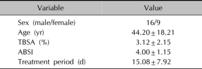
Fig. 1. (A) The heat shock protein (HSP) concentration variation within partial thickness burn blister. The HSP70 concentration was the highest on the first day (up to 12 hours after the damage). Then, the value drastically dropped down to near zero on the second day. There was a small increased on the third day and no significant amount was found on the fourth and fifth day. (B) The concentration of HSP70 within partial thickness burn blister of up to 12 hours after damage. The HSP70 concentration after one hour of the damage was measured to be the highest. After one hour, the concentration drops rapidly.
Table 1. Dermographic data
Variable Value
Sex (male/female) 16/9
Age (yr) 44.20±18.21
TBSA (%) 3.12±2.15
ABSI 4.00±1.15
Treatment period (d) 15.08±7.92
Values are presented as number only or mean±standard devaition. TBSA: total burn surface area, ABSI: abbreviated burn severity index.
tics 18.0 (IBM Co., Armonk, NY, USA). The results were ex- pressed as means±standard deviations or median values and interquartile range (IQR). A correlation analysis was performed to determine the relationship between the con- centration of HSP70 and IL-8 and the length of the treat- ment period. The correlation analysis was performed us- ing the Spearman test. We used the Mann-Whitney U-test to compare non-normally distributed variables.
RESULTS
Among 53 total cases (29 patients), 36 cases (25 patients) that met the inclusion criteria were analyzed. Twenty cas- es from the first day, 6 cases from the second day, 5 cases from the third day, 3 cases from the fourth day, and 2 cas- es from the fifth day were evaluated. Table 1 presents the demographic data of the patients. Sixteen patients were male, and 9 patients were female. The average age was 44.20 years old (minimum, 18 years old; maximum, 90
years old), the average TBSA was 3.12% (minimum, 1%;
maximum, 8%), and the average ABSI score was 4.00 (minimum, 2; maximum, 7). The average treatment period was 15.08 days (minimum, 6 days; maximum, 37 days).
Age, TBSA, ABSI score, and treatment period were not normally distributed, and there was no significant correla- tion between them and HSP70 and IL-8.
Heat shock protein 70
The median concentration of HSP70 was the highest on the first day (median, 5.555 ng/dl; IQR, 1.602∼12.164 ng/dl). On the second day and third day, the median val- ues were 0.038 ng/dl (IQR, 0.007∼0.058 ng/dl) and 0.170 ng/dl (IQR, 0.058∼0.552 ng/dl), respectively. HSP70 was not detected on the fourth and the fifth days (Fig. 1A). The majority of the HSP70 was found in the first 12 hours. The highest median HSP70 value was measured 1 hour after the damage, at 9.261 ng/dl (IQR, 7.347∼15.345 ng/dl).
After 1 hour, the concentration decreased rapidly: 7.285 ng/dl (IQR, 1.732∼14.515 ng/dl) after 2 hours, 2.038 ng/dl after 4 hours, 0 ng/dl after 5 hours, 1.264 ng/dl after 11 hours, and 0.182 ng/dl after 12 hours (Fig. 1B). There was no correlation between the concentration of HSP70 and treatment period during the first 12 hours (rho=−
0.316, p=0.175).
Interleukin-8
The concentration of IL-8 was measured from the first day to the fifth day. The median concentration of IL-8 was 0 pg/dl on the first day, 1,477.245 pg/dl (IQR, 733.622∼
2,302.500 pg/dl) on the second day, 511.429 pg/dl (IQR,
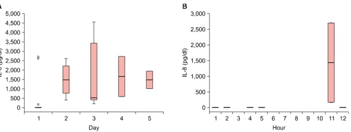
Fig. 2. (A) Variation of the interleukin-8 (IL-8) concentration within partial thickness burn blister over the time. The IL-8 concentration was measured to be the highest on the fourth day. (B) The IL-8 concentration within partial thickness burn blister up to 12 hours after damage. IL-8 was not observed up to 5 hours of the occurrence.
322.483∼4,021.122 pg/dl) on the third day, 1,653.010 pg/dl (IQR, 584.388∼2,187.322 pg/dl) on the fourth day, and 1,473.6735 pg/dl (IQR, 1,006.071∼1,707.477 pg/dl) on the fifth day (Fig. 2A). When the concentration of IL-8 within the first 12 hours was analyzed, IL-8 was not de- tected up to 5 hours after the damage (Fig. 2B). There was no correlation between the concentration of IL-8 and treat- ment period (rho=–0.326, p=0.053).
DISCUSSION
HSP70 is released in a passive manner in necrotic cells and is also secreted through an active mechanism involv- ing lysosomal vesicles and lipid rafts21. The functions of HSP70 include acting as a molecular chaperone to facili- tate the assembly of proteins, targeting misfolded and da- maged proteins for degradation, dissembling protein ag- gregates, acting as a regulatory molecule in protecting cells from thermal or oxidative proteins, and inhibiting apoptosis8. In addition, HSP70 is involved in inflammatory reactions and the healing process of the epidermis and dermis following thermal injury8-10. HSP70 has an ex- tracellular function that appears to potentiate the immune response, whereas the intracellular mechanisms appear to be anti-inflammatory. HSP70 can also interrupt the cleav- age of pro-matrix metalloproteinases to an active form, which in turn may reduce immune signaling22.
The concentration of HSP70 was found to be very high only on the first day. It was weakly detected on the sec- ond day. A small amount was found on the third day.
Finally, no detectable amount was found on the fourth and the fifth days. Because the concentration of HSP70 was the highest in the first 12 hours, the results from 1
hour to 12 hours after the burn injury were analyzed in detail. The concentration was the highest 1 hour after the injury (Fig. 1B). Then, the concentration decreased rapidly afterwards. It was weakly detected after 5 hours. Even though there were not many cases included in this study, the results indicated a pattern that HSP70 is present in blisters for the first few hours, after which the amount decreases. In this study, no correlation was found between the concentration of HSP70 during the first 12 hours and the length of the treatment period (rho=−0.316, p=0.175).
A major burn injury induces an inflammatory response, which is accompanied by the release of various cyto- kines23. The chemokine IL-8 induces chemotaxis in neu- trophil granulocytes (polymorphonuclear leukocytes, PMNs), causing them to adhere to endothelial cells and then move into tissue cells to induce PMN-mediated damage24. Therefore, many inflammatory mediators are involved in multiple steps of the systemic inflammatory response syn- drome to a trauma or severe burn, and they may be used as an index to estimate the extent of the inflammatory re- sponse or prognosis. IL-8 has been shown to play a pivotal role in the activation of the pro-inflammatory cascade in necrotizing enterocolitis (NEC)25,26, and it has been re- cently demonstrated that IL-8 may serve as a potential pre- dictive marker in diagnosing NEC. Hans-Oliver reported that IL-8 increases the amount of the intracellular integrin α6 subunit, keratinocytes proliferation, and the number of mitotic keratinocytes and enhances reepithelializa- tion17. Berney et al.27 reported that the serum IL-8 concen- tration increased after several days or the first day in acute pancreatitis, reaching a plateau between days 2 and 5, and also reported that the concentration of serum IL-8 in a severe case increased on the day the patient visited the
emergency department. Lee et al.28 reported that immuno- modulatory effect of interleukine expressed.
In this study, the changes in IL-8 levels in burn blister fluid were investigated. The IL-8 concentration in partial thick- ness burn cases was found to be the highest on the fourth day. IL-8 was not detected up to five hours, but it was de- tected after 11 hours. Cases for 6 to 10 hours are not in- cluded in this study. However, no relationship between the IL-8 concentration in burn blisters and the length of the treatment period was found.
Our results suggest that IL-8 is an important mediator in inflammatory changes following a burn injury but that var- ious factors may influence the concentrations of this cy- tokine. Luo et al.29 demonstrated a novel role for exoge- nous human HSP70 in suppressing the production of IL-6, IL-8, and monocyte chemoattractant protein-1 in fibro- blast-like synoviocytes that involves inhibiting the activa- tion of the mitogen-activated protein kinases and nuclear factor-κB signaling pathways.
There is still a debate regarding how to best treat the blis- ters of burn patients19-21. This study showed that HSP70 in- creased in the first few hours and decreased afterwards and that IL-8 was produced after a few hours and in- creased afterwards. It is speculated that HSP70 present in a blister has a pro-inflammatory effect outside the cell and a protective effect in the intracellular space of the ische- mic cell zone30. If this is the case, then the proper treat- ment would be to remove the blister fluid during the peri- od when HSP70 is present. In addition, a better under- standing of the changes in IL-8 concentration in partial thickness burn blisters would help to improve blister management.
Because this study was based on patient schedules, it was not possible to obtain all of the data on the cases for the targeted times. In addition, the number of cases was lim- ited because we counted the first 12 hours as one day;
thus, we likely lost some cases, and there were many cas- es for which we could not re-sample the fluid from the same blister.
This study had limitations and could not clearly define the healing mechanism in burn blister fluid, but the results demonstrate the changes in the concentrations of HSP70 and IL-8 and may help determine the guidelines for when blister fluid should be removed.
Various inflammatory mediators function during the heal- ing process. To better understand the immunological proc- esses during wound healing and how to best treat wounds, additional studies on the changes in HSP70 and IL-8 levels over time as well as those of other mediators are necessary.
ACKNOWLEDGMENT
This work was supported by National Research Foundation of Korea (NRF) funded by the Ministry of Science, ICT &
Future Planning (NRF-2014R1A1A3A04049491), a grant from the Hallym University Medical Center Research Fund (01-2007-13), Hallym University Hangang Research Fund (2013-068), and Hallym University Research Fund 2014 (HURF-2014-58).
CONFLICTS OF INTEREST
The authors have nothing to disclose.
REFERENCES
1. Pan SC, Wu LW, Chen CL, Shieh SJ, Chiu HY. Deep partial thickness burn blister fluid promotes neovascularization in the early stage of burn wound healing. Wound Repair Regen 2010;18:311-318.
2. Ono I, Gunji H, Zhang JZ, Maruyama K, Kaneko F. A study of cytokines in burn blister fluid related to wound healing.
Burns 1995;21:352-355.
3. Widgerow AD, Tussardi IT, Banyard DA, King K, Chiang R, Wirth G, et al. Burn wound fluid: an important diagnostic source. Wound Healing South Afr 2014;7:9-12.
4. Pan SC. Burn blister fluids in the neovascularization stage of burn wound healing: a comparison between superficial and deep partial-thickness burn wounds. Burns Trauma 2013;
1:27-31.
5. Srivastava P. Roles of heat-shock proteins in innate and adaptive immunity. Nat Rev Immunol 2002;2:185-194.
6. Giffard RG, Han RQ, Emery JF, Duan M, Pittet JF. Regulation of apoptotic and inflammatory cell signaling in cerebral ischemia: the complex roles of heat shock protein 70. Anes- thesiology 2008;109:339-348.
7. Henderson B. Integrating the cell stress response: a new view of molecular chaperones as immunological and phy- siological homeostatic regulators. Cell Biochem Funct 2010;
28:1-14.
8. Kennedy D, Mnich K, Samali A. Heat shock precon- ditioning protects against ER stress-induced apoptosis through the regulation of the BH3-only protein BIM. FEBS Open Bio 2014;4:813-821.
9. Sajjadi AY, Mitra K, Grace M. Expression of heat shock proteins 70 and 47 in tissues following short-pulse laser irra- diation: assessment of thermal damage and healing. Med Eng Phys 2013;35:1406-1414.
10. Kim S, Kwon J. Thymosin β4 has a major role in dermal burn wound healing that involves actin cytoskeletal remo- delling via heat-shock protein 70. J Tissue Eng Regen Med 2015. doi: 10.1002/term.2028. [Epub ahead of print]
11. Xiong L, Sun J, Hirche C, Yang J, Yang Y, Xia Y, et al. In vitro N-acetyl-L-cysteine promotes proliferation and sup- presses interleukin-8 expression in adipose-derived stem
cells. Aesthetic Plast Surg 2012;36:1260-1265.
12. Ashcroft GS, Mills SJ, Lei K, Gibbons L, Jeong MJ, Taniguchi M, et al. Estrogen modulates cutaneous wound healing by downregulating macrophage migration inhibitory factor. J Clin Invest 2003;111:1309-1318.
13. Nwariaku F, Sikes PJ, Lightfoot E Jr, Mileski WJ. Inhibition of selectin- and integrin-mediated inflammatory response after burn injury. J Surg Res 1996;63:355-358.
14. Iocono JA, Colleran KR, Remick DG, Gillespie BW, Ehrlich HP, Garner WL. Interleukin-8 levels and activity in delayed-healing human thermal wounds. Wound Repair Regen 2000;8:216-225.
15. Fujita T, Yoshimoto T, Matsuda S, Kajiya M, Kittaka M, Imai H, et al. Interleukin-8 induces DNA synthesis, migration and down-regulation of cleaved caspase-3 in cultured human gingival epithelial cells. J Periodontal Res 2015;50:
479-485.
16. Michel G, Kemény L, Peter RU, Beetz A, Ried C, Aren- berger P, et al. Interleukin-8 receptor-mediated chemotaxis of normal human epidermal cells. FEBS Lett 1992;305:
241-243.
17. Rennekampff HO, Hansbrough JF, Kiessig V, Doré C, Sticherling M, Schröder JM. Bioactive interleukin-8 is expressed in wounds and enhances wound healing. J Surg Res 2000;93:41-54.
18. Sargent RL. Management of blisters in the partial-thickness burn: an integrative research review. J Burn Care Res 2006;
27:66-81.
19. Cleland H. Thermal burns--assessment and acute mana- gement in the general practice setting. Aust Fam Physician 2012;41:372-375.
20. duKamp A. Deroofing minor burn blisters--what is the evidence? Accid Emerg Nurs 2001;9:217-221.
21. Ireland HE, Leoni F, Altaie O, Birch CS, Coleman RC, Hunter-Lavin C, et al. Measuring the secretion of heat shock proteins from cells. Methods 2007;43:176-183.
22. Kim JY, Yenari MA. The immune modulating properties of the heat shock proteins after brain injury. Anat Cell Biol 2013;46:1-7.
23. Weissman C. The metabolic response to stress: an overview and update. Anesthesiology 1990;73:308-327.
24. Ono SJ, Nakamura T, Miyazaki D, Ohbayashi M, Dawson M, Toda M. Chemokines: roles in leukocyte development, trafficking, and effector function. J Allergy Clin Immunol 2003;111:1185-1199; quiz 1200.
25. Benkoe T, Baumann S, Weninger M, Pones M, Reck C, Rebhandl W, et al. Comprehensive evaluation of 11 cytokines in premature infants with surgical necrotizing enterocolitis.
PLoS One 2013;8:e58720.
26. Chan KY, Leung FW, Lam HS, Tam YH, To KF, Cheung HM, et al. Immunoregulatory protein profiles of necrotizing enterocolitis versus spontaneous intestinal perforation in preterm infants. PLoS One 2012;7:e36977.
27. Berney T, Gasche Y, Robert J, Jenny A, Mensi N, Grau G, et al. Serum profiles of interleukin-6, interleukin-8, and inter- leukin-10 in patients with severe and mild acute pancrea- titis. Pancreas 1999;18:371-377.
28. Lee YB, Kim SJ, Park SM, Lee KH, Han HJ, Yu DS, et al.
Immunomodulatory effects of Deokgu thermomineral water balneotherapy on oxazolone-induced atopic dermatitis murine model. Ann Dermatol 2016;28:192-198.
29. Luo X, Zuo X, Zhou Y, Zhang B, Shi Y, Liu M, et al.
Extracellular heat shock protein 70 inhibits tumour necrosis factor-alpha induced proinflammatory mediator production in fibroblast-like synoviocytes. Arthritis Res Ther 2008;
10:R41.
30. Krause M, Heck TG, Bittencourt A, Scomazzon SP, New- sholme P, Curi R, et al. The chaperone balance hypothesis:
the importance of the extracellular to intracellular HSP70 ratio to inflammation-driven type 2 diabetes, the effect of exercise, and the implications for clinical management.
Mediators Inflamm 2015;2015:249205.