Received: March 3, 2017 Revised: April 6, 2017 Accepted: April 26, 2017
Correspondence to Byung-Jo Kim
Department of Neurology, Korea Univer- sity College of Medicine, 73 Inchon-ro, Seongbuk-gu, Seoul 02841, Korea Tel: +82-2-920-6619
Fax: +82-2-925-2472 E-mail: [email protected]
http://www.e-acn.org pISSN 2508-691X eISSN 2508-6960
Copyright © 2017 The Korean Society of Clinical Neurophysiology
This is an Open Access article distributed under the terms of the Creative Commons Attribution Non-Commercial License (http://
creativecommons.org/licenses/by-nc/4.0) which permits unrestricted non-commercial use, distribution, and reproduction in any medium, provided the original work is properly cited.
Elevated immunoglobulin E (IgE) can be detected in various conditions such as allergic asthma, atopic eczema, anaphylaxis, parasite infection, vasculitis, IgE myeloma, and hy- per-IgE syndrome (HIES).1 The binding of IgE to specific antigens causes mast-cell degranu- lation, resulting in an inflammatory reaction.2 Here we present a patient with Guillain-Barré syndrome (GBS) who exhibited elevated serum IgE.
CASE
A 72-year-old man was admitted for paresthesia in both feet and a gait disturbance that had progressed for the previous 3 days. He was generally healthy except for frequent skin abscesses and eczema. He denied any medical history of allergy or a family history of immune deficiency. A neurologic examination revealed symmetric muscle weakness (Medical Research Council [MRC] grade 4) in the distal lower limbs and decreased deep tendon reflexes. The findings of laboratory tests were normal except for creatinine kinase (390 IU/L), lactate dehydrogenase (448 IU/L), and myoglobin (244.5 ng/mL). Brain magnet- ic resonance imaging also produced normal results. Cerebrospinal fluid analysis revealed albuminocytologic dissociation, normal cell count and glucose level, but elevated protein (183.2 mg/dL). A nerve conduction study (NCS) and electromyography showed that the compound muscle action potentials were slightly low in all of the nerves examined on the ANNALS OF
CLINICAL
NEUROPHYSIOLOGY
CASE REPORT
Ann Clin Neurophysiol 2017;19(2):148-150 https://doi.org/10.14253/acn.2017.19.2.148
Guillain-Barré syndrome associated with hyper-IgE-emia
Jongsuk Choi, Jeong Hwa Rho, and Byung-Jo Kim
Department of Neurology, Korea University College of Medicine, Seoul, Korea
Peripheral neuropathy associated with hyper-IgE-emia have been rarely reported. Here we present a 72-year-old man with acute motor axonal neuropathy who had relatively poor prog- nosis. The serum was weakly positive for IgG GQ1b and GT1a, and serum IgE was significantly elevated. He was transferred to a rehabilitation center with Medical Research Council grade 3 lower extremity weakness on admission day 65. We would suggest that hyper-IgE-emia may increase the magnitude and rate of neural damage in this case.
Key words: Guillain-Barré syndrome; Hyper-IgE-emia; Hyper IgE syndrome
149
http://www.e-acn.org https://doi.org/10.14253/acn.2017.19.2.148
Jongsuk Choi, et al. GBS with hyper-IgE-emia
Guillain-Barré syndrome associated with hyper-IgE-emia
Jongsuk Choi, Jeong Hwa Rho, and Byung-Jo Kim
Department of Neurology, Korea University College of Medicine, Seoul, Korea
Peripheral neuropathy associated with hyper-IgE-emia have been rarely reported. Here we present a 72-year-old man with acute motor axonal neuropathy who had relatively poor prog- nosis. The serum was weakly positive for IgG GQ1b and GT1a, and serum IgE was significantly elevated. He was transferred to a rehabilitation center with Medical Research Council grade 3 lower extremity weakness on admission day 65. We would suggest that hyper-IgE-emia may increase the magnitude and rate of neural damage in this case.
Key words: Guillain-Barré syndrome; Hyper-IgE-emia; Hyper IgE syndrome
admission day. Somatosensory evoked potentials were nor- mal.
Following a diagnosis of GBS, intravenous immunoglobulin (IVIg) was started. The muscle weakness progressed rapidly in both lower limbs (MRC grade 2) and upper limbs (MRC grade 3) over the next few days. Pneumonia appeared on day 3, whose progression resulted in mechanical ventilation being started on day 5 after admission. Facial diplegia was noted on day 7. The serum was weakly positive for IgG GQ1b and GT1a.
Serum IgE was significantly elevated (5,715.7 IU/mL, normal range: 0–100 IU/mL), whereas IgA, IgM, and IgG were within the normal ranges. The serum eosinophil count was 191/µL, corresponding to 2.2% of whole blood cells. Serology tests for parasites, human immunodeficiency virus, hepatitis B and C, cytomegalovirus, Epstein-Barr virus, herpes simplex virus types 1 and 2, varicella zoster virus, and Campylobacter jejuni were all negative.
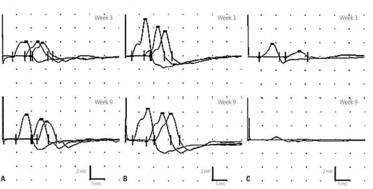
Two weeks after starting IVIg administration, improvements in upper extremity weakness, facial diplegia, and respiratory distress permitted ventilator weaning. However, lower ex- tremity weakness had not changed. Follow-up NCS findings
were compatible with acute motor axonal neuropathy (Fig.
1), and his follow-up serum IgE level had increased to 7,528.3 IU/mL. His IgE level decreased to 4,260.9, 2,645.1, and 2,223.4 IU/mL at weeks 1, 2, and 3 after administering high-dose IV methylprednisolone pulse therapy. On day 65 he was trans- ferred to a rehabilitation center with MRC grade 3 lower ex- tremity weakness.
DISCUSSION
Differential diagnosis of hyper-IgE-emia is very extensive, but the final diagnosis is mostly based on the clinical history and physical findings. The present patient did not have any problematic history except for his recurrent skin infection with eczema. However, the level of IgE was extremely high and beyond that expected in patients with pure skin allergy problems. The findings of extensive laboratory tests were all negative, including the eosinophil count and parasite panel.
Finally we considered HIES, which is a rare primary immuno- deficiency disease with a genetic association, but data from
A B C
Week 3
Week 9
2 mV 2 mV 2 mV
5 ms 5 ms 5 ms
Week 3
Week 9
Week 3
Week 9
Fig. 1. Data from nerve conduction studies. Motor nerve conduction studies revealed normal velocities but variable decreases in amplitude in the median (A), ulnar (B), and tibial (C) nerves. The compound muscle action potential amplitude in the median nerve had improved by week 9, while that in the tibial nerve had decreased.
150 https://doi.org/10.14253/acn.2017.19.2.148 http://www.e-acn.org
Annals of Clinical Neurophysiology Volume 19, Number 2, July 2017
a genetic evaluation were not available. Although peripheral nerve involvement is not a well-known complication, a few cases of hyper-IgE-emia associated with subacute or chronic polyneuropathy have been reported.3,4 However, to our knowledge this is the first case of GBS associated with hy- per-IgE-emia. It is possible that GBS and the hyper-IgE-emia in this case were coincidental. Nevertheless, the elevation in IgE might have played a role in the pathogenesis of this case with GBS.5,6 In the pathogenesis of GBS, multiple fac- tors including breakdown of blood–nerve barrier (BNB) and extent of inflammation are all relevant to producing auto- antibody-mediated nerve fiber injury.7 A healthy peripheral nervous system is tightly sealed by the BNB, which has a very low permeability to serum immunoglobulins.8 Therefore, in the absence of a local inflammatory response, diffusion of an antibody into the nerve is unable to initiate a functional deficit.8 Mast cells activated by IgE trigger disruption of the BNB as an initial GBS insult, and this is followed by macro- phage activation.9 Therefore, hyper-IgE-emia may increase the magnitude and rate of neural damage in early GBS. Fur- ther research regarding the role of IgE in GBS might help to identify the mechanisms underlying the induction of GBS.
Conflicts of Interest
The authors have no financial conflicts of interest.
REFERENCES
1. Johansson SG. The History of IgE: From discovery to 2010. Curr Allergy Asthma Rep 2011;11:173-177.
2. Amin K. The role of mast cells in allergic inflammation. Respir Med 2012;106:9-14.
3. Kimura A, Yoshino H, Yuasa T. Chronic inflammatory demyelinat- ing polyneuropathy in a patient with hyperIgEaemia. J Neurol Sci 2005;231:89-93.
4. Coutinho BM, Nascimento OJ, Freitas MR. Distal acquaried de- mylinating symmetric neuropathy in two patients with essential hiperIgEmia. Arq Neuropsiquiatr 2013;71:493-494.
5. Pate MB, Smith JK, Chi DS, Krishnaswamy G. Regulation and dys- regulation of immunoglobulin E: a molecular and clinical perspec- tive. Clin Mol Allergy 2010;8:3.
6. Walker ME, Hatfield JK, Brown MA. New insights into the role of mast cells in autoimmunity: evidence for a common mechanism of action? Biochim Biophys Acta 2012;1822:57-65.
7. He L, Zhang G, Liu W, Gao T, Sheikh KA. Anti-Ganglioside anti- bodies induce nodal and axonal injury via Fcγ receptor-mediated inflammation. J Neurosci 2015;35:6770-6785.
8. Mathey EK, Derfuss T, Storch MK, Williams KR, Hales K, Woolley DR, et al. Neurofascin as a novel target for autoantibody-mediated axonal injury. J Exp Med 2007;204:2363-2372.
9. Dines KC, Powell HC. Mast cell interactions with the nervous sys- tem: relationship to mechanisms of disease. J Neuropathol Exp Neurol 1997;56:627-640.