J Korean Soc Radiol 2017;77(1):27-31 https://doi.org/10.3348/jksr.2017.77.1.27
INTRODUCTION
Epithelial-myoepithelial lesions of the breast, also known as ad- enomyoepithelial lesions, consist of a heterogeneous group of en- tities that exhibit a wide spectrum of benign lesions to malignant ones (1). Adenomyoepithelioma is a biphasic neoplasm composed of ductal and myoepithelial cells, and exhibits potential for ma- lignant progression (1). When malignant progression occurs, the malignant component can be epithelial, myoepithelial, or both and can exhibit a wide histological grade spectrum ranging from low-to-high (1). Due to the rare occurrence of these tumors, there
is very limited literature describing them, especially their radio- logical appearance. In the present study a case of myoepithelial carcinoma arising within an adenomyoepithelioma of the breast is being reported. This study also discusses the imaging findings of the case obtained by a mammography, sonography, and mag- netic resonance imaging (MRI).
CASE REPORT
A 55-year-old woman visited our hospital for breast cancer screening. She had no significant medical history, and no histo-
Myoepithelial Carcinoma Arising within an Adenomyoepithelioma of the Breast: A Case Report
유방의 선근상피종에서 발생한 근상피암의 증례 보고
Youyeon Kim, MD
1, Kyu Ran Cho, MD
1*, Sung Eun Song, MD
1, Bo Kyoung Seo, MD
2, Ok Hee Woo, MD
3, Jeonghyun Lee, MD
4Departments of 1Radiology, 4Pathology, Anam Hospital, Korea University College of Medicine, Seoul, Korea
2Department of Radiology, Ansan Hospital, Korea University College of Medicine, Ansan, Korea
3Department of Radiology, Guro Hospital, Korea University College of Medicine, Seoul, Korea
Adenomyoepithelioma of the breast is a rare tumor. A myoepithelial carcinoma aris- ing within an adenomyoepithelioma is even more unusual. There are a limited num- ber of reports discussing myoepithelial carcinoma; most of them describe patholog- ical findings, but not imaging findings. We present a case of a 55-year-old woman who had a screen-detected myoepithelial carcinoma arising within an adenomyo- epithelioma in her right breast. Upon the completion of a mammography and so- nography an oval shaped mass with an indistinct margin in the upper portion of the right breast had been seen. It as appeared to be a spiculated, irregular-shaped, pe- ripheral-enhancing mass on an MRI. On sonography-guided biopsy, an epithelial- myothelial tumor was confirmed, and the possibility of myoepithelial carcinoma was suggested. Breast-conserving surgery with a sentinel lymph node dissection was performed, and a pathological examination revealed a myoepithelial carcinoma arising within an adenomyoepithelioma.
Index terms Breast Neoplasms Adenomyoepithelioma Myoepithelial Tumor Mammography
Magnetic Resonance Imaging
Received August 4, 2016 Revised September 12, 2016 Accepted October 18, 2016
*Corresponding author: Kyu Ran Cho, MD Department of Radiology, Anam Hospital, Korea University College of Medicine, 73 Inchon-ro, Seongbuk-gu, Seoul 02841, Korea.
Tel. 82-2-920-5578 Fax. 82-2-929-3796 E-mail: [email protected]
This is an Open Access article distributed under the terms of the Creative Commons Attribution Non-Commercial License (http://creativecommons.org/licenses/by-nc/4.0) which permits unrestricted non-commercial use, distri- bution, and reproduction in any medium, provided the original work is properly cited.
ry of personal or familial breast cancer. Physical examination of the right breast revealed a painless small mass in the right upper breast. There was no skin retraction, nipple discharge, or palpable axillary-lymph-node seen during the physical examination.
Mammography was performed using Lorad Selenia (Hologic Inc., Bedford, MA, USA). On the screening mammography (Fig.
1A), an oval-shaped, isodense mass with indistinct margins and a long-diameter of 2 cm was seen in the 12 o’clock direction of right breast. There was no associated calcification or axillary- lymphadenopathy observed. Breast sonography was performed using a LOGIQ 9 (GE Healthcare, Milwaukee, WI, USA) with a broad-bandwidth 14–5 MHz linear probe. On breast sonogra-
phy (Fig. 1B), the lesion presented as an oval, hypoechoic mass with indistinct margins in the half past 12 o’clock direction of the right breast, 8 cm from the nipple. The legion was classified as a category 4 per the American College of Radiology Breast Imag- ing-Reporting and Data System (ACR BI-RADS) criteria (2). A breast MRI was performed using a 3.0-T Scanner (Achieva 3.0T TX; Philips Healthcare, Best, the Netherlands) with a breast coil (MRI Devices; InVivo Research, Orlando, FL, USA), with the patient maintained in the prone position. Images were acquired in the axial plane. The interpretation of the degree and patterns of enhancement were performed using CAD stream TM (Merge Health Care, Chicago, IL, USA). An irregular mass with spicu-
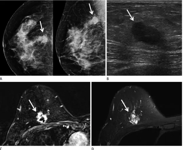
Fig. 1. A 55-year-old woman with myoepithelial carcinoma arising within adenomyoepithelioma.
A. Screening mammography shows a 2 cm-sized, oval-shaped, isodense mass with indistinct margins (arrows) in the 12 o’clock direction of the right breast.
B. Breast sonography shows an indistinct, oval, hypoechoic mass (arrow) seen in the right upper breast. There is no observed abnormal axillary lymphadenopathy.
C. On MRI, axial fat-saturated T1-weighted subtraction image with post-contrast gadolinium injection shows an irregular mass with spiculated margins and rim enhancement (arrow) in the right upper mid-to-outer portion.
D. On axial fat-saturated T2-weighted image, the mass was hyperintense (arrow). On dynamic study (not shown), this mass shows initial fast-en- hancement and delayed plateau pattern.
A
C
B
D
lated margins and rim enhancement was seen in the right upper mid-to-outer portion (Fig. 1C). It was hyperintense on the fat- saturated T2-weighted image (Fig. 1D) and isointense on the T1- weighted image. Upon a kinetic curve assessment, it initially showed fast-enhancement with a delayed plateau pattern. It also showed diffusion restriction on a diffusion-weighted image and apparent diffusion coefficient map. There were not any signifi- cantly enlarged lymph nodes in either axillae.
Sonography-guided core needle biopsy was done for the right breast mass. It was confirmed to be an epithelial-myothelial tu- mor with myoepithelial overgrowth, moderate degree of atypism and frequent mitoses. The possibility of myoepithelial carcinoma was suggested and a complete excision was recommended. There- fore, breast-conserving surgery was performed. On gross exami- nation, the cut surface of the specimen showed a well-demarcated, ovoid, grayish mass with multifocal hemorrhagic changes. Micro- scopic examination (Fig. 1E, F) revealed a myoepithelial carcino- ma in an adenomyoepithelioma. The tumor was 1.8 × 1.4 cm in size, and was composed of a ductal and spindle cell component.
The ductal component was small, tubular and cystically dilated, with two cell components, epithelial and myoepithelial cells. The spindle cells were epithelioid, vaguely clustered, and fibroblastic or smooth muscle-like. On immunostaining, the spindle cell com- ponents were positive for smooth muscle markers, epithelial markers (CK, HMW-CK), and a myoepithelial cell marker (p63).
The stromal cells showed frequent mitoses (10/10 HPF), necro-
sis, and 30–40% Ki-67 labeling. The surgical margin was free of carcinoma. An 18F-fludeoxyglucose positron emission tomogra- phy-CT scan was performed as a part of the metastatic work up;
no distant metastasis was found.
DISCUSSION
Myoepithelial cells are the normal components of breast lob- ules and ducts (3, 4). These cells have the characteristics of both epithelial and smooth muscle cells. Thus, tumors that arise from these cells show characteristics of epithelial and smooth muscle cells (4). They can arise in the salivary glands, skin, soft tissue, retroperitoneum, breast, and lungs (4). Epithelial-myoepithelial lesions of the breast are also known as adenomyoepithelial le- sions (1). These are a heterogeneous group of lesions, exhibiting a wide spectrum from benign to malignant (1).
Adenomyoepithelioma is an extremely rare neoplasm of the breast, and occurs almost exclusively in women (5). It is generally benign and has a low potential for malignancy and local recur- rence (6, 7). However, malignant progression can occur, and be associated with distant metastasis, mainly via a hematogenous spread (5, 6). In more than 150 case reports studied, about 40 le- sions were found to be malignant or potentially malignant, ac- cording to Tavassoli (8). Malignant degeneration of adenomyo- epithelioma typically presents as a sudden, rapid growth of a long-standing, often-large breast mass (1). Adenomyoepithelioma
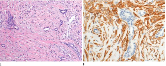
Fig. 1. A 55-year-old woman with myoepithelial carcinoma arising within adenomyoepithelioma.
E. On histological examination (hematoxylin and eosin stain, × 100), the tumor shows a relatively uniform admixture of scattered, glandular, epi- thelial-lined spaces, and surrounding spindle and epithelioid myoepithelial-cell proliferation.
F. On immunostaining (SMA, × 200), the myoepithelial cells are immunoreactive for SMA.
SMA = smooth muscle actin
E F
is common after the fifth decade of life, and malignant degener- ation may occur in older age (1). However, the 55-year-old pa- tient presented in this study had no symptoms because of the relatively small size of her breast mass which measured less than 2 cm.
There are several reports of adenomyoepithelioma and myo- epithelial carcinoma of the breast, but the imaging findings in these reports are not well-described. According to the results of previous studies, these imaging only states that the features may mimic radiological findings of breast cancer (5, 7, 9, 10), but oth- er details were not provided. The most common mammographic finding is an irregular, non-calcified mass with variable margins (5, 7, 9, 10). Calcification and parenchymal distortions are uncom- mon findings, and are usually associated with malignancy (3, 7).
Upon sonography, a hypoechoic, irregular or oval mass with mi- crolobulated or indistinct margins is the most common finding (5, 7, 10). Lesions may also show posterior acoustic enhancement (3, 7). Although there are a very limited number of reports that de- scribe MRI findings, the most common are of an enhancing mass with an irregular or spiculated margin, and delayed washout or plateau kinetics (5, 7). In the present case, on the basis of mammo- graphic and sonographic findings, the mass was classified as being suspiciously abnormal (BI-RADS 4) per the ACR classification.
The MRI features were consistent with a diagnosis of malignancy.
Overall, the imaging features of the mass in our present case were consistent with those seen in previous studies.
Adenomyoepitheliomas have more prominent proliferative image features than do other common benign lesions (10) that adenomyoepitheliomas considered to be almost benign were cat- egorized in BI-RADS classifications as 4 or higher in previous studies (5, 7, 9, 10). Since there are no definite differences in im- aging features between benign and malignant adenomyoepitheli- al tumors, a sonography-guided core needle biopsy may be re- quired for diagnosis and future treatment planning (5). Irrespective of whether a lesion is benign or malignant, a wide local surgical excision is recommended. If the lesion is confirmed to be malig- nant a lymph node dissection may be done in addition to the aforementioned wide excision (4, 5). Additional adjuvant chemo- therapy or radiotherapy can be considered, even though the pre- cise role of both these in the management of adenomyoepithelial tumors is unknown (1, 4). In this case, the patient underwent breast-conserving surgery with a sentinel lymph node biopsy.
After surgery, she was treated with adjuvant chemotherapy and radiotherapy to reduce the possibility of tumor recurrence.
Myoepithelial carcinoma arising within an adenomyoepithe- lioma of the breast is extremely rare. There are few reports about the radiological features of this tumor, especially describing MRI findings. Understanding the radiological findings of this rare entity can be helpful in making an accurate diagnosis, appropri- ate treatment planning and follow-up.
REFERENCES
1. Ali RH, Hayes MM. Combined epithelial-myoepithelial le- sions of the breast. Surg Pathol Clin 2012;5:661-699 2. American College of Radiology. ACR BI-RADS atlas: breast
imaging reporting and data system. Reston, VA: American College of Radiology, 2013
3. Howlett DC, Mason CH, Biswas S, Sangle PD, Rubin G, Al- lan SM. Adenomyoepithelioma of the breast: spectrum of disease with associated imaging and pathology. AJR Am J Roentgenol 2003;180:799-803
4. Endo Y, Sugiura H, Yamashita H, Takahashi S, Yoshimoto N, Iwasa M, et al. Myoepithelial carcinoma of the breast treat- ed with surgery and chemotherapy. Case Rep Oncol Med 2013;2013:164761
5. Ruiz-Delgado ML, López-Ruiz JA, Eizaguirre B, Saiz A, As- tigarraga E, Fernández-Temprano Z. Benign adenomyoepi- thelioma of the breast: imaging findings mimicking malig- nancy and histopathological features. Acta Radiol 2007;48:
27-29
6. Petrozza V, Pasciuti G, Pacchiarotti A, Tomao F, Zoratto F, Rossi L, et al. Breast adenomyoepithelioma: a case report with malignant proliferation of epithelial and myoepithelial elements. World J Surg Oncol 2013;11:285
7. Adejolu M, Wu Y, Santiago L, Yang WT. Adenomyoepithelial tumors of the breast: imaging findings with histopathologic correlation. AJR Am J Roentgenol 2011;197:W184-W190 8. Tavassoli FA. Myoepithelial lesions of the breast. Myoepi-
theliosis, adenomyoepithelioma, and myoepithelial carci- noma. Am J Surg Pathol 1991;15:554-568
9. Moritz AW, Wiedenhoefer JF, Profit AP, Jagirdar J. Breast adenomyoepithelioma and adenomyoepithelioma with carcinoma (malignant adenomyoepithelioma) with associat-
유방의 선근상피종에서 발생한 근상피암의 증례 보고
김유연
1· 조규란
1* · 송성은
1· 서보경
2· 우옥희
3· 이정현
4유방의 선근상피종은 매우 드문 종양이며, 이로부터 발생한 근상피암은 더욱 흔치 않다. 유방에서 발생한 근상피암에 대한 보고들은 대부분이 병리학적 소견만을 다루고 있으며, 영상의학적 소견을 다룬 보고들은 거의 볼 수 없다. 본 증례는 검진 상 우측 유방의 선근상피종 내부에 발생한 근상피암이 발견되었던 55세 여자 환자의 예로, 유방촬영술 및 초음파 검사상 병변은 불분명한 경계를 가진 난원형의 결절로 관찰되었다. 유방 자기공명촬영에서는 침상의 비정형의 결절로 변연부 조영 증강을 동반하였다. 초음파 유도하에 시행한 조직검사상 선근상피종으로 확인되었으며, 동반된 악성 가능성이 제시되어 유 방보존술 및 감시림프절 곽청술을 시행하였다.
고려대학교 의과대학 안암병원 1영상의학과, 4병리과, 2고려대학교 의과대학 안산병원 영상의학과,
3고려대학교 의과대학 구로병원 영상의학과
ed breast malignancies: a case series emphasizing histologic, radiologic, and clinical correlation. Breast 2016;29:132-139 10. Park YM, Park JS, Jung HS, Yoon HK, Yang WT. Imaging
features of benign adenomyoepithelioma of the breast. J Clin Ultrasound 2013;41:218-223