© Copyright The Korean Academy of Asthma, Allergy and Clinical Immunology • The Korean Academy of Pediatric Allergy and Respiratory Disease http://e-aair.org 573 INTRODUCTION
It is well known that upper airway cough syndrome (UACS), asthma, and gastroesophageal reflux disease (GERD) common- ly induce chronic cough, which persists longer than 8 weeks.1,2 Chronic cough caused by GERD is usually diagnosed by esoph- agogastroduodenoscopy (EGD) and 24-hour pH monitoring, and it may respond to proton pump inhibitors (PPIs), antacids, or surgery.1
Achalasia is a rare esophageal disorder caused by failure of the lower esophageal sphincter to relax.3,4 The most common symp- toms of achalasia are gastrointestinal symptoms including dys- phagia and regurgitation of undigested food. However, in many cases, it is accompanied by respiratory symptoms including cough and wheezing.5,6 Achalasia has been reported in unusual causes of chronic cough in pediatric patients,7,8 but it has not been reported as a cause of chronic cough in adult patients who present with chronic cough but without typical gastrointestinal symptoms. In addition, achalasia can cause symptoms similar to GERD, thus may be misdiagnosed as GERD. Here, we report a case of achalasia misdiagnosed as GERD in an adult patient with chronic cough.
CASE REPORT
A previously healthy 40-year-old woman was admitted to the
A Case of Chronic Cough Caused by Achalasia Misconceived as Gastroesophageal Reflux Disease
Hea Yoon Kwon, Jun Hyeok Lim, Yong Woon Shin, Cheol-Woo Kim*
Department of Internal Medicine, Inha University School of Medicine, Incheon, Korea
Gastrointestinal Center for cough and heartburn, which were aggravated at night. Her symptoms had been ongoing for over 4 months. She also complained of rhinorrhea and salivation dur- ing sleep, acid reflux during coughing, and intermittent dys- phagia. On her first visit, her vital signs were stable and white blood cell (WBC) count was 14,440/mm3 (neutrophil 82.0%, lymphocyte 13.8%). All other laboratory data were unremark- able. Chest X-ray revealed haziness in the right middle and low- er lobe, suggesting community acquired pneumonia (Fig. 1).
EGD revealed multiple acute ulcers on the esophagus and chronic superficial gastritis (Fig. 2). She was treated with levo- floxacin for pneumonia and with a PPI and calcium channel blocker for esophageal ulcers and GERD.
Although the chest X-ray findings for pneumonia improved, she continued to complain of nocturnal cough and rhinorrhea for 4 months after discharge. She was referred to the Allergy Clin- ic for further evaluation of cough and rhinorrhea.
Laboratory studies revealed that her complete blood count Cough is one of the most common symptoms that causes patients to seek outpatient medical care. If cough persists longer than 8 weeks, common causes of chronic cough, such as upper airway cough syndrome, asthma, and gastroesophageal reflux disease (GERD), should be considered. Although not a common cause of chronic cough, achalasia may cause symptoms very similar to reflux that can lead to its misdiagnosis as GERD. In this report, a 40-year-old woman presenting with chronic cough was initially diagnosed with GERD; however, her symptoms were refractory to conventional GERD treatment. Finally, she was diagnosed with achalasia. Her cough improved completely after pneumatic dilatation. Achalasia is a rare disease accompanied by dysphagia or regurgitation. If cough presumably due to GERD does not respond to treatment, or if the cause of chronic cough is un- certain, physicians should suspect achalasia.
Key Words: Chronic cough; achalasia; gastroesophageal reflux disease
This is an Open Access article distributed under the terms of the Creative Commons Attribution Non-Commercial License (http://creativecommons.org/licenses/by-nc/3.0/) which permits unrestricted non-commercial use, distribution, and reproduction in any medium, provided the original work is properly cited.
Correspondence to: Cheol-Woo Kim, MD, PhD, Department of Internal Medicine, Inha University School of Medicine, 27 Inhang-ro, Jung-gu, Incheon 400-711, Korea.
Tel: +82-32-890-3495; Fax: +82-32-882-6578; E-mail: [email protected] Received: August 18, 2013; Revised: November 11, 2013
Accepted: December 2, 2013
•There are no financial or other issues that might lead to conflict of interest.
Case Report
Allergy Asthma Immunol Res. 2014 November;6(6):573-576.
http://dx.doi.org/10.4168/aair.2014.6.6.573 pISSN 2092-7355 • eISSN 2092-7363
Kwon et al.
Allergy Asthma Immunol Res. 2014 November;6(6):573-576. http://dx.doi.org/10.4168/aair.2014.6.6.573 Volume 6, Number 6, November 2014
574 http://e-aair.org
and differential were normal without peripheral blood eosino- philia (WBC 9,150/mm3, total eosinophil count 200/mm3). Her total serum IgE concentration was 65.1 IU/mL, and skin tests for 55 common aeroallergens were negative. A chest X-ray and paranasal sinus films did not show any abnormalities. A pulmo- nary function test (PFT) showed unremarkable findings (FEV1/
FVC 80.7%, FEV1 2.26 L [70%], FVC 2.80 L), and a methacholine challenge test was negative.
Because she had a history of esophageal ulcers and symptoms of cough and rhinorrhea, GERD and UACS were considered to be the main causes of her symptoms. However, her symptoms did not improve and, in fact, worsened upon use of the PPI, in-
tranasal corticosteroids, decongestants, and anti-histamines.
She was readmitted for further evaluation.
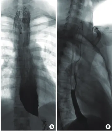
On second admission, her blood pressure was 139/93 mmHg, heart rate was 93/min, respiration rate was 20/min, and body temperature was 36.9°C. Her peripheral blood WBC count was 15,600/mm3 (neutrophils 71.6%, lymphocytes 23.1%), C-reactive protein was 4.83 mg/dL (normal 0-0.3 mg/dL), and erythrocyte sedimentation rate was 37 mm/h (normal 0-22 mm/h). EGD was performed to reexamine the esophageal ulcers and to eval- uate gastroesophageal reflux severity. However, the ulcers were completely healed, and there was no evidence of GERD. Plain chest X-ray showed multiple consolidations on bilateral lung parenchyma, and chest CT exhibited multiple patchy ground glass opacities on both lungs. The dilated esophagus was dis- tended with fluid-filled material, which suggested achalasia with aspiration pneumonia (Fig. 1). Esophagography showed symmetric esophageal narrowing and beaking appearance of the esophago-gastric junction level and mild dilatation of the proximal esophageal portion, which were consistent with char- acteristic findings of achalasia (Fig. 3A).
To treat achalasia, graded pneumatic dilatation was performed.
A follow-up esophagography was performed 4 days after pneu- matic dilatation, and the beak-shaped narrowing segment of the distal esophagus was dilated and showed good passage of the oral contrast media (Fig. 3B). Aspiration pneumonia gradu- A
B
C Fig. 1. Chest X-ray taken during the patient’s first admission demonstrates opac- ities in the right middle lobe and right lower lobe, suggesting pneumonia (A).
Chest CT taken during the second admission shows dilated esophagus with re- tained food materials consistent with achalasia (B), and multiple ground glass opacities on both lungs, suggesting aspiration pneumonia associated with acha- lasia (C).
Fig. 2. Multiple acute esophageal ulcers were noted on the first esophagogas- troduodenoscopic examination for cough. This was misinterpreted as esopha- geal ulcers and chronic cough associated with gastroesophageal reflux disease.
Fig. 3. Esophagography shows the typical beaking appearance of the esophago- gastric junction and dilatation of the proximal esophagus (A). After pneumatic dilatation, the contrast media showed good passage (B).
A B
Chronic Cough Caused by Achalasia AAIR
Allergy Asthma Immunol Res. 2014 November;6(6):573-576. http://dx.doi.org/10.4168/aair.2014.6.6.573 http://e-aair.org 575 ally improved with third generation cephalosporin and metro-
nidazole treatment and completely resolved soon after pneu- matic dilatation. After pneumatic dilatation, she did not com- plain of cough or other symptoms during the 1-year follow-up period.
DISCUSSION
Cough is one of the most common symptoms for which pa- tients seek medical care. UACS, asthma, and GERD are the most common causes of chronic cough, but other causes including laryngopharyngeal reflux disease, eosinophilic bronchitis, and the use of ACE inhibitors, should be suspected if previous treat- ment for such diseases do not improve the patient’s symptoms.1,2 Achalasia is an uncommon cause of chronic cough. It is ob- served in both genders at the same frequency and occurs both in children and the elderly.3 The average age at diagnosis is dur- ing the sixth decade, and its incidence and prevalence were re- ported as 1.6 per 100,000 and 10.8 per 100,000 individuals, re- spectively, in the United States.3,9 Gastrointestinal symptoms are the primary symptoms of achalasia, and respiratory symp- toms occur less frequently.6 According to previous reports,5,6,10 30%-50% of patients with achalasia have respiratory symptoms, such as cough, aspiration, and wheezing. However, most of these symptoms are accompanied by gastrointestinal symptoms, and respiratory symptoms are unlikely to precede the more typical gastrointestinal symptoms of achalasia.5,6
Cough as a primary presenting symptom of achalasia has rarely been reported and only so in the pediatric population.7,8 Furthermore, a few cases of achalasia misdiagnosed as uncon- trolled asthma or as an aggravating factor for existing asthma have been reported, again predominantly in pediatric patients.11-13 Although cases of acute airway obstruction due to achalasia have been reported in the elderly,14 achalasia has not been re- ported as the primary cause of cough in adult patients who pre- sented with chronic cough but did not have gastrointestinal symptoms.
Achalasia can cause cough and respiratory symptoms sec- ondary to food retention in the dilated esophagus with regurgi- tation, and to the mass effect of the dilated esophagus that compresses trachea.5,6 Several symptoms suggest that chronic cough in our patient might be caused mainly by retained food or gastric juice regurgitation/aspiration. First, the patient expe- rienced 2 episodes of radiologically proven aspiration pneumo- nia. Second, the patient’s symptoms worsened at night, and re- flux and aspiration occurred more readily at night. Finally, there was no evidence of intra- or extra-thoracic obstruction, suggest- ing a mass effect of the dilated esophagus in flow-volume curves of PFT. However, it took approximately 10 months to diagnose achalasia, since her main symptoms were cough, heartburn, and intermittent dysphagia. In addition, the initial EGD showed esophageal ulcers, which were misinterpreted as the patient’s
symptoms were caused by gastroesophageal regurgitation.
Although esophageal manometry is the key test for achalasia diagnosis, it may be unavailable depending on the clinical situ- ation. In contrast, EGD has been used widely for the evaluation of chronic cough due to GERD. Endoscopic findings of achala- sia may show esophageal dilatation and retention of food or se- cretions. However, findings might be normal in approximately 44% of patients with achalasia.15 In patients with achalasia, pro- gressive dysphagia, regurgitation, and heartburn are the usual symptoms and the reasons for seeking medical care. Because these symptoms are very similar to GERD symptoms, achalasia is often first misdiagnosed as the more common GERD.16 There- fore, it is difficult to diagnose achalasia using EGD. When such an uncommon disease is suspected or when cough assumed to be caused by GERD does not improve upon treatment, esopha- gography and chest CT should be performed to exclude acha- lasia or other related diseases. Even if first diagnosed as GERD, another combined disease that may provoke chronic cough should be considered if the patient does not respond to GERD treatment.
Here we report a case of achalasia-induced chronic cough without typical gastrointestinal symptoms in an adult patient.
This case suggests that it is important to suspect achalasia, par- ticularly when the symptoms do not improve despite treatment for GERD. Finally, use of esophagography, chest CT, or esopha- geal manometry should be considered in the diagnosis of acha- lasia or other uncommon diseases.
ACKNOWLEDGMENTS
This study was supported by the Inha University Research Grant.
REFERENCES
1. Chung KF, Pavord ID. Prevalence, pathogenesis, and causes of chronic cough. Lancet 2008;371:1364-74.
2. Dicpinigaitis PV. Thoughts on one thousand chronic cough patients.
Lung 2012;190:593-6.
3. Beck WC, Sharp KW. Achalasia. Surg Clin North Am 2011;91:1031- 7.
4. Richter JE. Achalasia - an update. J Neurogastroenterol Motil 2010;
16:232-42.
5. Sinan H, Tatum RP, Soares RV, Martin AV, Pellegrini CA, Oelschlager BK. Prevalence of respiratory symptoms in patients with achalasia.
Dis Esophagus 2011;24:224-8.
6. Parshad R, Devana SK, Panchanatheeswaran K, Saraya A, Makhar- ia GK, Sharma SK, Bhalla AS. Clinical, radiological and functional assessment of pulmonary status in patients with achalasia cardia before and after treatment. Eur J Cardiothorac Surg 2012;42:e90-5.
7. Mehdi NF, Weinberger MM, Abu-Hasan MN. Achalasia: unusual cause of chronic cough in children. Cough 2008;4:6.
8. Kugelman A, Berkowitz D, Best LA, Bentur L. Upper airway ob- struction as a presenting sign of achalasia in childhood. Acta Pae-
Kwon et al.
Allergy Asthma Immunol Res. 2014 November;6(6):573-576. http://dx.doi.org/10.4168/aair.2014.6.6.573 Volume 6, Number 6, November 2014
576 http://e-aair.org diatr 2000;89:356-8.
9. Sadowski DC, Ackah F, Jiang B, Svenson LW. Achalasia: incidence, prevalence and survival. A population-based study. Neurogastro- enterol Motil 2010;22:e256-61.
10. Makharia GK, Seith A, Sharma SK, Sinha A, Goswami P, Aggarwal A, Puri K, Sreenivas V. Structural and functional abnormalities in lungs in patients with achalasia. Neurogastroenterol Motil 2009;21:
603-8, e20.
11. Kwok SC, O’Loughlin EV, Kakakios AM, van Asperen PP. Wheezy swallow: poorly responsive ‘asthma’. J Paediatr Child Health 2008;
44:74-7.
12. Al-Abdoulsalam T, Anselmo MA. An 11-year-old male patient with refractory asthma and heartburn. Can Respir J 2011;18:81-3.
13. Chung HM, Keum KH, Park HJ, Lee KH, Lee GH, Choi EJ, Kim JK, Kim WT, Chung HL. A case of esophageal achalasia in a 10-year- old girl with persistent asthma. Pediatr Allergy Respir Dis 2008;18:
97-103.
14. Khan AA, Shah SW, Alam A, Butt AK, Shafqat E, Malik K, Amin J.
Achalasia esophagus; presenting as acute air way obstruction. J Pak Med Assoc 2007;57:423-5.
15. Howard PJ, Maher L, Pryde A, Cameron EW, Heading RC. Five year prospective study of the incidence, clinical features, and diagnosis of achalasia in Edinburgh. Gut 1992;33:1011-5.
16. Fisichella PM, Raz D, Palazzo F, Niponmick I, Patti MG. Clinical, ra- diological, and manometric profile in 145 patients with untreated achalasia. World J Surg 2008;32:1974-9.