© 2013 The Korean Academy of Medical Sciences.
This is an Open Access article distributed under the terms of the Creative Commons Attribution Non-Commercial License (http://creativecommons.org/licenses/by-nc/3.0) which permits unrestricted non-commercial use, distribution, and reproduction in any medium, provided the original work is properly cited.
pISSN 1011-8934 eISSN 1598-6357
Muscular Sarcoidosis Detected by F-18 FDG PET/CT in a Hypercalcemic Patient
Sarcoidosis is a systemic granulomatous disease of unknown etiology that involves many organs, occasionally mimicking malignancy. We herein report a 50-yr-old woman of muscular sarcoidosis of chronic myopathic type, manifested by hypercalcemia and muscle wasting. Besides insignificant hilar lymphadenopathy, her sarcoidosis was confined to generalized atrophic muscles and therefore, F-18 FDG PET/CT alone among conventional imaging studies provided diagnostic clues for the non-parathyroid-related hypercalcemia.
On follow-up PET/CT during low-dose steroid treatment, FDG uptake in the muscles disappeared whereas that in the hilar lymph nodes remained. PET/CT may be useful in the evaluation of unexpected disease extent and monitoring treatment response in suspected or known sarcoidosis patients.
Key Words: Muscular Disease; Sarcoidosis; Hypercalcemia; Fluorine-18 Fluorodeoxyglucose; Positron Emission Tomography
Eun Ji Han,1 Yi Sun Jang,2 In Suk Lee,2 Jong Min Lee,2 Siwon Kang,1 and Hye Soo Kim2
Departments of 1Radiology, 2Internal Medicine, College of Medicine, The Catholic University of Korea, Seoul, Korea
Received: 17 September 2012 Accepted: 23 April 2013 Address for Correspondence:
Hye Soo Kim, MD
Department of Internal Medicine, Daejeon St. Mary’s Hospital, College of Medicine, The Catholic University of Korea, 64 Daeheung-ro, Jung-gu, Daejeon 301-723, Korea Tel: +82.42-220-9297, Fax: +82.42-220-9087 E-mail: [email protected]
http://dx.doi.org/10.3346/jkms.2013.28.9.1399 • J Korean Med Sci 2013; 28: 1399-1402
CASE REPORT
Medical Imaging
INTRODUCTION
Sarcoidosis is a systemic granulomatous disease of unknown etiology that involves many organs. The manifestations vary by involved site and grade, occasionally mimicking malignancy.
Isolated muscular involvement is known as common in sarcoi- dosis patients, but painless or impalpable cases may be undiag- nosed (1, 2). We report a case of generalized muscular sarcoid- osis detected only by fluorine-18 fluorodeoxyglucose (F-18 FDG) positron emission tomography (PET)/computed tomography (CT) performed during the evaluation of non-parathyroid-re- lated hypercalcemia.
CASE DESCRIPTION
In November 2009, a 50-yr-old female was admitted to our hos- pital with idiopathic transient pulmonary edema. Her weight was 49 kg, creatinine clearance 33.8 mL/min and calcium 9.9 mg/dL. Her echocardiogram showed mild diastolic dysfunc- tion with ejection fraction of 55%. Her pulmonary function was moderately restricted with 51% of forced vital capacity. Her pul- monary edema was rapidly improved with diuretics therapy. In August 2010, she was re-admitted with hypercalcemia. She had underlying diabetes for 20 yr. Her diabetes was complicated with proliferative retinopathy, chronic renal impairment, and a chronic foot ulcer. At the time of her recent admission, her wei- ght was 37 kg and she appeared chronically ill and emaciated.
She reported 2 weeks of general weakness. We could not find
any abnormal mass or tenderness except generalized mus cle atrophy at the beginning. Creatinine clearance was 19.4 mL/
min, calcium 14.5 mg/dL, phosphate 5.0 mg/dL, and ionized calcium 1.78 mM/L. Her 24-hr urine calcium level was 186.2 mg/day, and intact parathyroid hormone (PTH) of 15.77 pg/mL.
These ambiguous laboratory findings prompted further studies of non-parathyroid-related hypercalcemia. Her 25-hydroxy Vi- tamin D level was 21.3 ng/mL, 1,25-dihydroxy Vitamin D was 43.1 pg/mL (normal, 25.1-66.1), and parathyroid hormone re- lated peptide (PTHrP) was under 1.1 pM/L. The thyroid hor- mone level, serum electrophoresis and tumor markers were normal. The angiotensin-converting enzyme (ACE) level was elevated to 353.1 IU/L (normal, 20-70), and muscle enzymes (CPK/LDH/AST) were normal. The chest radiograph, neck ul- trasound, and whole body bone scan were unremarkable. F-18 FDG PET/CT was performed for the evaluation of possible hid- den malignancy. The PET/CT showed increased uptake of small lymph nodes in the subcarina and both hila. In addition, multi- ple streaky and dotted muscular uptakes were noted along whole body including the back and extremities (Fig. 1). We reexam- ined her whole body and found a 2 cm-sized soft and non-ten- der mass in the left gastrocnemius muscle. Excisional biopsy was performed of the mass, and the microscopic finding dem- onstrated non-necrotizing granulomas with multinucleated gi- ant cells and without acid-fast bacilli or fungi (Fig. 2) consistent with muscular sarcoidosis. Imaging studies also revealed gall- stones and bilateral renal stones, and we performed extracor- poreal shock wave lithotripsy of the ureteral stone successfully.
Han EJ, et al. • Muscular Sarcoidosis Detected by FDG PET/CT
1400 http://jkms.org http://dx.doi.org/10.3346/jkms.2013.28.9.1399 However, some renal stones remained.
Acute severe hypercalcemia was relieved temporarily by hy- dration and furosemide. Prednisolone was started with 20 mg per day and then rapidly tapered to 5 mg per day within 1 week.
The serum calcium level was normalized and maintained with prednisolone 5 mg a day, but ACE level continued to fluctuate.
Her follow-up clinical data were laid out in the Table 1. Her glu-
cose levels were controlled with insulin, and her weight increas- ed to 50 kg with muscle gain. On follow-up PET/CT at one and half year later, muscular uptake was no longer seen. However, increased uptake in the mediastinal and hilar lymph nodes per- sisted (Fig. 3).
DISCUSSION
Muscular involvement is a common, usually asymptomatic fea- ture of sarcoidosis. The presence of noncaseating epithelioid cell granulomas was detected in 50%-80% of sarcoidosis patients on random muscle biopsy (3). However, symptomatic muscu- Table 1. Follow-up clinical data of the patient
November, 2009
(previous admission) August, 2010 (this admission) March,
2011 (OPD)May, 2012 (OPD)
Weight (kg) 49 37 47 50
Serum Cr (mg/dL) 1.63 2.43 1.44 1.58
ECC (mL/min) 33.8 19.4 36.1 33.2
Serum Ca (mg/dL) 9.9 14.5 9.2 10.0
Serum P (mg/dL) 3.8 5.0 4.0 3.9
Hemoglobin (g/dL) 9.5 9.8 11.2 11.9
HbA1c (%) 6.5 7.8 7.5 9.4
ACR (µg/mg) 144 553 - 521
ACE (IU/L) - 353.1 117.6 > 150
1,25(OH)2D3 (pg/mL) - 43.1 22.9 -
OPD, outpatient department; Cr, creatinine; ECC, estimated creatinine clearance cal- culated using the Cockcroft and Gault formula; Ca, calcium; P, phosphate; HbA1c, hemoglobin A1c; ACR, urine albumin creatinine ratio; ACE, serum angiotensin con- verting enzyme; 1,25(OH)2D3, 1,25-dihydroxy Vitamin D.
Fig. 2. Histologic result. Biopsy specimen was obtained from muscular mass of left lower leg. Microscopic findings (hematoxylin-eosin stain) demonstrated non-necrotiz- ing granulomas (arrows) with multinucleated giant cell (arrowhead) and without acid- fast bacilli or fungi. The histology was consistent with muscular sarcoidosis.
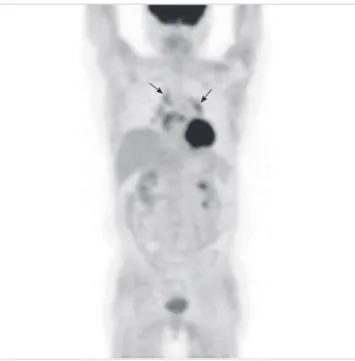
Fig. 3. Follow-up PET/CT finding. In MIP image, previously noted multiple muscular uptakes were no longer seen. However, increased uptake in mediastinal and bilateral hilar lymph nodes persisted (arrows).
Fig. 1. F-18 FDG PET/CT findings. In maximum intensity projection (MIP) (A) and cor- onal (B, C) images of PET/CT, increased uptake was noted in mediastinal and bilateral hilar lymph nodes (arrows). In addition, multiple streaky and dotted muscular uptakes were noted along whole body. Nodular uptake was seen in left lower lateral leg (ar- rowhead) in MIP (A) and transaxial (D) images, and excisional biopsy was performed in this mass.
B C
D
A
Han EJ, et al. • Muscular Sarcoidosis Detected by FDG PET/CT
http://jkms.org 1401
http://dx.doi.org/10.3346/jkms.2013.28.9.1399
lar sarcoidosis is noted in only 0.5%-2.3%, and is more common in patients with extensive systemic involvement. There are three clinical subtypes of muscular sarcoidosis: acute myositic, chro- nic myopathic, and palpable nodular (1, 4, 5). The acute myosi- tic type is accompanied by myalgia, fevers and elevated muscle enzymes, whereas the chronic myopathic type shows muscle atrophy and muscle weakness (6). The palpable nodular type presents as non-tender muscular masses requiring differential diagnosis of nodular muscular diseases (5).
Diagnosis of sarcoidosis is based on the combination of clini- cal, radiologic and histologic findings. Because of its unknown etiology, sarcoidosis remains a diagnosis of exclusion. The dif- ferential diagnosis includes neoplastic diseases such as lympho- ma and various granulomatous diseases (7). Clinical symptoms and signs vary based on the organ affected.
No laboratory tests are diagnostic of sarcoidosis. The ACE level is elevated in approximately two-thirds of patients with sarcoidosis, but this finding is not diagnostic. Hypercalcemia is manifested in 5%-11% of patients with sarcoidosis because of overproduction of active Vitamin D by macrophages in the sar- coid granuloma as the enzyme 1α-hydroxylase is activated for immune modulation without feedback regulation (8). Accord- ing to another hypothesis, hypovitaminosis D may causes sar- coidosis (9). In contrast to parathyroid- or PTHrP-related hy- percalcemia, active Vitamin D-induced hypercalcemia usually accompanies hyperphosphatemia as noted in this case. Hyper- calcemia can induce hypercalciuria and nephrolithiasis, and even renal function impairment. In this patient, in addition to underlying diabetic nephropathy, renal function was aggravat- ed transiently due to hypercalcemia and ureteral stone-related obstructive uropathy. However, the patient’s laboratory findings were not diagnostic of hypervitaminosis D. Although her PTH level was not the cause of this patient’s hypercalcemia, it was not appropriately depressed. The accompanying hyperphos- phatemia might stimulate the parathyroid gland in this case.
The 1,25-dihydroxy Vitamin D level was in a relatively high nor- mal range compared with the 25-hydroxy Vitamin D and the serum calcium levels. The diagnostic clue for hypercalcemia among her laboratory findings was only high ACE level.
Imaging abnormalities are commonly noted in sarcoidosis patients. Most of patients with sarcoidosis demonstrate bilater- al hilar lymphadenopathy on chest radiograph or CT (7, 10, 11).
In this patient, enlargement of lymph nodes in the mediastinum and hila were subtle. She had sarcopenia and general weakness but did not manifest myalgia. Her muscle enzymes were also normal. If we first performed CT in our search for a hidden ma- lignancy, we would not have discovered the presence of muscle sarcoidosis, and therefore the cause of her hypercalcemia. This type of muscular sarcoidosis seems to be chronic myopathy with muscle wasting.
FDG PET/CT has been used worldwide for staging and re-
staging various malignancies. Because FDG uptake reflects tis- sue metabolism, PET/CT can detect some infectious and in- flammatory diseases as well as malignancy. Recently, PET/CT has been used in evaluation of fever of unknown origin and hy- percalcemia (12-14). Hypercalcemia is a common clinical find- ing, mainly related to hyperparathyroidism and malignancy. In hypercalcemic patients with unremarkable PTH level, anatom- ical and functional imaging studies are necessary to investigate the possibility of occult malignancy (14). Sarcoidosis is a well- known granulomatous disease with increased FDG uptake (15).
Traditionally, gallium-67 scan has been used in the detection and evaluation of extent of sarcoidosis. Recent study reported that FDG PET demonstrates higher sensitivity in detection of sarcoidosis involvement compared to the gallium-67 scan (79%
vs 58%). Another study reported that FDG PET contributes to a better evaluation of sarcoidosis especially in extrapulmonary involvement. In addition, FDG PET requires a shorter imaging time and delivers a lower radiation dose compared to the galli- um-67 scan (16-18). Magnetic resonance imaging (MRI) has been used in the diagnosis of sarcoidosis. In muscular sarcoid- osis patients, MRI is useful to assess degree of invasion as well as extent of disease. However, any type of muscular sarcoidosis may not be detected in MRI and regional MRI is difficult to ap- ply to a patient with uncertain symptom that is not localized (19). Because the whole body is evaluated in FDG PET, location of involvement can be discovered, and biopsy site can be deter- mined in suspected sarcoidosis patients (4, 16, 17). In our pa- tient, the left lower leg was selected as a proper biopsy site be- cause this lesion was the only palpable nodule, was located in a relatively superficial area and showed intense FDG uptake in PET/CT.
FDG PET is useful for monitoring treatment response in known sarcoidosis patients. Several reports have demonstrated that changes in FDG uptake before and after treatment correlate with clinical symptoms and laboratory tests (4, 17, 20). Braum et al. reported a patient with disease progression that showed increased FDG uptake in post-treatment PET (16). In our pa- tient, abnormal muscular uptake was no longer visualized in follow-up PET/CT, but increased uptake in mediastinal and bi- lateral hilar lymph nodes persisted. Although clinical improve- ment was maintained with continuity of low dose steroid thera- py, uptake in lymph nodes may be related to the remaining ac- tivity of the disease.
In summary, we report a case of a woman who presented with hypercalcemia and renal stones and was found to have atypical sarcoidosis mainly involving generalized skeletal muscles with minimal hilar lymph node involvement. FDG PET/CT secured the diagnosis. In conclusion, FDG PET/CT can be useful to eval- uate unexpected disease extent and to monitor treatment re- sponse in suspected or known sarcoidosis patients.
Han EJ, et al. • Muscular Sarcoidosis Detected by FDG PET/CT
1402 http://jkms.org http://dx.doi.org/10.3346/jkms.2013.28.9.1399
REFERENCES
1. Vardhanabhuti V, Venkatanarasimha N, Bhatnagar G, Maviki M, Iyen- gar S, Adams WM, Suresh P. Extra-pulmonary manifestations of sarcoi- dosis. Clin Radiol 2012; 67: 263-76.
2. Spagnolo P, Luppi F, Roversi P, Cerri S, Fabbri LM, Richeldi L. Sarcoid- osis: challenging diagnostic aspects of an old disease. Am J Med 2012;
125: 118-25.
3. Silverstein A, Siltzbach LE. Muscle involvement in sarcoidosis: asymp- tomatic, myositis, and myopathy. Arch Neurol 1969; 21: 235-41.
4. Marie I, Lahaxe L, Vera P, Edet-Samson A. Follow-up of muscular sar- coidosis using fluorodeoxyglucose positron emission tomography. QJM 2010; 103: 1000-2.
5. Tohme-Noun C, Le Breton C, Sobotka A, Boumenir ZE, Milleron B, Car- ette MF, Khalil A. Imaging findings in three cases of the nodular type of muscular sarcoidosis. AJR Am J Roentgenol 2004; 183: 995-9.
6. Costabel U. Skeletal muscle weakness, fatigue and sarcoidosis. Thorax 2005; 60: 1-2.
7. Newman LS, Rose CS, Maier LA. Sarcoidosis. N Engl J Med 1997; 336:
1224-34.
8. Conron M, Young C, Beynon HL. Calcium metabolism in sarcoidosis and its clinical implications. Rheumatology (Oxford) 2000; 39: 707-13.
9. Richmond BW, Drake WP. Vitamin D, innate immunity, and sarcoid- osis granulomatous inflammation: insights from mycobacterial research.
Curr Opin Pulm Med 2010; 16: 461-4.
10. Baughman RP, Lower EE, du Bois RM. Sarcoidosis. Lancet 2003; 361:
1111-8.
11. Studdy PR, Lapworth R, Bird R. Angiotensin-converting enzyme and its
clinical significance: a review. J Clin Pathol 1983; 36: 938-47.
12. Meller J, Sahlmann CO, Scheel AK. 18F-FDG PET and PET/CT in fever of unknown origin. J Nucl Med 2007; 48: 35-45.
13. Solav SV. FDG PET/CT in evaluation of pyrexia of unknown origin. Clin Nucl Med 2011; 36: e81-6.
14. Cecchin D, Motta R, Zucchetta P, Bui F, Basso SM, Lumachi F. Imaging studies in hypercalcemia. Curr Med Chem 2011; 18: 3485-93.
15. Culverwell AD, Scarsbrook AF, Chowdhury FU. False-positive uptake on 2-[¹⁸F]-fluoro-2-deoxy-D-glucose (FDG) positron-emission tomogra- phy/computed tomography (PET/CT) in oncological imaging. Clin Ra- diol 2011; 66: 366-82.
16. Braun JJ, Kessler R, Constantinesco A, Imperiale A. 18F-FDG PET/CT in sarcoidosis management: review and report of 20 cases. Eur J Nucl Med Mol Imaging 2008; 35: 1537-43.
17. Aide N, Benayoun M, Kerrou K, Khalil A, Cadranel J, Talbot JN. Impact of [18F]-fluorodeoxyglucose ([18F]-FDG) imaging in sarcoidosis: unsus- pected neurosarcoidosis discovered by [18F]-FDG PET and early meta- bolic response to corticosteroid therapy. Br J Radiol 2007; 80: e67-71.
18. Nishiyama Y, Yamamoto Y, Fukunaga K, Takinami H, Iwado Y, Satoh K, Ohkawa M. Comparative evaluation of 18F-FDG PET and 67Ga scintig- raphy in patients with sarcoidosis. J Nucl Med 2006; 47: 1571-6.
19. Sohn HS, Kim EN. A case of muscular sarcoidosis diagnosed by gallium-67 scintigraphy and magnetic resonance imaging. Korean J Nucl Med 1999;
33: 543-8.
20. Lee JH, Lim YJ, Lee S, Joo KB, Choi YY, Park CK, Lee YH. Early-onset childhood sarcoidosis with incidental multiple enchondromatosis. J Ko- rean Med Sci 2012; 27: 96-100.