Identification of Outer Membrane Vesicles Derived from Orientia tsutsugamushi
Orientia tsutsugamushi, a causative pathogen of Scrub typhus, is a gram-negative intracellular bacterium. Outer membrane vesicles (OMVs) are produced from the membrane of bacteria and play many roles related to the survival of the pathogen. However, there have been no reports confirming whether O. tsutsugamushi indeed produce OMVs.
O. tsutsugamushi boryong was cultured in ECV-304 cells for the purification of OMVs.
Western blot analysis and immunoenrichment using anti-O. tsutsugamushi monoclonal antibody and electron microscopy were employed for identification and characterization of OMVs. We confirm the presence of OMVs derived from O. tsutsugamushi, and also found that those OMVs contain a major surface antigen of 56-kDa protein and variant
immunogenic antigens.
Keywords: Orientia tsutsugamushi; Scrub Typhus; Outer Membrane Vesicles; Antigens Sun-Myoung Lee,1 Hea Yoon Kwon,2
Jae-Hyong Im,2 Ji Hyeon Baek,2 Jae-Seung Kang,3 and Jin-Soo Lee2
1Translation Research Center, 2Department of Internal Medicine, 3Department of Microbiology, Inha University School of Medicine, Incheon, Korea Received: 17 October 2014
Accepted: 1 April 2015 Address for Correspondence:
Jin-Soo Lee, MD
Department of Internal Medicine, Inha University School of Medicine, 27 Inhang-ro, Jung-gu, Incheon 400-711, Korea Tel: +82.32-890-2819, Fax: +82.32-882-6578 E-mail: [email protected]
http://dx.doi.org/10.3346/jkms.2015.30.7.866 • J Korean Med Sci 2015; 30: 866-870
INTRODUCTION
Orientia tsutsugamushi is a rickettsial organism and a causative pathogen for Scrub typhus (1). It causes acute and chronic in
fection, leading to symptoms of fever, rash, pneumonitis, ence
phalitis, and ultimately death (2). It is found endemically main
ly in Northeast Asian regions, China, India, and Australia. Scrub typhus is one of the most common vector borne diseases occur
ring in Korea and the Asia pacific regions (3). More than 5,000 cases are reported annually in Korea, with incidents currently on the rise (4).
Gramnegative bacteria produce OMVs (outer membrane vesicles) of 50250 nm in diameter from the outer membrane (5). To date, OMVs originating from many bacteria, including Escherichia coli, Neisseria meningitides, and Pseudomonas ae- ruginosa have been documented (4). OMVs are secreted from the bacterial surface membrane, and therefore consist of outer membrane proteins (OMP), lipopolysaccharides (LPS), phos
pholipids and other periplasmic components (6). OMVs have been reported to play various roles associated with the functions of secretion and delivery, supporting the survival and patho
genesis of bacteria (7). OMVs have also been observed in intra
cellular gramnegative bacteria of Salmonella spp., Franciella spp. and Chlamydia spp. (810). However, no report has yet con
firmed whether OMVs are produced by O. tsutsugamushi, an obligate intracellular bacterium. In this regard, we intend to in
vestigate whether O. tsutsugamushi produces OMVs and puri
fies microvesicles by immunoprecipitation.
MATERIALS AND METHODS Preparation of O. tsutsugamushi
O. tsutsugamushi Boryong strain was propagated in ECV304 cells (CLS, Germany) cultivated in M199 (WelGENE, Korea) with 10% (v/v) fetal bovine serum (Corning Cellgro, USA). Con
fluency of bacteria in ECV304 was confirmed by immunofluo
rescence assay (IFA). When ECV304 cells were heavily infected, they were gathered and used for electron microscopic observa
tion of O. tsutsugamushi in cytosol of host cells. Heavily infected cells were disrupted with glass beads (diameter, 1.0 mm) to re
lease bacteria from the cells and bacteria were purified with 40%
percoll density solution utilizing the same method of Tamura et al. (11). Purified bacteria were also observed by electron micro
scope.
Purification of OMVs
ECV304 cells, heavily infected with O. tsutsugamushi, were cen
trifuged at 13,000 rpm for 30 min at 4°C, after which the centrif
ugal media was passed through a 0.22 μm pore size filter system (Corning, MA, USA). The cell free medium was concentrated to 50 mL by ultrafiltration using a QuixStand Benchtop system (GE Healthcare BioSciences, Uppsala, Sweden) with a 100 kDa hollow fiber membrane and harvested by ultracentrifugation at 150,000 g for 3 hr at 4°C. The pelleted OMVs were resuspended in 1 mL of PBS and then purified as described previously with modifications (12). After ultracentrifugation in a sucrose gradi
ent solution, each fraction was analyzed by SDSPAGE, Cooma
ssie Brilliant Blue staining, and western blotting. The fractions
of sucrose density gradient showing protein profiles correspond
ing to O. tsutsugamushi in immunoblot bands were collected and centrifuged at 150,000 g for 3 hr at 4°C. The resulting pellets of purified OMVs were resuspended in PBS containing protease inhibitor cocktail (SigmaAldrich Co., MO, USA). The suspend
ed OMVs were observed using an electron microscope. The pu
rified OMVs were quantified using DC protein assay reagents (BioRad Laboratories Inc., Hercules, CA, USA) and aliquots of the OMVs were stored at 70°C. Purified OMVs were taken for immunoenrichment and immunoblot analysis.
Immunoenrichment of O. tsutsugamushi derived OMVs For enrichment of O. tsutsugamuhsi derived OMVs from a mix
ed population of vesicles, FS15 mouse monoclonal antibody reacting against 56 kDa protein of O. tsutsugamushi Boryong strain was combined with 10 μL of protein G magnetic beads (NEW ENGLAND BioLabs., MA, USA) and incubated at room temperature for 1 hr while rotating (25). The resultant was wash
ed three times with IP buffer (25 mM Tris pH 7.5, 150 mM NaCl, 2.5 mM EDTA, 0.05% Triton X100) and then combined with appropriate concentrations of purified OMVs overnight while rotating at 4°C. The mixture was washed four times with IP buf
fer and the final wash was performed with PBS. Pellets in re
ducing sample buffer (50 mM TrisCl pH 6.8, 100 mM dithioth
reitol (DTT), 2% SDS, 0.1% bromophenol blue, 10% glycerol) were solubilized by boiling for 10 min at 100°C. The solubilized samples were loaded on a 10% polyacryl amide gel. The proteins from the OMVs were transferred to a PVDF (Millipore, Darm
stadt, Germany) membrane. The membrane was blocked with 5% nonfat dry milk in PBST (0.1% tween20 in PBS) for 1 hr at room temperature and then incubated overnight at 4°C with O.
tsutsugamushi polyclo nal antibody. The membrane was washed three times with PBST and incubated with HRPconjugated secondary antibody (Jackson Immunoresearch Laboratories, PA, USA) for 1 hr at room temperature. The membrane was washed again three times with PBST and developed with en
hanced chemiluminescence (ECL) solution (GE Healthcare LifeSciences, Uppsala, Sweden). Antibody used for the western blot assay, which was purified from the serum of a patient in
fected with O. tsutsugamushi Boryong, was confirmed by nest
ed PCR amplifying the 56 kDa region. The two pairs of primers used were as follows: outer primers, 1F (5´ATAATTAATGTATT
TTCGAACG3´) and 2R (5´CCTKCA AAGGACTTTTAGCT3´), and inner primers, 1Fn (5´AACACA GTGTTTTATAGATTGT
TTA3´), and 2Rn (5´RCATTAATTGCTACACCAAGT3´). The amplified length was 1,562 bp. The PCR product was purified and sequenced by GenoTech Corp. (Daejeon, Korea). The re
sulting sequence was identified as 56kDa TSA gene using BLAST (http://ncbi.nlm.nih.gov/blastn).
Transmission electron microscopy (TEM)
To observe the blebbing of O. tsutsugamushi in infected ECV304 cells and purified bacteria, each pellet was fixed with 2.5% glu
taraldehyde in 0.1 M phosphate (pH 7.4) for 2 hr. The samples were washed and placed in 1% osmium tetroxide for 30 min, dehydrated in ethanol and embedded in epoxy resin (Epon 812, Electron Microscope Sciences, UK) in Beam capsules. Ultrathin sections were cut using an ultramicrotome (UltraE, Reichert
Jung, USA) and stained with uranyl acetate and citrate. For ob
servation of purified OMVs structure, samples were placed on 200 mesh carboncoated copper grids until settled on the film for 5 min. The grids were stained for 5 min with 1% uranyl ace
tate. After air drying, the samples were analyzed under a CM200 transmission electron microscope (Phillips, Netherlands).
RESULTS
Electron microscopic observation of OMVs derived from O. tsutsugamushi
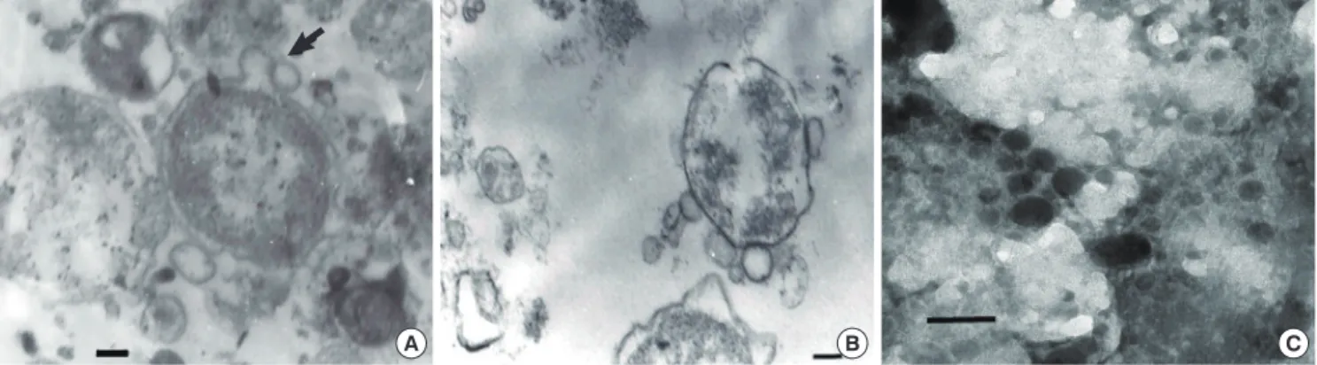
Electron microscopic examination showed that OMVs were se
creted from the bacterial surface membrane in cytosol of host cells (Fig. 1A). Several microvesicles that might have been de
rived from O. tsutsugamushi were observed near the bacteria.
The budding OMVs had a monolayer membrane and were ap
proximately 130 nm in diameter. Microvesicles were observed in the vicinity of purified O. tsutsugamushi (Fig. 1B). Purified OMVs were observed as round or spherical vesicles of relatively diverse size ranging from 50150 nm in diameter (Fig. 1C).
Identification of O. tsutsugamushi derived OMVs
Western blot analysis of purified OMVs with FS15 monoclonal antibody, specific to O. tsutsugamushi 56kDa protein showed the presence of proteins corresponding to O. tsutsugamushi (Fig.
2A). In control experiments, ECV 304 cell lysates used for cell culture did not react with either FS15 monoclonal antibody or polyclonal antibody. This result validates that microvesicles ob
served in the electron microscopic examination were derived from O. tsutsugamushi. Western blot analysis with O. tsutsuga- mushi polyclonal antibody show ed that a 56kDa protein band was present as a prominent band (Fig. 2B).
Immunoenrichment of O. tsutsugamushi derived OMVs To further purify O. tsutsugamushi derived OMVs from mixed populations with host derived microvesicles, we enriched bac
terial OMVs by immunoprecipitation with FS15 monoclonal antibody. Western blot analysis of enriched OMVs showed 42, 46, and 56kDa protein bands, corresponding to the surface an
tigens of O. tsutsugamushi (Fig. 2C). As expected, the 56kDa protein was the most abundant protein band. ECV304 cell ly
sates used for cell cultures did not have a responding band in the western blot analysis using either FS15 monoclonal or poly
clonal antibody. This result shows that OMVs from O. tsutsuga- mushi could be enriched with immunoprecipitation.
DISCUSSION
Scrub typhus is an important cause of febrile diseases in ende
mic regions. In Korea scrub typhus occurs mainly in the autumn and the incidence is increasing (4). While most patients respond favorably to antibiotic treatment some may experience severe complications and others may suffer chronic infection even af
ter recovery from the disease (2). In this regard, a good under
standing of hostpathogen interaction in this disease is needed for development of effective treatment and prevention.
In the current study, we were able to demonstrate that OMVs
are derived from Orientia tsutsugamushi, an intracellular patho
gen. OMVs derived from the outer membrane layer of bacteria are related to the survival of pathogens inside host cells (7, 13).
The secretion of OMVs is dependent on growth conditions or environmental factors. OMVs produced from O. tsutsugamushi might play roles for their survival. Many of the components constituting OMVs might be attributed to those found abundant
ly in the bacteria (13). The protein profile of OMVs may be simi
lar, but not entirely identical, to that of bacterial outer membrane.
The major antigens for O. tsutsugamushi are 110, 80, 70, 56, 46, and 25kDa in size (14). The most abundant and important sur
face antigen of O. tsutsugamushi is the 56kDa protein, which is also the major antigen isolated from OMVs of the current study.
Being one of the most important antigens of O. tsutsugamushi, Fig. 1. Transmission electron micrographs of outer membrane vesicles of O. tsutsugamushi. (A) Blebs of vesicles (arrow) before they are liberated from O. tsutsugamushi (arrow).
(B) Microvesicles are shown on the surface of purified O. tsutsugamushi. (C) Clusters of purified microvesicles are observed. They vary in size ranging from 50 to 150 nm and are made of monolayer membranes. Scale bars indicate 0.1 µm.
A B C
Fig. 2. Western blot analysis of purified and enriched OMVs derived from O. tsutsugamushi. (A) Blots were revealed with FS15 monoclonal antibody against OMVs and had simi- lar patterns with blots of bacterial lysates. (B) Immunoblot of OMVs with polyclonal antibody showed a prominent 56-kDa protein band. (C) OMVs immunoenriched with FS15 mono clonal antibody revealed major surface antigens. ECV304, lysates of host cells; OT, lysates of purified O. tsutsugamushi; OMVs, outer membrane vesicles; FS15, mouse monoclonal antibody of O. tsutsugamushi; poly Ab, human polyclonal antibody purified from scrub typhus patient`s serum; IP, immunoprecipitation; IB, Immuno blot.
ECV304 OT OMV
IB: FS15
kDa 130
100 70 55
35
25
ECV304 OT OMV
IB: poly Ab
kDa 200 130 100 70 55
35
25
OMV
IB: poly Ab
IP: - + (FS15) kDa 250 170 130 100 75
50
40
35
25
A B C
the 56kDa protein has a role in the attachment on and pene
tration into the host cell (15). The immune response to 56kDa protein has an important role in prevention of the disease. The 56kDa protein has been shown to induce neutralizing antibo
dies in animals immunized using the 56kDa protein (16).
O. tsutsugamushi requires host cells for their growth and pro
liferation. Hence, OMVs obtained from such cell culture may contain vesicles not only derived from the pathogen but also from the host cell used for survival. As a result, demonstration or isolation of vesicles derived from intracellular pathogens is not easy. In our experiment, we isolated OMVs from the culture media of heavily infected cells. Frohlich et al. (17) successfully demonstrated that vesicles were derived from bacteria using an immunoenrichment method in Chlamydia, which is an intra
cellular organism. In our study, we also isolated bacterial micro
vesicles using antibody against O. tsutsugamushi. OMVs with a high purity could contribute towards the understanding of host
pathogen interactions or further studies to delineate the func
tion of microvesicles.
An OMV derived from the outer membrane layer measures 20200 nm in diameter, and it characteristically represents the serotype of the pathogen as it contains the pathogen’s outer mem
brane proteins and surface antigens (7). Consequently, the po
tential of OMVs to function as a vaccine or adjuvant has been suggested since OMVs have similar properties as found in vari
ous pathogens and can be recognized effectively by antigen pre
senting cells to induce immune reactions (18). It is also expect
ed that OMVs could be modified by combining with heteroge
neous antigens (19). The OMVconjugate vaccine created by the unified genetic fusion method proved to have desirable ef
fects (20). In fact, a Meningococcus group B vaccine using OMVs has been used successfully to control an epidemic (21). Vaccine research for O. tsutsugamushi had been limited by the fact that vaccines induced only strain specific immune responses and that a durable protection period was not provided (3, 22). OMVs obtained in this study contain a 56kDa, which is the most im
portant antigen for O. tsutsugamushi and other surface antigens.
Likewise, OMVs derived from O. tsutsugamushi could be utiliz
ed to induce immune reaction against various antigens or rein
force immunogenicity as an adjuvant.
Since the outer membrane of most Gram negative strains con
sists of LPS, OMVs, having been derived from that source, also consist of LPS (7). However, despite being a gramnegative path
ogen, the outer membrane of O. tsutsugamushi is known not to consist of LPS or peptidoglycans (23). If OMVs obtained from other gramnegative pathogens are used as immune adjuvants, the safety of LPS comes into question since it works precisely due to endotoxin contained in OMVs. Some safety measures are based on alteration of the OMVs structure or use of nonpa
thogens to lower LPS endotoxicity (24). OMVs derived from O.
tsutsugamushi, conversely, would be clinically safer since they
do not contain LPS.
To summarize, we demonstrated that OMVs were produced from Orientia tsutsugamushi, which were purified with ultra
centrifugation and immunoprecipitation. We also found that OMVs contain a 56kDa protein which is a neutralizing antigen.
Further studies are required to examine the function of OMVs and methods for development of vaccines or adjuvants based on such findings.
ACKNOWLEDGEMENTS
We would like to deeply appreciate Dr. SangHyun Kim, from Gyeongsang National University, for valuable discussion and advice to this study.
DISCLOSURE
The authors have no potential conflicts of interest.
AUTHOR CONTRIBUTION
Study design: Lee JS, Baek JH, Kang JS. Research and data col
lection & analysis: Lee SM, Kwon HY, Im JH. Writing: Lee JS, Kang JS, Lee SM. Approval of final manuscript: All authors.
ORCID
JinSoo Lee http://orcid.org/0000-0001-7862-5519 Ji Hyeon Baek http://orcid.org/0000-0002-1783-6950 Hea Yoon Kwon http://orcid.org/0000-0003-4548-1496 JaeHyong Im http://orcid.org/0000-0001-9395-0221 SunMyoung Lee http://orcid.org/0000-0002-1327-0689 Jaeseung Kang http://orcid.org/0000-0003-2703-3709 REFERENCES
1. Tamura A, Ohashi N, Urakami H, Miyamura S. Classification of Rickett- sia tsutsugamushi in a new genus, Orientia gen. nov., as Orientia tsutsu- gamushi comb. nov. Int J Syst Bacteriol 1995; 45: 589-91.
2. Chung MH, Lee JS, Baek JH, Kim M, Kang JS. Persistence of Orientia tsu- tsugamushi in humans. J Korean Med Sci 2012; 27: 231-5.
3. Seong SY, Choi MS, Kim IS. Orientia tsutsugamushi infection: overview and immune responses. Microbes Infect 2001; 3: 11-21.
4. Korean Centers for Diseases Control and Prevention. Current status of selected infectious diseases. Public Health Wkly Rep 2014; 27: 585-91.
5. Collins BS. Gram-negative outer membrane vesicles in vaccine develop- ment. Discov Med 2011; 12: 7-15.
6. Kuehn MJ, Kesty NC. Bacterial outer membrane vesicles and the host- pathogen interaction. Genes Dev 2005; 19: 2645-55.
7. Kulp A, Kuehn MJ. Biological functions and biogenesis of secreted bacte- rial outer membrane vesicles. Annu Rev Microbiol 2010; 64: 163-84.
8. Garciadel Portillo F, Stein MA, Finlay BB. Release of lipopolysaccharide
from intracellular compartments containing Salmonella typhimurium to vesicles of the host epithelial cell. Infect Immun 1997; 65: 24-34.
9. Anthony LD, Burke RD, Nano FE. Growth of Francisella spp. in rodent macrophages. Infect Immun 1991; 59: 3291-6.
10. Stirling P, Richmond SJ. Production of outer membrane blebs during chlamydial replication. FEMS Microbiol Lett 1980; 9: 103-5.
11. Tamura A, Urakami H, Tsuruhara T. Purification of Rickettsia tsutsuga- mushi by Percoll density gradient centrifugation. Microbiol Immunol 1982; 26: 321-8.
12. Lee EY, Bang JY, Park GW, Choi DS, Kang JS, Kim HJ, Park KS, Lee JO, Kim YK, Kwon KH, et al. Global proteomic profiling of native outer mem- brane vesicles derived from Escherichia coli. Proteomics 2007; 7: 3143-53.
13. Schwechheimer C, Sullivan CJ, Kuehn MJ. Envelope control of outer mem- brane vesicle production in Gram-negative bacteria. Biochemistry 2013;
52: 3031-40.
14. Tamura A, Ohashi N, Urakami H, Takahashi K, Oyanagi M. Analysis of polypeptide composition and antigenic components of Rickettsia tsutsu- gamushi by polyacrylamide gel electrophoresis and immunoblotting.
Infect Immun 1985; 48: 671-5.
15. Seong SY, Huh MS, Jang WJ, Park SG, Kim JG, Woo SG, Choi MS, Kim IS, Chang WH. Induction of homologous immune response to Rickettsia tsutsugamushi Boryong with a partial 56-kilodalton recombinant anti- gen fused with the maltose-binding protein MBP-Bor56. Infect Immun 1997; 65: 1541-5.
16. Seong SY, Kim HR, Huh MS, Park SG, Kang JS, Han TH, Choi MS, Chang WH, Kim IS. Induction of neutralizing antibody in mice by immuniza- tion with recombinant 56 kDa protein of Orientia tsutsugamushi. Vac- cine 1997; 15: 1741-7.
17. Frohlich K, Hua Z, Wang J, Shen L. Isolation of Chlamydia trachomatis
and membrane vesicles derived from host and bacteria. J Microbiol Meth- ods 2012; 91: 222-30.
18. Lee DH, Kim SH, Kang W, Choi YS, Lee SH, Lee SR, You S, Lee HK, Chang KT, Shin EC. Adjuvant effect of bacterial outer membrane vesicles with penta-acylated lipopolysaccharide on antigen-specific T cell priming.
Vaccine 2011; 29: 8293-301.
19. Bosma T, Kanninga R, Neef J, Audouy SA, van Roosmalen ML, Steen A, Buist G, Kok J, Kuipers OP, Robillard G, et al. Novel surface display sys- tem for proteins on non-genetically modified gram-positive bacteria. Appl Environ Microbiol 2006; 72: 880-9.
20. Chen DJ, Osterrieder N, Metzger SM, Buckles E, Doody AM, DeLisa MP, Putnam D. Delivery of foreign antigens by engineered outer membrane vesicle vaccines. Proc Natl Acad Sci U S A 2010; 107: 3099-104.
21. Sadarangani M, Pollard AJ. Serogroup B meningococcal vaccines-an un- finished story. Lancet Infect Dis 2010; 10: 112-24.
22. Chattopadhyay S, Richards AL. Scrub typhus vaccines: past history and recent developments. Hum Vaccin 2007; 3: 73-80.
23. Amano K, Tamura A, Ohashi N, Urakami H, Kaya S, Fukushi K. Deficien- cy of peptidoglycan and lipopolysaccharide components in Rickettsia tsutsugamushi. Infect Immun 1987; 55: 2290-2.
24. Kim SH, Kim KS, Lee SR, Kim E, Kim MS, Lee EY, Gho YS, Kim JW, Bish
op RE, Chang KT. Structural modifications of outer membrane vesicles to refine them as vaccine delivery vehicles. Biochim Biophys Acta 2009;
1788: 2150-9.
25. Seong SY, Kim MK, Lee SM, Odgerel Z, Choi MS, Han TH, Kim IS, Kang JS, Lim BU. Neutralization epitopes on the antigenic domain II of the Orientia tsutsugamushi 56-kDa protein revealed by monoclonal anti- bodies. Vaccine 2000; 19: 2-9.