태아의 골 성장은 임신 마지막 3개월 동안에 골에 급속한 무 기질 침착을 통해 이루어지며, 이 시기에 정상 만삭아 무기질 함량의 80%가 침착된다 (1-4). 미숙아의 경우 모체로부터 무 기질 및 칼슘, 인 등을 제공받는 기간이 적기 때문에, 이로 인 한 골감소증과 골절 등이 쉽게 생긴다. 미숙아의 골감소증 진 단을 위해 이용되는 방법으로는 손목의 단순 촬영과 혈중 생 화학 지표, 광자 감마선 측정법(photon absortiometry), 정량 적 전산화 단층 촬영법(Quantitative CT, QCT), 정량적 초음 파(Quantitative US), 정량적 자기 공명 영상 측정법 (Quantitative MR) 등이 있었다 (3-11). 최근에는 이중 에너 지 방사선 흡수법(Dual Energy X-ray Absorptiometry, 이하 DEXA)을 이용한 골밀도 측정이 소아에서 유용하다고 보고되 고 있다 (1, 2, 8, 11, 12). 국내에서는 DEXA를 이용하여 전
신 골밀도를 측정하고 미숙아와 만삭아의 골밀도에 차이가 있 었다는 보고는 있지만 (12), 요추와 손목의 골밀도치를 측정 한 보고는 없었다. 이에 저자들은 DEXA를 이용하여 미숙아와 만삭아에서 요추와 손목의 골밀도를 측정하고 이들 간에 차이 가 있는지 알아보고자 하였다.
대상과 방법
미숙아 68명(남:여=46:22)과 만삭아 31명(남:여=13:18)을 대상으로 하였으며, 미숙아군의 수태 나이는 26주에서 36주, 만삭아군은 38주에서 42주였다. 미숙아군의 출생체중은 1666
±402 g(1035-2620 g) 이었고, 만삭아군은 3476±521 g (2390-5000 g)였다. 미숙아군은 무호흡, 신생아 호흡 곤란 증 후군, 고빌리루빈혈증, 폐혈증, 기관지 폐 이형성증, 신생아 궤 사성 장염, 동맥관 개존등의 다양한 주산기 문제로 신생아 집 중 치료실에서 치료를 받았던 환자들로서 선천성 질환 및 기 형, 대사성 장애와 신장질환을 지닌 환아나 부당 경량아(small
이중 에너지 방사선 흡수법을 이용한 미숙아와 만삭아에서 요추와 손목의 골밀도 차이
1차민정・김승철・이영석・장영표2・박진영3
목적: 수태 나이 40주로 교정한 미숙아와 정상 만삭아의 요추와 손목의 골밀도 치에 차이가 있 는지 알아보고자 하였다.
대상과 방법: 수태 나이 26주에서 36주에 태어난 미숙아 68명과 38주에서 42주에 태어난 정
상 만삭아 31명을 대상으로 하였다. 골밀도 측정은 이중 에너지 방사선 흡수법(DEXA)을 이용 하였고, 요추(제 2요추에서 제 4요추까지)와 손목에서 측정하였다. 미숙아군은 수태 나이(con- ceptional age)가 40주 되는 시점을 교정 나이 0개월로 정하여 이 시점에서 시행했고, 대조군 인 만삭아는 태어난 지 3일 이내에 시행하였다. 미숙아군과 만삭아군 사이에 부위별 골밀도 평 균을 비교하였고, 미숙아군 내에서 출생 체중, 수태 기간과 골밀도의 관계를 보았다. 통계학적 유의성은 student t-test 및 상관분석(Pearson’s Correlation)을 사용하였고, 유의 수준은 p=0.05로 정하였다.
결과: 미숙아군의 부위별 골밀도치는 요추에서 0.137±0.018 g/cm2(0.061-0.202 g/cm2), 손 목은 0.089±0.013 g/cm2(0.065-0.123 g/cm2)였고, 만삭아의 골밀도 평균치는 요추에서 0.214±0.030 g/cm2(0.160-0.296 g/cm2), 손목은 0.118±0.014 g/cm2(0.096-0.162 g/cm2) 이었다. 미숙아군의 요추 골밀도는 수태 나이 (r=0.384, p<0.05)및 출생 체중 (r=0.438, p<0.05)과 정상관 관계를 보였다. 미숙아군의 손목 골밀도는 출생 체중과는 정상관 관계가 있 었으며 (r=0.281, p<0.05), 수태 나이와는 의의 있는 상관 관계가 없었다 (r=0.223, p>0.05).
결론: 교정 나이 0개월인 시점에서 미숙아군의 요추와 손목의 골밀도는 만삭아군보다 낮았다.
미숙아군의 요추 골밀도는 출생 체중이 적을수록, 수태 기간이 짧을수록 낮았다.
1단국대학교 병원 진단방사선과
2단국대학교 병원 소아과
3단국대학교 병원 정형외과
이 논문은 1999년 12월 15일 접수하여 2000년 8월 3일에 채택되었음.
나이(corrected age) 0개월로 정하여 골밀도를 측정하였고, 대 조군인 만삭아는 태어난 지 3일 이내에 시행하였다.
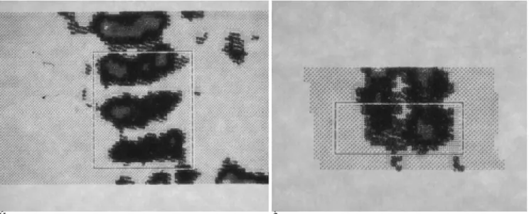
기기는 이중 에너지 방사선 흡수법을 이용한 골밀도 측정기 로 XR-26 MARK II (NORLAND, Baarn, Netherlands)를 사 용하였다. 측정 범위는 요추는 횡경 2.7 cm으로 통일하였고, 장경은 제 2요추에서 제 4요추까지를 포함하였다. 손목은 횡 경 2.5 cm, 장경은 요골 말단부에서 근위부 1 cm 까지 포함 시켰다. 임상적으로 안정된 상태로 공복 후 수유함으로써 수면 을 유도하여 앙와위로 실시하였고, 요추는 2분30초, 손목은 1 분 30초 동안 스캔하였다 (Fig. 1).
생화학적 표지자는 미숙아군의 55명에서 골밀도 측정 시점 에 혈청 칼슘, 인, alkaline phosphatase를 검사하였다.
미숙아군과 만삭아군 사이에 부위별 골밀도 평균에 차이가 있는지를 알아보았고, 미숙아군 내에서 수태 나이, 출생 체중 에 따라 골밀도의 차이가 있는지를 알아보았다. 또한 생화학적 표지자와 골밀도가 상관 관계를 보이는지 알아보았다. 통계학 적 유의성은 student t-test 및 상관분석(correlation analy- sis)을 사용하였고, 유의 수준은 p=0.05로 정하였다.
결 과
부위별 골밀도 평균치는 미숙아군은 요추에서 0.137±0.018 g/cm2 (0.061-0.202 g/cm2), 손목에서 0.089±0.013 g/cm2
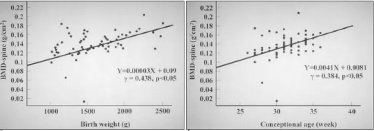
는 경향을 보였다 (Fig. 2). 손목의 골밀도와 출생 체중과는 정 상관 관계가 있었으며(r=0.281, p < 0.05), 수태 나이와는 통 계학적으로 유의한 상관 관계가 없었다(r=0.223, p > 0.05) (Table 2, Fig. 3).
Table 1. Comparison of Bone Mineral Densities (BMD) Using Dual Energy X-ray Absorptiometry on Preterm and Full-term Infants
preterm (n=68) full-term (n=31) p-value BMD-spine 0.137±0.018 0.214±0.030 p<0.05 (g/cm2)
BMD-wrist 0.089±0.013 0.118±0.014 p<0.05 (g/cm2)
student t- test
Table 2. Correlation Coefficients of Bone Mineral Density versus Conceptional Age and Birth Weight on Preterm Infants
Conceptional age Birth weight at birth
BMD-spine r value 0.384** 0.438**
p value 0.001 0.000
BMD-wrist r value 0.223 0.281*
p value 0.068 0.020
* correlation is significant at the 0.05 level
** correlation is significant at the 0.01 level
A B
Fig. 1. Representative scan image of lumbar spine (A) and wrist (B) in preterm infant. The subject is a female infant (conceptional age 31weeks, birth weight 2250 g) with corrected age 0. Bone mineral density of lumbar spine is 0.135 g/cm2and that of wrist is 0.085 g/cm2.
그러나 미숙아군 중 55명에서 골밀도 측정과 같은 날 시행 한 혈청 칼슘(r=0.167, p=0.223; r=0.080, p=0.563), 인 (r=0.064, p=0.642; r=-0.029, p=0.835), alkaline phos- phatase (r=-0.158, p=0.248; r=-0.098, p=0.447)와 요추 및 손목의 골밀도 사이에는 통계학적으로 유의한 상관 관계가 없었다.
고 찰
미숙아에서 골감소증의 빈도는 13-32%로 보고되고 있으 며, 진단은 혈청 내 생화학적 표지자 및 다양한 방사선학적 검 사를 사용한다 (5, 7, 13-15). 본 연구에서는 미숙아와 만삭 아의 골밀도를 측정을 위해 DEXA를 사용하였고, 이는 다른 검 사법에 비해 방사선 피폭량이 적고, 짧은 시간에 시행할 수 있 으며, 객관적인 수치를 얻을 수 있다는 장점이 있어 소아의 골 밀도 측정에 가장 유용할 것으로 생각하였다 (1, 8, 12, 16-
19).
측정부위로 피질골이 많은 사지골 보다는 해면질이 풍부한 척추가 외부 자극에 대해 반응성이 크고 골대사 회전이 빨라, 척추에서의 골밀도 측정이 골감소증의 진단 및 치료에 대한 반 응을 더욱 예민하게 반영할 것이라 알려져 있다 (4, 6, 9). 실 제 임상에서 미숙아 골감소증의 진단에 흔히 쓰는 방법은 혈 청 칼슘, 인, alkaline phosphatase 등의 생화학적 표지자와 손 목 부위의 단순 촬영을 통한 수근 골격 변화를 보기 때문에 (1, 10), 요추와 손목의 골밀도를 측정하게 되었다.
과거 다른 연구에서 사춘기 이전의 만삭아 및 미숙아 모두 에서 남녀의 골밀도 차는 거의 없다고 하여 (1), 남녀의 구분 은 하지 않았다.
본 연구의 정상 만삭아에서 요추 골밀도치는 평균 0.214±
0.030 g/cm2 (0.160-0.296 g/cm2)로 Braillon (2) 등이 만삭 아의 요추에서 DEXA를 이용하여 얻은 골밀도치인 평균 0.268
±0.030 g/cm2 (0.227-0.307 g/cm2)와 Tsukahara (4) 등의
A B
Fig. 2. Correlation of lumbar spine-BMD with birth weight (A) and conceptional age (B). The lumbar spinal BMD correlated signifi- cantly with birth weight (r=0.438, p<0.05) and conceptional age (r=0.384, p<0.05).
A B
Fig. 3. Correlation of wrist-BMD with birth weight (A) and conceptional age (B). The wrist BMD correlated significantly with birth weight (r=0.281, p<0.05) but did not correlate significantly with conceptional age (r=0.223, p>0.05).
찾을 수 없어 비교가 불가능하였다.
본 연구의 교정 나이 0개월에 시행한 미숙아군의 전체적인 평균 골밀도치는 요추및 손목에서 0.137±0.018 g/cm2, 0.089
±0.013 g/cm2로 만삭아에 비해 의의 있게 낮았다 (p<0.05).
미숙아군 내에서 수태 기간이 짧을수록, 출생시 체중이 적을 수록 요추의 골밀도치가 낮아서 다른 문헌의 연구와 같은 결 과를 얻었다(1, 2, 8, 12). 전 (12) 등이 미숙아에서 전신골밀 도를 측정하여 출생 체중, 수태 나이와 전신골밀도가 정상관 관계(linear relationship)를 보인다고 한 것과 일치하였다. 손 목의 골밀도는 출생 체중이 클수록 증가했고 (r=0.281, p <
0.05), 수태 기간과는 통계학적으로 유의한 상관관계가 없었다 (r=0.223, p > 0.05).
교정나이 0개월에 시행한 혈청 칼슘, 인, alkaline phos- phatase와 요추 및 손목의 골밀도는 통계학적으로 유의한 상 관관계를 보이지 않아, James (20) 등이 혈청 alkaline phos- phatase와 골밀도간에 상관관계가 없었다고 한것과 같은 결과 를 보였다.
미숙아군에서 골밀도가 전반적으로 낮은 데는 다양한 원인 요소가 있다. 태아의 골 성장은 정상 만삭아 무기질 함량의 80%가 임신 마지막 삼 개월 동안 급속한 골 기질화를 통해 이 루어지는데, 미숙아의 경우 모체로부터 무기질 및 칼슘, 인 등 을 제공받을 시기를 상대적으로 제한 받음으로 골감소증의 위 험이 만삭아에 비해 높다. 생후 모유나 우유만으로는 무기질 공급이 대개 불충분하여 특히 태어나서 처음 몇 개월 동안은 같은 수태기간에 해당되는 정상 만삭아에 비해 골밀도도 낮고, 현성 구루병이나 골절 등이 흔하다 (1-4, 17, 20-22). 또한 만성 이뇨제 사용 등 신생아 집중치료실 처치 중 무기질 소실 도 원인이 될 것이다. 이 연구에서는 미숙아군은 모두 같은 미 숙아 분유를 섭취하였지만, 무기질 식이량 및 흡수량에 개개인 의 차이가 있을수 있으며, 전비경구적 영양(total parenteral nutrition), 이뇨제 사용 등의 외적 요건이 고려되지 않았다는 제한점이 있었다. 그러나 향후 미숙아 골감소증의 진단 및 치 료 후의 추적 관찰에 객관적인 지표로서 DEXA를 이용한 골 밀도 측정이 도움을 줄 것으로 기대된다.
또한 저자들의 연구 결과로 만삭아의 요추 골밀도 평균인 0.214±0.030 g/cm2 (0.160-0.296 g/cm2)와 손목의 골밀도 인 0.118±0.014 g/cm2 (0.096-0.162 g/cm2)가 절대값으로 보기에는 성급하다. 그러나 미숙아의 경우 교정 나이가 0개월 이 되었을 때 골밀도는 만삭아 보다 요추에서는 약 2/3에 지 나지 않았고, 손목에서는 3/4에 지나지 않아 미숙아의 골밀도 가 낮은 것은 확실하다.
결론적으로 교정나이 0개월에 측정한 미숙아의 골밀도 측정
Measurement of bone mineral content of spine by dual energy X- ray absorptiometry in normal children: correlations with growth parameters. J Clin Endocrinol Metab 1990;70:1330-1333
2. Braillon PM, Salle BL, Brunet J, Glorieux FH, Delmas PD, Meunier PJ. Dual energy X-ray absorptiometry measurement of bone mineral content in newborns : validation of the technique.
Pediatr Res 1992;32:77-80
3. Henderson RC. Assessment of bone mineral content in children. J Pediatr Orthop 1991;11:314-317
4. Tsukahara H, Sudo M, Umezaki M, et al. Measurement of lumbar spinal bone mineral density in preterm infants by dual energy X- ray absorptiometry. Biol Neonate 1993;64:96-103
5. Behrman RE, Kligman RM, Arvin AM. Rickets of vitamin D defi- ciency in nutritional disorders. In Nelson WE. Textbook of pediatrics, 15th ed. Philadelphia : Saunders, 1996:179-183
6. Lang P, Steiger P, Faulkner K, Gluer C, Genant HK. Osteoporosis.
Radiol Clin North Am 1991;29:49-76
7. Koo WWK, Gupta JM, Nayanar VV, Wilkinson M, Posen S.
Skeletal changes in preterm infants. Arch Dis Child 1982;57:447- 452
8. Grampp S, Jergas M, Gluer CC, Lang P, Brastow P, Gennant HK.
Radiologic diagnosis of osteoporosis. Radiol Clin North Am 1993;
31:1133-1145
9. Faulkner KG, Gluer CC, Majumdar S, Lang P, Engelke KE, Gennant HK. Noninvasive measurements of bone mass, structure and strength : current methods and experimental techniques. AJR Am J Roentgenol 1991;157:1229-1237
10. Yochum TR, Rowe LJ. Nutritional, metabolic and endocrine disor- ders. Essentials of skeletal radiology 2nd ed. Baltimore: Williams &
Wilkins, 1996:1327-1370
11. Steichen JJ, Gratton TL, Tsang RC. Osteopenia of prematurity: the cause and possible treatment. J Pediatr 1980;96:528-534
12. 전봉진, 허진도, 신상범 등. 미숙아의 골감소증 진단에 있어서 전신 골밀도 검사의 유용성 : 단 순촬영 및 생화학적 표지자와의 비교분 석. 대한방사선의학회지 1997;36:337-342
13. Kovar I, Mayne P. Plasma alkaline phosphatase activity in the preterm neonate. Acta Paediatr Scand 1981;70:501-506
14. Pittard WB, Geddes KM, Hursely TC, Hollis BW. Osteocalcin, skeletal alkaline phosphatase, and bone mineral content in very low birth weight infants : a longitudinal assessment. Pediatr Res 1992;31:181-185
15. Kroger H, Kotaniemi A, Vainio P, Alhava E. Bone densitometry of the spine and femur in children by dual energy X-ray absorptiome- try. Bone Miner 1992;17:75-85
16. Hansen MA, Hassager C, Overgaard K, Marslew U, Riis BJ, Christiansen C. Dual energy X-ray absorptiometry: A precise method of measuring bone mineral density in the lumbar spine. J Nucl Med 1990;31:1156-1162
17. Forbes GB. Some remarks on bone mineralization. J Pediatr 1988;
113:167-171
18. Sartoris DJ, Resnick D. Dual energy radiographic absorptiometry for bone densitometry: current status and perspective. AJR Am J Roentgenol 1989;152:241-246
19. Barden HS, Mazess RB. Bone densitometry in infants. J Pediatr
1988; 113(suppl):172-177
20. James JR, Congdon PJ, Truscott J, Horsman A, Arthur R.
Osteopenia of prematurity. Arch Dis Child 1986;61:871-876 21. Kulkarni PB, Hall RT, Rhodes PG, et al. Rickets in very low birth
weight infants. J Pediatr 1980;96:249-252
22. Callenbach JC, Sheehan MB, Abramson SJ, Hall RT. Etiologic fac- tors in rickets of very low-birth-weight infants. J Pediatr 1981;98:
800-805
J Korean Radiol Soc 2000;43:371-375
Address reprint requests to : Seung Cheol Kim, M.D., Department of Diagnostic Radiology, Dankook University Hospital, College of Medicine, 29 Anseodong Chonan Choongnam, 330-715, Korea.
Tel. 82-41-550-6921 Fax. 82-41-552-9674 E-mail: [email protected]
The Difference of Bone Mineral Density of Lumbar Spine and Wrist in the Preterm and Full-term Infants:
Using Dual Energy X-ray Absorptiometry
1Min Jung Cha, M.D., Seung Cheol Kim, M.D., Young Seok Lee, M.D., Young Pyo Chang, M.D.2, Jin-Young Park, M.D.3
1Department of Diagnostic Radiology, Dankook University Hospital, College of Medicine
2Department of Pediatrics, Dankook University Hospital, College of Medicine
3Department of Orthopedic Surgery, Dankook University Hospital, College of Medicine
Purpose: To assess the differences in bone mineral density (BMD) of lumbar spine and wrist between preterm infants of postconceptional age 40 weeks and normal full-term infants.
Materials and Methods: Sixty-eight preterm infants born at conceptional age 26-36 weeks and 31 normal full- term infants born at 38-42 weeks were investigated. Bone mineral densities of the lumbar spine (from the sec- ond to the fourth segment) and wrist were measured by dual energy X-ray absorptiometry. In preterm infants, the corrected age of 0 month was defined as postconceptional 40 weeks. Full-term infants were evaluated within three days of birth, and the average bone mineral densities of preterm and full-term infants were com- pared. In the preterm group, birth weight and conceptional age were correlated with lumbar spinal and wrist bone mineral densities. Data were analyzed by student’s t-test and Pearson’s correlation coefficient, and a p value of less than 0.05 was considered significant.
Results: In preterm in fants, the values of bone mineral densities of the lumbar spine and wrist were 0.137±
0.018 g/cm2(0.061-0.202 g/cm2) and 0.089±0.013 g/cm2(0.065-0.123 g/cm2), respectively, while the respec- tive values for full-term infants were 0.214±0.030 g/cm2(0.160-0.296 g/cm2) and 0.118±0.014 g/cm2(0.096- 0.162 g/cm2). In the preterm group, lumbar spinal BMD correlated significantly with conceptional age (r=0.384, p<0.05) and birth weight (r=0.438, p<0.05). While wrist BMD correlated significantly with birth weight (r=0.281, p<0.05), its correlation with conceptional age was not significant (r=0.223, p>0.05).
Conclusion: The lumbar spinal and wrist BMDs of preterm infants at corrected age 0 were lower than those of normal full-term infants. In the preterm group, BMD values for the lumbar spine were lower in infants of low- er conceptional age and birth weight.
Index words : Bones, absorptiometry Bones, mineralization Bone density
Infants, newborn, skeletal system
E-mail: [email protected])
■ 7TH ANNUAL MEETING EUROPEAN SOCIETY OF MUSCULO-SKELETAL RADIOLOGY (ESSR) (2000년 10월 27-28일)
venue: Leiden, The Netherlands.
contact: Dr. W. R. Obermann, Prof. J. L. Bloem, Leiden University Medical Ctr.,
Dept. of Radiology, C2-S, Albinusdreef 2, NL-2333 AZ Leiden, The Netherlands.
(tel: 31-71-5261913; fax: 31-71-5248256;
E-mail: [email protected])
■ FIFTH INTERNATIONAL SYMPOSIUM ON CARDIOVASCULAR AND INTERVENTIONAL RADIOLOGY (2000년 11월 5-9일)
venue: Seaport Boston Hotel, Boston, MA, USA.
contact: Danielle Pokorski, Dept. of Radiology, 75 Francis Street, Boston, MA 02115, USA.
(tel: 1-617-7326265; fax: 1-617-7326509)
■ PRACTICAL TRAINING IN INTERVENTIONAL RADIOLOGY COURSE (2000년 11월 6-10일) venue: Univ. Hosp. Sart Tilman, Lie`ge, Belgium.
contact: Prof. R. F. Dondelinger, Univ. Hosp. Sart Tilman, Domaine du Sart Tilman B35, B-4000 Lie`ge 1, Belgium.
(tel: 32-4-3667259; fax: 32-4-3667224;
E-mail: [email protected])
■ 86TH MEETING RADIOLOGICAL SOCIETY OF NORTH AMERICA (RSNA) (2000년 11월 26일-12월1일) venue: McCormick Place, Chicago, USA.
contact: Steven T. Drew, Ass. Executive Director, 820 Jorie Boulevard, Oak Brook, IL 60523-2251, USA.
(tel: 1-630-5717879; fax: 1-630-5717837;
E-mail: [email protected])
■ 32ND ANNUAL SCIENTIFIC CONFERENCE AND EXHIBITION OF THE BRITISH MEDICAL ULTRASOUND SOCIETY (BMUS) (2000년 12월 6-8일)
venue: Devonshire Park Centre, Eastbourne, United Kingdom.
contact: Mrs. Elaine Brown, General Secretary BMUS, 36 Portland Place, London WIN 3DG, United Kingdom.
(tel: 44-171-6363714; fax: 44-171-3232175;
E-mail: [email protected])
■ INTERNATIONAL ENDOVASCULAR SYMPOSIUM (2000년 12월 10-12일)
venue: Sydney, Australia.
contact: Ms Margaret Blackwell, Abacus Management, P. O. Box 77, Pymble NSW 2073, Australia.