One would expect the size of vertebral bodies to in- crease gradually in the lower direction to the level of L5-S1 (1, 2). But in fact some variation occurs, For ex- ample, the lower vertebral bodies may be smaller than the upper ones. In this situation, changes in vertical stress can be a causative factor in disc degeneration (3).
As far as we are aware, however, no previous report has described the different types of vertebral hypoplasia and the relationship between vertebral hypoplasia and re- gional disc abnormality. In this paper we therefore clas- sify the types of vertebral hypoplasia and investigate the prevalence and the patterns of associated disc degenera- tion.
Materials and Methods
We retrospectively reviewed the sagittal and axial MR imaging findings of the spine in an initial series of 58 pa-
Regional Disc Change in Segmental Hypoplasia of the Lumbosacral Vertebral Bodies: MR Findings1
Sung-Kyu Kim, M.D., Seung-Ro Lee, M.D., Won-Jin Moon, M.D., Dong-Woo Park, M.D., Chang-Kok Hahm, M.D.
Purpose: To classify types of vertebral hypoplasia and to investigate the prevalence and patterns of associated disc degeneration.
Materials and Methods: Defining vertebral hypoplasia as occurring when the AP diam- eter of a lower vertebral body is smaller than that of an upper ones, we retrospectively reviewed the MR images obtained in 34 cases of this condition invloving young adults.
Two major types and two subtypes, a total of four different entities were classified as follows; type I: hypoplasia involving a single vertebral body; type II: hypoplasia in- volving serial lower segmental vertebral bodies; subtype a: hypoplastic body located anteriorly along the anterior spinal line; subtype b: hypoplastic body located posterior- ly along the posterior spinal line. We also investigated each type of vertebral hypopla- sia and patterns of associated disc changes.
Results: Three different types were observed. In type IIa(n= 29), posterior disc oc- curred in 8/29 cases, diffuse degeneration in 21/29 patients, and posterior disc hernia- tion in all. All type Ia cases(3/3) showed diffuse disc degeneration at both upper and lower disc levels, with posterior disc herniation, while both type IIb cases(2/2) showed diffuse disc degeneration, with bidirectional disc herniation.
Conclusion: By identifying the exact patterns of vertebral hypoplasia, we were able to predict which portion of the disc was likely to degenerate.
Index words : Spine, MR
Spine, intervertebral disks Spine, abnormalities
1Department of Radiology, Hanyang University Hospital Received January 17, 2000 ; Accepted May 30, 2000
Address reprint requests to : Seung-Ro Lee, M.D., Department of Diagnostic Radiology, College of Medicine, Hanyang University.
17, Haengdang-Dong, Sungdong-Gu, Seoul 133-792, Korea.
Tel. 82-2-2290-9164 Fax. 82-2-2293-3111
tients with vertebral hypoplasia. Exclusion criteria in- cluded 1) previous spinal disc surgery, 2) the existence of spondylolysis or spondylolisthesis, 3) age over 35years (it is well known that beyond this age, disc changes are most clearly associated with the aging process), 4) incomplete imaging(lost films). The final def- inite series consisted of 34 patients (M:F = 26:8) aged between 17 and 35 (mean, 27.1) years. Images of the spine were obtained using a 1.5T unit (GE Medical Systems, Milwaukee, Wis., U.S.A.) and a sequence which included sagittal T1-weighted(TR/TE: 600/39), T2-weighted (TR/TE: 2000/80) and proton density- weighted(TR/TE: 2000/30) spin echo imaging of the en- tire lumbar spine with the following sequence parame- ters: 256×192 matrix, 260 mm field of view, 5 mm sec- tion thickness, and 0.5 mm intersectional gap. In addi- tion, axial T1-weighted (TR/TE: 600/39) and T2-weight- ed (TR/TE: 2000/80) spin echo images of all lumbar in- tervertebral spaces (three sections per disc level from L1 to S1) were obtained with the following sequence para- meters: 256×128 matrix, 160 mm field of view, 4 mm section thickness, 0.5 mm intersectional gap. Vertebral hypoplasia was defined as occuring when the AP diame- ter of a lower vertebral body was less than that of an up- per one, with abrupt transition of the anterior or posteri- or spinal line, as seen on three consecutive middle sliced sagittal MR images. The AP diameters of adjacent end
plates of vertebral bodies as seen on mid sagittal MR im- ages, were measured and the findings were expressed as the ratio(in percentage). On the basis of the radiographic findings, the following four different entities were class- fied two major types and two subtypes (Figure. 1). Type I: hypoplasia involving a single vertebral body; Type II:
hypoplasia involving serial lower segmental vertebral bodies; Subtype a: hypoplastic body located anteriorly along the anterior spinal line, Subtype b: hypoplastic body located posteriorly along the posterior spinal line.
We also investigated the prevalence and location of disc degeneration(anterior, posterior or diffuse) and the di- rection of disc herniation(anterior, posterior, or bilateral- ly) at the level of hypoplastic variation. Through which axial cuts were taken at the upper and lower levels.
Both axial and sagittal cuts were used to assess the loca- tion of disc degeneration and direction of disc hernia- tion. The signal intensity observed on T2-weighted im- ages in the central four-fifths of the T11-T12, T12-L1, or L1-L2 disc was considered the normal standard for each given patient. Discs showing a detectable decrease with respect to that standard were considered to have abnor- mal signal intensity and disc extension beyond the inter- space was considered to be disc herniation. All these im- ages were retrospectively analysed by three neuroradi- ologists who reached a consensus.
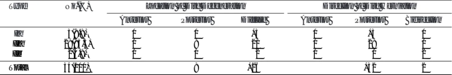
Table 1. Prevalence of Each Type of Vertebral Hypoplasia and Patterns of Associated Disc Changes
Type No.(%) Location of Disc Degeneration Direction of Disc Herniation Anterior Posterior Diffuse Anterior Posterior Bidirection
Ia 03(8.8)0 0 0 0*3 0 0*3 0
IIa 29(85.3) 0 8 *21 0 *29 0
IIb 02(5.9)0 0 0 *02 0 000 2
Total 34(100). 8 *26 *32 2
* Incidence at upper and lower disc level
Fig. 1. Diagram illustrates four types of vertebral hypoplasia.
Results
In all 34 patients, hypoplastic lower vertebral bodies had a smaller AP diameter than upper ones, and the ra- tio of AP diameter between adjacent end plates varied from 83:100 to 93:100(mean 91:100). After reviewing the radiographic imaging findings, four different types of vertebral hypoplasia were classified. Because we had used exclusion creteria in order to reduce bias, only
three different types were included in our study group, However. Type IIa occurred in 85.3% of cases (29/34), type Ia in 8.8% (3/34), and type IIb in 5.9% (2/34). Levels at which segmental hypoplasia was commonly involved were L4-5 (20/34) and L5-S1 (13/34). Among the 29 type IIa patients, anterior disc degeneration was not found, through posterior disc degeneration was noted in 8/29, diffuse disc degeneration in 21/29 and posterior disc her- niation in all 29. In all three type Ia patient, diffuse disc degeneration was found at both upper and lower disc
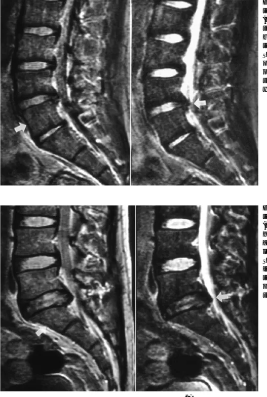
Fig. 2. 23-year-old man of Type IIa ver- tebral hypoplasia between L4 and L5.
(left) Sagittal proton-weighted image shows hypoplasia below L5 vertebral body and hypoplasic bodies located an- teriorly along anterior spinal line (right) Sagittal T2-weighted image shows loss of normal signal intensity at the posteri- or portion of the L4-5 disc with exten- sion of the posterior disc margin be- yond the interpace (arrow).
Fig. 3. 34-year-old man of Type IIa ver- tebral hypoplasia between L5 and S1.
(left) Sagittal T1-weighted image shows hypoplasia below S1 vertebral body and hypoplasic bodies located anterior- ly along anterior spinal line (right) Sagittal T2-weighted image shows dif- fuse loss of normal signal intensity of the L5-S1 disc with extension of the posterior disc margin beyond the inter- space (arrow).
levels and all three showed posterior disc herniation. In both type IIb patients, diffuse disc degeneration and
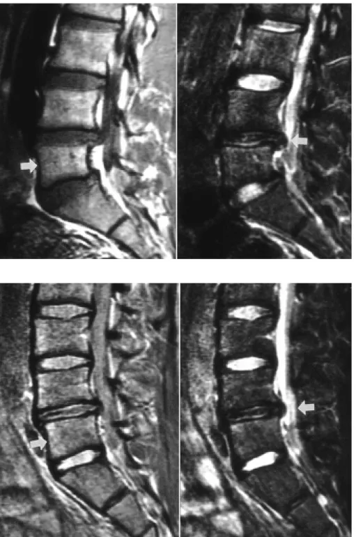
intervertebral discs and mechanical considerations, indi- cate that these weights subject the chain mainly to verti- Fig. 5. 20-year-old man of Type IIb ver- tebral hypoplasia between L4 and L5.
(left) Sagittal T1-weighted image shows hypoplasia below L5 vertebral body and hypoplasic bodies located posteri- orly along posterior spinal line (right) Sagittal T2-weighted image shows loss of normal signal intensity of the L5-S1 disc with bi-directional disc extension beyond the interspace (arrow).
Fig. 4. 27-year-old man of Type Ia ver- tebral hypoplasia involving L5 verte- bral body. (left) Sagittal T1-weighted image shows hypoplasia involving only L5 vertebral body and hypoplastic body located anteriorly along anterior spinal line (right) Sagittal T2-weighted image shows loss of normal signal in- tensity of L4-5 and L5-S1 discs with ex- tension of the posterior disc margin be- yond the interspace (arrow).
vertebral bodies, for example, are more hypoplastic than upper ones. In this situation abrupt reduction in size of a lower vertebra leads to the exertion of more compressive force per unit area upon a disc surface than in a case where the lower vertebal body is its usual larg- er size. It is now generally accepted that in addition to intervertebral disc degeneration, chronic mechanical compressive stress plays an important role in the devel- opment of intervertebral disc herniation (6-8). It may thus be assumed that hypoplasia of a lower vertebral body can be a causative factor in disc degeneration(in- cluding disc herniation). Though many distinct types of vertebral hypoplasia have been proposed, no previous report has examined the relationship between these and regional disc abnormality. The types proposed include hypoplasia involving a single vertebral body, the type involving serial lower segmental vertebral bodies, the type involving a series of vertebral bodies and hypopla- sia of an upper segmental vertebral body with abrupt enlargement of a lower body. After a review of the radi- ographic imaging findings, four different types of verte- bral hypoplasia were identified, but because of our ex- clusion criteria applied in order to reduce bias, only three types were included in our study. Most cases of vertebral hypoplasia occurred at the level of L4-5(20/34) and L5-S1(13/34). The most common form is type IIa, in which all associated disc degenerations was posterior or diffuse and in which posterior directional disc hernia- tion had occurred in all cases. In type Ia hypoplasia, all cases showed diffuse disc degeneration and posterior disc herniation at both upper and lower disc levels.
While in type IIb, diffuse disc degeneration and bidirec- tional disc herniation were found. It appears that for
each type of vertebral hypoplasia, the disc involved has a more vulnerable portion: for each of these types, an understanding of the basic pathogenesis of disc degener- ation will require the use of biomechanical models.
In conclusion, vertebral hypoplasia can be a causative factor of early onset degenerative disc disease. By identi- fying the exact pattern of vertebral hypoplasia, we can therefore, predict which portion of disc is likely to de- generate. The findings we have described may be useful for the clinician who needs to focus on those areas sus- ceptible to disc degeneration.
References
1. Davis PR. Human lower lumbar vertebrae: some mechanical and osteological considerations. J Anat 1966;95:337-344
2. Wang TM & Shih C. Morphometric variations of the lumbar verte- brae between Chinese and Indian adults. Acta Anat 1992;144:23- 29
3. Lotz JC & Colliou OK, Chin JR, Duncan NA, Liebenberg E.
Compression-induced degeneration of the intervertebral disc: an in vivo mouse model and finite-element study. Spine 1998;23:2493- 506
4. Pal GP & Routal RV. Transmission of weight through the lower thoracic and lumbar regions of the vertebral column in man. J Anat 1987;152:93-105
5. Pal GP& Routal RV. A study of weight transmission through the cervical and upper thoracic regions of the vertebral column in man. J Anat 1986;148:245-61
6. Ishihara H, Matsui H, Ryusuke O, et al. Facet joint asymmetry as a radiologic feature of lumbar intervertebral disc herniation in chil- dren and adolescents. Spine 1997;22:2001-2004
7. Hutton WC, Toribatake Y, Elmer WA et al. The effect of compres- sive force applied to the intervertebral disc in vivo. Spine 1998;23:2524-2537
8. Iatridis JC, Mente PL, Stokes IA, Aronsson DD, Alini M.
Compression-induced changes in intervertebral disc properties in a rat tail model. Spine 1999;24:996-1002
대한방사선의학회지 2000;43:25-30
분절성 요천추체 형성부전증과 추간판 국소변화: 자기공명영상소견1
1한양대학교 의과대학 진단방사선과학교실
김성규・이승로・문원진・박동우・함창곡
목적: 분절성 척추체 형성부전증의 유형을 분류하고 각 유형별로 동반되는 추간판의 퇴행성 변화양상을 연구하고자 하였다.
대상과 방법: 하부 척추골의 전후 직경이 상부 척추골의 전후 직경보다 작은 경우를 분절성 척추체 형성부전증으로
정의하였고 분절성 척추체 형성부전증을 보이는 34명의 척추 자기공명영상을 후향적으로 분석하였다. 4가지 유형의 분절성 척추체 형성부전증(유형I: 단일 척추체의 형성부전, II: 연속된 하부 척추체의 형성부전, 아유형a: 형성부전의 척추체가 전척추선을 따라 전방으로 위치하고 있는 경우, 아유형b: 형성부전의 척추체가 후척추선을 따라 후방으로 위치하고 있는 경우)을 가정하였고 각 유형별로 변이가 있는 위치에서 동반되는 추간판의 퇴행성 변화양상을 연구 하였다.
결과: 3가지 다른 유형의 분절성 척추체 형성부전증을 볼 수 있었다. 유형 IIb(29예)에서 추간판 후측에만 퇴행성 변 화가 있었던 경우가 8예, 전반적인 퇴행성 변화를 보인 경우가 21예 였고 모든 경우에서 후방 추간판 탈출증을 보 였다. 유형 Ia(3예)의 경우 형성부전을 보이는 추체 상하부 추간판의 모든 예에서 전반적인 추간판 퇴행성 변화와 후 방 추간판 탈출증을 보였다. 유형 IIb(2예)의 모든 예에서 추간판의 전반적인 퇴행성 변화와 전후방 추간판 탈출증 을 보였다.
결론: 분절성 척추체 형성부전증의 유형을 분류함으로써 이러한 환자에서 생길 수 있는 추간판 퇴행성 변화의 위치 와 유형을 예측할 수 있을 것이다.