Spasticity is the main feature of the many symptoms of cerebral palsy (CP). Continuous spasticity in CP causes injury to the muscles and tendons, leading to their subsequent regeneration and degeneration. Among the cytokines and growth factors participating in these regeneration and degen- eration processes, the transforming growth factor- (TGF- ) plays an important role in the formation of fibrosis while upregulating wound healing at the time of a muscle injury
1,2,4,7,8). TGF- induces cellular proliferation and increases
the rate of synthesis of both collagen and the cell matrix by stimulating various cells9,11). This study examined the expres- sion of the TGF- isoforms and collagen composition with- in the Achilles tendon of CP patients with an equinovarus deformity.
MATERIALS AND METHODS
From March 2001 to March 2002, 15 CP patients under- went an Achilles tendon lengthening for spastic equinovarus A 2×2×5 mm3specimen of the Achilles tendon was obtain- ed from the mid-substance. As a control, the same samples were obtained from healthy persons who had undergone surgery for a traumatic rupture of the tendon (Table 1). The expression level of the TGF- isoform and the collagen com- position were determined using histological studies, immu- nohistochemical staining (IHS) and the mRNA expression level was examined using a reverse transcriptase polymerase chain reaction (RT-PCR).
1. Immunohistochemistry
Immunohistochemical analyses of TGF- 1, TGF- 2, and TGF- 3 as well as, type I and III collagen were performed using the avidin-biotin complex (ABC) method. The speci- mens were frozen and stored at -70℃prior to immunos- taining. Four-micrometer parallel sections were cut on a cryo- stat, mounted on a probe on plus slides, and air-dried. The
531 531
Increased Expression of the TGF- Isoform and Changed Contents of Collagen in Tendon of Cerebral Palsy Patients
Sung Taek Jung, M.D., Hyoung Yeon Seo, M.D., Jae Joon Lee, M.D., Myung Sun Kim, M.D., Yang Kyung Kim, M.D.
and Gye Jin Kim, M.D.
Department of Orthopaedic Sugery, Chonnam National University Medical School, Gwangju, Korea
531 531 Address reprint requests to
Sung-Taek Jung, M.D.
Department of Orthopaedic Surgery, Chonnam National University Hospital, 8 Hak-dong, Dong-gu, Gwangju 501-757, Korea
Tel: +82.62-220-6336, Fax: +82.62-225-7794 E-mail: [email protected]
*This work was financially supported by Chonnam National University in the program, 2001.
Purpose: This study measured the expression level of the transforming growth factor- (TGF- ) isoform expression and the collagen composition within the Achilles tendon from cerebral palsy (CP) patients.
Materials and Methods: The Achilles tendons were obtained from 15 CP patients with spastic equino- varus. The presence of the TGF- isoform and the composition of the collagen were examined histologi- cally, performing by immunohistochemical staining (IHS) and determining the mRNA expression level using a reverse transcriptase polymerase chain reaction (RT-PCR).
Results: IHS revealed the presence of TGF- 1 and TGF- 2 expression in 2/15 cases and 4/15 cases respectively, and weak TGF- 3 expression in 7/15 cases. The TGF- 1 and TGF- 2 expression levels were uniform in all 15 cases according to RT-PCR, while TGF- 3 expression was observed in 8 out of 15 cases.
IHS and RT-PCR showed strong TGF- 3 expression in 6/7 non-ambulatory patients. An immature form of collagen, type III collagen, was observed more abundantly in the non-ambulatory patients.
Conclusion: These results suggest that contracture in CP may induce changes in the type of collagen via growth factors such as TGF- .
Key Words: Cerebral palsy, Achilles tendon, Transforming growth factor- (TGF- ), Collagen
sections were incubated with the primary antibodies against TGF- 1, TGF- 2 and TGF- 3 for 2 hours, and type I and III collagen for 1 hour in a humidity chamber at room tem- perature (listed in Table 1). Biotin-labeled secondary antibod- ies (Zymed Laboratories, USA) were utilized for 7 minutes at 45℃. The streptavidin-horseradish peroxidase (Zymed Laboratories, USA) detection system was then applied to the capillary channels, followed by 7 minutes of incubation at 45℃. After drainage, the tissue sections were ready for the chromogen reaction with 3-amino-9-ethyl carvazole (AEC, Zymed Laboratories, USA). The sections were counterstained with hematoxylin and mounted on a Universal Mount (Re- search Genetics, USA).
2. RNA isolation and RT-PCR
The specimen was freshly frozen in liquid nitrogen. The total RNA from the tissue was extracted using the method- ology for the TriZol reagent (Gibco BRL, Life Technologies).
The RNA (1 g) was reverse-transcribed using the Super- script First-Strand Synthesis System for RT-PCR (Gibco BRL, Life Technologies).
The primers for TGF- 1, - 2, - 3, type I, type III col- lagen, and actin, which is a constitutively expressed house- keeping gene, were designed from a reference paper. Table 2 shows the primer sequences. The PCR reaction mixture con- tained 2 Lof each cDNA sample, 10 pM each of sense and antisense primers, and the other PCR reagents in a final vol-
ume of 20 L. The PCR reagents, dNTP, Taq DNA poly- merase, 10×reaction buffer (40 mM KCl, 10 mM Tris-HCl pH 9.0, 1.5 mM MgCl2, stabilizer and tracking dye) were obtained from Accupower�PCR PreMix (Bioneer, Korea).
The PCR cycles were at 94℃for 5 minutes, and 35 cycles of denaturation at 94℃for 1 minute. The annealing temper- ature was set for 1 minute, and polymerization was performed at 72℃for 2 minutes followed by 72℃for 10 minutes.
The PCR products were electrophoresed on 1.0 percent agarose gel, visualized by ethidium bromide staining, and photographed under UV light. The TGF- 1 cDNA, - 2 cDNA, - 3 cDNA, the type I cDNA collagen, type III cDNA collagen, and -actin cDNA were semi-quantified by IMAGERTM & 1D MAIN (Bioneer, Korea)
The mRNA integrity and amplification efficiency of the TGF- isoforms, as well as the type I and type III collagen transcripts were evaluated by amplifying the -actin sequence from the equivalent amounts of the total RNA from each sample.
RESULTS
1. Clinical characteristics
The average age of the 15 patients (10 males and 5 females) was 10 years (6-17 years). Seven patients were spastic para- plegic and 8 were hemiplegic. All 7 paraplegics were non- ambulatory and the 8 hemiplegics were ambulatory.
2. Histological findings
The control specimens collected from a 38-year-old female
None -: 0-25%, Mild +: 26-50%, Moderate ++: 51-75%, Severe +++:
76-100%.
1 2 3
TGF- isoform No. Age Sex Type
1 9 G Whole body Nonambulatory - ++ + 2 11 B Whole body Nonambulatory - + + 3 11 B Whole body Nonambulatory - - - 4 7 B Whole body Nonambulatory - - + 5 12 B Hemiplegia Ambulatory - - + 6 11 G Hemiplegia Ambulatory - + - 7 17 G Hemiplegia Ambulatory + + - 8 6 B Hemiplegia Ambulatory - - - 9 7 G Whole body Nonambulatory - - + 10 12 B Hemiplegia Ambulatory + - - 11 11 B Whole body Nonambulatory - - + 12 9 B Hemiplegia Ambulatory - - - 13 8 B Whole body Nonambulatory - - + 14 11 B Hemiplegia Ambulatory - - - 15 8 G Hemiplegia Ambulatory - - - Table 1.Demographic data
Primer sequence (Sense/Antisense)
PCR Product
size
Anneal- ing Temper-
ature mRNA
Template
-actin 5 -CTGGAGCATGCCCGTATTTA-3 280 bp 54℃
5 -TTTGGTCTTGCCACTTTTCC-3
TGF- 1 5 -CCAACTATTGCTTCAGCTCCA-3 196 bp 54℃
5 -TTATGCTGGTTGTACAGGGC-3
TGF- 2 5 -CTGGAGCATGCCCGTATTTA-3 233 bp 54℃
5 -TTTGGTCTTGCCACTTTTCC-3
TGF- 3 5 -CCAATTACTGCTTCCGCAACT-3 211 bp 54℃
5 -GCAGATGCTTCAGGGTTCAG-3
Type I 5 -CCCCCTCCCCAGCCACAAAGA-3 360 bp 51℃
Collagen 5 -TCTTGGTCGGTGGGTGACTCT-3
Type III 5 -CCAAACTCTATCTGAA-3 449 bp 34℃
Collagen 5 -GGACTCATAGAATACA-3 Table 2.Primer sequences
′
′
′
′
′
′
′
′
′
′
′
′
′
′
′
′
′
′
′
′
′
′
′
′
3 days after the trauma showed that the Achilles tendon was composed of fibroblasts and matrix including collagen fibers.
The collagen fibers were parallel to the longitudinal axis of
the tendon with fibroblasts located between the fibers. Com- pared with the control, no difference was observed in the ambulatory CP. However, tendinolipomatosis and decreased cellular distribution were observed in 2 cases of nonambu- latory CP (Fig. 1).
3. Immunohistochemical staining (IHS) 1) TGF- isoforms
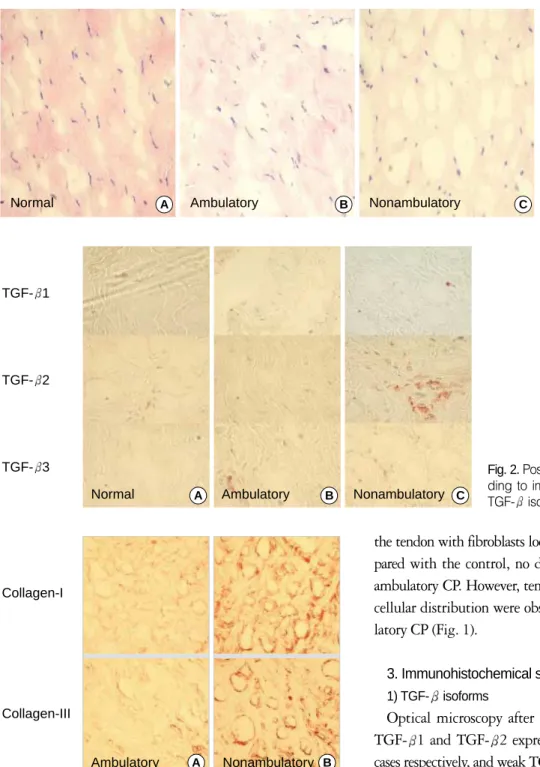
Optical microscopy after IHS revealed the presence of TGF- 1 and TGF- 2 expression in 2/15 cases and 4/15 cases respectively, and weak TGF- 3 expression in 7/15 cases (Fig. 2). It was also noted that TGF expression was observed only in the tenocytes but not in the matrix. This study did not determine the matrix composition and the level of teno- cyte proliferation according to the type of paralysis and the degree of TGF- expression.
2) Collagen staining
Type I collagen showed relatively dark stain and type III collagen light stain in the ambulatory CP patients. How-
Fig. 1.The histologic findings from the Achilles tendon; (A) The normal tendon shows the parallel matrix with abundant fibroblasts present within the matrix. (B, C) Mucoid regener- ation is seen between fibrob- lasts in the CP Achilles tendon (Hematoxylin & eosin, ×200).
A B C
Normal Ambulatory Nonambulatory
Fig. 2.Positive and negative findings accor- ding to immunohistochemical staining of TGF- isoforms.
A B C
TGF- 1
TGF- 2
TGF- 3
Normal Ambulatory Nonambulatory
Fig. 3.Findings of immunohistochemical staining of Type I and Type III collagens: (A) Type I collagen with relatively abundant bundles is seen. Type III collagen has collagen bundle between fibroblasts. (B) Increased Type I and Type III collagens are seen in non-ambulatory CP patients.
B Collagen-I
Collagen-III
Ambulatory A Nonambulatory
ever, type I and type III collagen were stained the same in the nonambulatory CP patients with a relative increase in the immature type III collagen level (Fig. 3). No correla- tion was found between the expression level of the TGF- isoforms and the degree of collagen staining.
4. The level of mRNA according to RT-PCR
TGF- 1 and TGF- 2 expression were observed in all cases, whereas TGF- 3 expression was observed only in 8 out of 15 cases. The expression of the TGF- isoform was strong in 6 out of the 7 non-ambulatory patients and weak in all 8 hemiplegic patients. The type III collagen level was upregulated in all 7 cases showing TGF- 3 upregulation.
Regarding the collagen composition, immature type III col- lagen was observed in all 15 CP cases. The distribution of type III collagen was more prominent in the non-ambula- tory CP cases than that of the type I collagen. However, the expression level of the TGF- isoform increased evenly and the level of collagen synthesis was also increased in the trau- ma group. These results are similar to those of the spastic non-ambulatory CP cases, which might coincide with the phase of the active inflammatory repair process.
5. TGF- isoform expression and collagen composition According to the IHS, the expression level of the TGF- isoform was not uniform. Therefore, no correlation was found between the degree of collagen staining and TGF expression.
However, the mRNA level according to RT-PCR showed increased synthesis of the TGF- isoform and collagen in the non-ambulatory CP. It was also increased in the control
group (Fig. 4).
DISCUSSION
Muscle spasticity in CP induces muscle and tendon injury.
However, the complete regeneration of the injured tissues is hindered as a result of repetitive spasms, eventually lead- ing to muscle contracture and a secondary joint deformity6). A normal tendon is composed of fibroblasts and collagen fibers. Collagen fibers form a triple-stranded helix and are composed of type I collagen in 95% of cases. On the other hand, type III collagen is present in an immature form and is usually present during the healing stage5,10,13-17,19-21). There are few reports on the growth factor or cytokines associated with muscle damage that occurs after CP, which is the main subject of this study.
Chang et al.9)reported, after experimenting with TGF- 1 antibodies in rabbits, that TGF- 1 participates in the for- mation of excessive scarring by improving the tendon excur- sion. Shin et al.16)reported that the expression of type I col- lagen mRNA and collagen formation, as well as cellular pro- liferation in the tendon cells are promoted with the intro- duction of TGF- 1 alone or in combination with other growth factors, indicating that TGF- actively participates in the healing process of injured tendons inducing fibrosis.
In our CP patients, it is believed that the continued spasms were probably the cause of the increase in the TGF- level rather than the uncontrollable action of the TGF- isoform according to the clinical findings.
The increase in the type III collagen level in the rupture site of the human Achilles tendon is probably due to con-
Fig. 4.The mRNA level according to RT- PCR showed an upregulation of TGF- and Type III collagen within the Achilles tendon of non-ambulatory CP patients.
N, Normal site; I, Injured site.
TGF- 1 TGF- 2 TGF- 3 -actin
Collagen-I Collagen-III
-actin
Nonambulatory Ambulatory TraumaN I
Trauma N I
Nonambulatory Ambulatory
tinued microtrauma and the subsequent healing process3). The quantitative measurement of the type I to type III col- lagen ratio was measured in the tendon using IHS18). This study was also able to determine a difference in collagen staining and the mRNA level. The injured tendons produce type III collagen during the inflammatory phase and the type I collagen during the reparative phase. In addition, col- lagen formation is reduced and the collagens is arranged lon- gitudinally in the remodeling phase, which can last for 20 weeks13). This study using IHS and RT-PCR also confirmed the increase in the type III collagen level within the CP ten- don. A continued spasm in CP patients would initiate a microtrauma and induce an injury, which may increase the collagen III level via the participation of TGF- . It is pos- sible that the type III collagen level would decrease when further tissue damage is prevented by controlling the spasms in CP patients. The histological examination revealed hypox- ic degenerative tendinopathy, mucoid degeneration, ten- dolipomatosis or combination of these in the injured ten- dons12). In the fresh trauma which was used as the control, the TGF- expression level was increased in the rupture site.
However, the alignment of the collagen fibers was regular and the major composition was collagen I. This study also observed similar findings in the Achilles tendon of the severe CP cases. Alioto et al.1)experimented with the palma fascia of Dupuytren’s disease and predicted that the contracture could be prevented by inhibiting the TGF- and collagen receptors. However, in CP, the tendon injury induced by a spasm would produce growth factors such as TGF- to repair the injury, which in turn would increase the level of collagen synthesis. The initial pulse would repeat and con- tinue with time, which is in contrast to traumatic rupture.
Therefore, it is important to continue with physical thera- py to decrease the spasm and prevent the trauma from spas- ticity. The early and active rehabilitation will help in the formation of a mature matrix.
The Type III collagen mRNA level was upregulated in the tendons of the non-ambulatory patients as result of the severe spasm at the time of the upregulated TGF- mRNA level.
However, this study could not determine whether or not the presence of cellular proliferation is related to the upreg- ulation of TGF- mRNA.
A further study with a larger number of subjects and the
standardization of objective measurements in the spasticity in CP and in vivo studies will clarify the direct relationship between TGF- and collagen formation.
CONCLUSION
This study focused on a histological study of the Achilles tendon of spastic CP patients, the degree and pattern of TGF- expression, and the changes in the collagen types.
The results of this study can provide the basic data for under- standing the functions of the growth factors not only in CP but also in those diseases related to tendon contracture. It can also be used to implement the logical treatments and to improve the clinical evaluations of the disease.
REFERENCES
1. Alioto RJ, Rosier RN, Burton RI and Puzas JE: Comparative effects of growth factors on fibroblasts of Dupuytren’s tissue and normal palmar fascia. J Hand Surg, 19-A: 442-452, 1994.
2. Aronson D, Wojtaszewski JF, Thorell A, et al: Extracellu- lar-regulated protein kinase cascades are activated in response to injury in human skeletal muscle. Am J Physiol, 275: C555-C561, 1998.
3. Bakou S, Cherel Y, Gabinaud B, Guigand L and Wyers M:
Type-specific changes in fibre size and satellite cell activation fol- lowing muscle denervation in two strains of turkey. J Anat, 188:
677-691, 1996.
4. Bernasconi P, Di Blasi C, Mora M, et al: Transforming growth factor-beta1 and fibrosis in congenital muscular dystrophies. Neu- romuscul Disord, 9: 28-33, 1999.
5. Billeter R, Weber H, Lutz H, Howald H, Eppenberger HM and Jenny E:Myosin types in human skeletal muscle fiber. His- tochemistry, 65: 249-259, 1980.
6. Bleck EE: Orthopedic management in cerebral palsy. London, Lippincott: 121-141, 1987.
7. Border WA and Noble NA: Transforming growth factor beta in tissue fibrosis. N Engl J Med, 331: 1286-1292, 1994.
8. Carlson BM and Faulkner JA: The regeneration of skeletal mus- cle fibers following injury: a review. Med Sci Sports Exerc, 15: 187- 198, 1983.
9. Chang J, Most D, Stelnicki E, et al: Gene expression of trans- forming growth factor beta-1 in rabbit zone II flexor tendon wound healing: evidence for dual mechanisms of repair. Plast Reconstr Surg, 100: 937-944, 1997.
10. Eriksen HA, Pajala A, Leppilahti J and Risteli J: Increased
content of type III collagen at the rupture site of human Achilles tendon. J Orthop Res, 20: 1352-1357, 2002.
11. Hurme T, Kalimo H, Lehto M and Jarvinen M: Healing of skeletal muscle injury: an ultrastructural and immunohistochem- ical study. Med Sci Sports Exerc, 23: 801-810, 1991.
12. Kannus P and Jozsa L: Histopathological changes preceding spontaneous rupture of a tendon. A controlled study of 891 patients.
J Bone Joint Surg, 73-A: 1507-1525, 1991.
13. Liu SH, Yang RS, al-Shaikh R and Lane JM: Collagen in ten- don, ligament, and bone healing. A current review. Clin Orthop, 318: 265-278, 1995.
14. Maffulli N, Ewen SW, Waterston SW, Reaper J and Bar- rass V:Tenocytes from ruptured and tendinopathic achilles ten- dons produce greater quantities of type III collagen than tenocytes from normal achilles tendons. An in vitro model of human tendon healing. Am J Sports Med, 28: 499-505, 2000.
15. Ngo M, Pham H, Longaker MT and Chang J: Differential expression of transforming growth factor-beta receptors in a rab- bit zone II flexor tendon wound healing model. Plast Reconstru Surg, 108: 1260-1267, 2001.
16. Shin DE, Kang HJ, Kim HW, et al: Effect of growth factors on
type I collagen synthesis in cultured rabbit’s deep flexor tendon cell. J Korean Orthop Assoc, 37: 288-294, 2002.
17. Silver RL, de la Garza J and Rang M: The myth of muscle balance. A study of relative strengths and excursions of normal muscles about the foot and ankle. J Bone Joint Surg, 67-B: 432- 437, 1985.
18. van der Loos CM, Marijianowski MM and Becker AE:
Quantification in immunohistochemistry: the measurement of the ratios of collagen types I and III. Histochemical J, 26: 347-354, 1994.
19. Waggett AD, Ralphs JR, Kwan AP, Woodnutt D and Ben- jamin M:Characterization of collagens and proteoglycans at the insertion of the human Achilles tendon. Matrix Biology, 16: 457- 470, 1998.
20. Warren GL, Hayes DA, Lowe DA and Armstong RB: Mecha- nical factors in the initiation of eccentric contraction-induced injury in rat soleus muscle. J Physiol, 464: 457-475, 1993.
21. Woo SLY, An KN, Frank CB, et al: Anatomy, biology, and biomechanics of tendon and ligament. In: Orthopaedic basic sci- ence: biology and biomechanics of the musculoskeletal system. 2nd ed. Rosemont, American Academy of Orthopaedic Surgeons: 582- 595, 2000.
목 적: 뇌성마비 환자의 아킬레스건 내 TGF- isoform의 발현 정도와 콜라겐 구성을 알고자 하였다.
대상 및 방법: 뇌성마비로 인한 경직성 첨족마비 환자 중 수술적 연장술을 시행하였던 15명 환자의 아킬레스건 중앙부를 약 2×2×5 mm3크기로 채취한 후, 면역조직화학 염색과 RT-PCR을 이용하여 TGF- 와 콜라겐 구성을 조사하였다.
결 과: 아킬레스건 중앙부의 구성은 섬유모세포와 콜라겐 섬유 등의 기질로 구성되어 있었다. 면역조직화학 염색상 TGF- 1과 TGF- 2는 15예 중 각각 2예, 4예에서만 발현되었으며, TGF- 3의 경우는 7예에서 약하게 발현되었다. 경직성이 심 한 비보행성 마비를 보인 7예 중 6예에서 TGF- 3의 양성 소견을 보였다. 콜라겐 염색상 경직성이 심한 비보행성 마비 환 자에서는 상대적으로 미성숙형태인 제3형의 콜라겐이 증가됨을 알 수 있었다. 면역조직화학 염색 및 RT-PCR상 TGF- 3 가 중등도 이상 발현된 7예에서 제3형 콜라겐이 증가되어 있었다.
결 론: 뇌성마비로 인한 지속적인 근 구축은 건내 콜라겐 성분의 변화를 초래하고 이 과정중 TGF- 가 관여할 것으로 사료 된다.
색인 단어: 뇌성 마비, 아킬레스 건, TGF- , 콜라겐
뇌성마비 환자의 아킬레스건에서 TGF- 발현 및 콜라겐 조성의 변화
정성택∙서형연∙이재준∙김명선∙김양경∙김계진
전남대학교 의과대학 정형외과학교실