Plate Cage of Benezech Implant를 이용한 경추골유합술에서 척추 간격의 높이와 전굴 각도의 변화
동국대학교 의과대학 경주병원 신경외과학교실
목진호·이영배·이규춘·박용석
Changes of Intervertebral Height and Lordotic Curvature in Anterior Cervical Fusion with the Plate Cage of Benezech Implant
Jin Ho Mok, MD, Young Bae Lee, MD, Kyu Chun Lee, MD and Yong Seok Park, MD
Department of Neurosurgery, College of Medicine, Dongguk University Gyongju Hospital, Gyeongju, Korea
Objective: The anterior cervical interbody fusion may not provide adequately immediate stabilization, whereas anterior cervical spine plating has overcome this main disadvantage. But various complications related to cervical spine plating system were also reported. In variable cervical pathologic conditions, the adequate intervertebral disc space height and lordotic curvature are the major factor for satisfactory outcome. The authors investigated the changes of the intervertebral height and lordotic curvature in anterior cervical fusion with the Plate Cage of Benezech (PCB) implant. Methods: From May 1999 to April 2001, 15 patients with cervical spine disorders [herniated nucleus pulposus (HNP), spondylosis &
trauma] were underwent anterior cervical spine fusion with the PCB implant (cervical plate cage system). Single level fusion was accomplished in 9 patients and two level fusion in 6 patients. The radiographic data (height of intervertebtal disc and lordotic angles) were obtained retrospectively from routine clinical radiographes preoperatively, immediate- postoperatively, and at 18-25 months postoperatively. The mean follow-up period was 20 months. Results: All patients show some improvement of their symptoms. The height of intervertebral disc was significantly increased from 5.2 mm preoperatively to 8.2 mm at last follow-up. The average increasement of lordotic angle of the entire cervical spine was 12.3°and Cobb angle was 7.8°. The increased height of intervertebral disc and lordotic angles were maintained th- roughout the follow-up period. Conclusion: We think that maintenance of the intervertebral disc height and cervical lordosis is one of the important factors for good prognosis, and that PCB implant can be considered to be one of the useful hardware for anterior cervical spine fusion in variable cervical spine disorders. (J Kor Neurotraumatol Soc 2007;3:29-33) KEY WORDS: Anterior cervical fusion·PCB implant·Intervertebral disc height·Lordotic curvature.
서 론
1955년 Smith와 Robinson16)이 장골 능선에서 얻은 골 이식편을 이용한 전방 경추 디스크 제거술에 대한 발 표 이후 이 방법이 경부 동통을 동반한 척추증, 경추 신
경근 병증과 경추 척수증에 대한 치료로서 널리 시행되 어 왔다. 그러나 이식 골편 돌출이나 이탈, 골유합 부전, 각 변형 등이 문제점으로 보고되었고14,18,20,24) 이러한 문 제점을 해결하기 위하여 금속판 고정 기구를 이용한 방 법이 시도되어 왔다. 금속판 고정 수술은 수술 초기의 불 안정성 및 골편 이동을 방지할 수 있는 장점이 있으나 금속판과 나사못이 빠지거나 골절되는 경우, 식도 천공, 연하곤란 등의 또 다른 합병증이 발생할 수 있고 골유합 과정에서 척추 간격이 감소되는 상황이 발생하는 것으로 보고되고 있다.7,8,12,18,21,22) 이에 저자는 요추부에서 사용 Address for correspondence: Young Bae Lee, MD
Department of Neurosurgery, College of Medicine, Dongguk Uni- versity Gyongju Hospital, 1090-1 Seokjang-dong, Gyeongju 780-350, Korea
Tel: +82-54-770-8230, Fax: +82-54-770-8234 E-mail: [email protected]
되는 cage system과 금속판을 병합한 plate cage of be- nezech (PCB) implant를 이용한 경추 전방 유합술을 시 행하여 좋은 결과를 얻었기에 문헌고찰과 함께 보고하는 바이다.
대상 및 방법
1999년 5월부터 2001년 4월까지 본원 신경외과에 경 추 질환으로 입원한 후 PCB implant를 사용하여 전방경 유 경추체 골유합술을 시행 받은 환자를 대상으로 임상기 록 및 방사선학적 자료에 대한 후향적 조사를 하였다. 환 자는 남자 9명, 여자 6명이었으며 이들의 평균 연령은 48.18세였다. 수술 적응증은 수술 전 신경근 병증이 뚜 렷하고 자기공명영상(MRI) 촬영 사진에서 임상 증상과 잘 맞는 병소가 확인된 경우로 하였다. 수술 방법은 전신 마취하에 앙와위에서 두부를 약 10도 정도 신전시키고 피 부 절개를 정중앙부에서 횡으로 약 4~6 cm정도의 길이 로 우측에 만들었다. 경동맥초를 외측으로, 식도와 기관지 를 내측으로 견인하면서 박리를 하여 경추 전방 부위를 노출하였다. 수술 현미경하에서 디스크를 제거하고 척추증 이 동반된 환자에서는 고속 드릴과 큐렛을 이용하여 골극 을 제거하여 신경근 감압을 하였다. 골유합을 유도하기 위 하여 고속 드릴과 큐렛으로 종판을 제거하고 PCB im- plant를 삽입하고 두 개의 나사못으로 수술 부위 상-하 추체에 고정하였다. 수술 후 모든 환자에서 Philadelphia 경추 보조기를 착용하도록 하였고 수술 후 2~3일째 보행 을 허용하였다. 수술 전, 수술 직후 및 마지막 추적 관찰 (3~12개월 사이, 평균 6개월) 때 시행한 경추 측면 단순
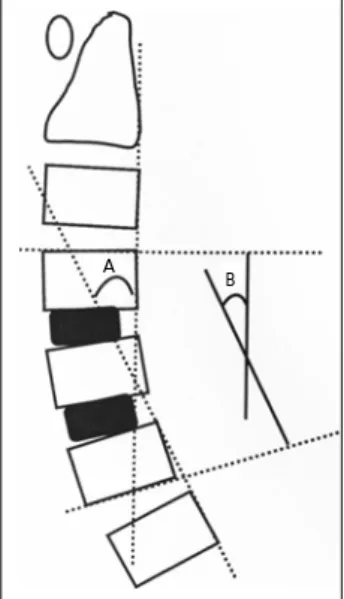
방사선 사진에서 척추 간격의 높이를 측정하고 일정한 촬 영 조건을 위해 제 3 경추체의 전후방 길이를 측정하여 촬 영 시마다 발생할 수 있는 오차에 대한 보정을 하였다. 경 추의 전굴 각도는 Katssura 방식10)에 따라 전체 경추의 전굴 각도 (angle A)와 골유합 분절의 전굴 각도 (angle B)를 측정하여 분석하였다 (Figure 1). 수술 후 임상 증 상은 Odom의 분류에 따라 분석하였다.
결 과
병소 부위는 하나 (single level)인 경우가 9명이었고 다발성 (multiple level)인 경우가 6명으로 모두 2 level 이었다. 각 부위별로는 C3~4가 2예, C4~5가 5예, C5~6 이 9예, C6~7이 5예 모두 21 level이었다 (Table 1).
신경학적 소견으로는 13명에서 신경근 병증을 보였고 2 명에서 척수 병증을 보였다. 신경근 병증을 보인 13명 모 두 주 증상이 경부 동통 및 상지 방사통이었고 이들 중 3 명에서 grade III-IV의 상지 운동 약화를 보였다. 척수 병증을 보인 2명에서는 하지의 심부건 반사 항진, 경도 의 사지 부전 마비에 의한 보행 장애를 보였다. PCB im- plant는 8 level에서 PCB 1 (높이: 5.5 mm, 길이: 16 mm)을 사용하였고 13 level에서 PCB 2 (높이: 7.0 mm, 길이: 16 mm)를 사용하였으며 나사못은 3 병소에서 14 mm를 사용하고 나머지 18 병소 부위에서는 16 mm를 사용하였다. 추적 관찰 기간은 18개월에서 25개월까지 평균 20개월이었다. 모든 예에서 수술 후 증상의 호전을 보였으며 Odom 분류에 따른 결과는 excellent가 7명, good이 5명, satisfactory가 3명이었다. 신경근 병증에 의한 상지 운동 약화를 보였던 3명은 모두 정상 근력으 로 회복되었고, 5명에서 수술 후 일시적인 연하곤란을 호소하였으나 3~5일 이내에 자연 소실되었고 기구와 연 관된 합병증은 없었다. 수술 후 모든 환자에서 척추 간격 의 증가를 보였으며 수술 전에 비하여 수술 직후에는 평 균 2.7 mm 증가, 마지막 추적 사진에서는 평균 3.0 mm 증가를 보여 추적기간 중 증가된 척추 간격이 잘 유지되
TABLE 1. Lesion level with the number of cases Level No. of cases (%)
C3-4 02 (009.5)
C4-5 05 (023.8)
C5-6 09 (042.9)
C6-7 05 (023.8)
Total 21 (100.0)
FIGURE 1. Measurement of the angle of the cervical spine.
Angle A is the supplementary angle between the line paral- lel with the dorsal border of the second vertebra and seventh vertebra. Angle B is formed by the upper plane and the lower plane of the fused segment.
A B
었다 (Table 2). 전체 경추의 전굴 각도 (angle A)는 수 술 전에 11.5도였고 수술 직후가 13.6도, 최종적으로 20.4도를 나타내었으며 골유합 분절의 전굴 각도 (angle B)는 수술 전에 0.3도, 수술 직후 5.4도, 최종적으로 8.1 도를 나타내었다 (Table 3). 수술 전에 angle A가 평균 11.5도이고 angle B가 0도에 가까운 것은 경추의 전만곡 이 심하게 감소하였거나 전만곡이 반전되었던 경우가 많 음을 의미하며 수술 후에는 이러한 비정상적인 전만곡의 감소가 교정되고 증가된 경추 전굴 각도가 지속적으로 유지되는 것으로 관찰되었다 (Figures 2, 3).
고 찰
경추 질환의 치료에 있어 전방 경유법을 이용한 디스크
TABLE 2. Serial changes of height of intervertebral disc space (mean value)
Height (mm) Level
Pre-op Post-op Last
C3-4 5.5 9.0 9.4
C4-5† 4.4 7.4 8.0
C5-6* 5.3 7.7 7.9
C6-7* 6.0 8.5 8.5
Total 5.2 7.9 8.2
*p<0.05, by Friedman’s oneway analysis of variance (ANOVA),
†p<0.01, by Friedman’s oneway ANOVA
TABLE 3. Serial changes of angle A and angle B (mean value) Angle Pre-op Post-op Last Angle A* 11.5° 13.6° 20.4°
Angle B* 00.3° 05.4° 08.1°
*p<0.01, by Friedman’s oneway analysis of variance (ANOVA)
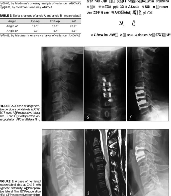
FIGURE 2. A case of degenera- tive cervical spondylosis at C5- 6-7 level. A: Preoperative lateral film. B and C: Postoperative an-
teroposterior (AP) and lateral film. A B C
FIGURE 3. A case of herniated intervertebral disc at C4-5 with kyphotic deformity. A: Preopera- tive lateral film. B: Preoperative
MRI. C: Postoperative lateral film. A B C
제거술과 장골편을 이용한 골유합술에 대하여 1955년 Robinson16)이 처음 기술한 이후 Bailey와 Baldgley 등1) 은 원뿔꼴 모양의 중첩골이식 방법을 경추골절 환자에 적용하면서 획기적인 발판이 마련되었다. 경추 전방 접 근법은 체위 변경으로 인한 신경 손상의 위험을 줄이고 전 방 감압술과 함께 견고한 골유합을 실시함으로써 경추 안 정성을 조기에 얻을 수 있는 장점이 있으나 수술 후 가 관절, 골이식편의 전위나 탈출, 각변형 등의 합병증이 발 생할 수 있다. 이에 1980년 Bohler와 Gaudernak2)이 최 초로 하경추부 골절-전위 환자에서 전방 금속판으로 고 정술을 시행하여 전방 경유 경추 유합술로 초래될 수 있 는 여러 단점을 보완하였으나 이 또한 수술 중 금속판 고 정 시에 척수 손상을 줄 수 있고 금속판과 나사에 의한 이 차적 합병증의 가능성이 있어 사용에 제한이 있었다. 1986 년 Caspar가 능형 금속판을 이용하여 경추 손상 환자 60 명에 대한 임상 연구를 통하여 자가 골편 이식과 금속판 을 동시에 고정하는 복합적인 방법으로 좋은 결과를 발 표한 이후 각광을 받기 시작하였다.4) Caspar plate sys- tem은 양피질 나사 고정을 통하여 생체 공학적 안정성을 확보하였으나5) 후방 피질골 관통 시 경수 손상이 유발될 수 있어 수술 중 세심한 주의를 요하며 특히 후방 피질골 의 나사관통이 1~2 mm 이상인 경우에는 재수술을 시행 하여야 하는 단점이 보고되었다.9) 1986년 Morscher가 개발한 경추 잠금 금속판 고정 기구는 티타니움 재질을 사용함으로써 방사선학적 적합성을 높였을 뿐 아니라 일 측 피질골 관통 나사를 채택함으로써 후방 피질골 관통 으로 인한 신경 손상의 위험성을 감소시킨 것으로 보고 되었다.15) 그러나 이들 잠금 나사 금속판에서 아직도 나 사의 풀림, 골절 등의 합병증이 보고되며 몇몇 생체 공학 적 실험에서 양피질 나사에 비하여 나사의 후퇴력(pull- out strength)이 약하여 안정성을 확보하지 못한 것으로 보고되고 있다.17,19) 그 외에 여러 형태의 금속판-나사 시스템이 개발되었고 현재 국내에서도 많은 종류의 금속 판-나사 시스템이 사용되고 있다.11,23) 하지만 이와 같은 금속판 고정술은 수술 후 초기에 불안정성 및 이식 골편 의 이동을 방지할 수 있는 장점이 있는 이외에 금속판과 나사의 이완이나 골절에 의한 불안정성의 재발, 식도 천 공, 연하곤란 등의 합병증이 초래될 수 있으며 특히 금속 판-나사 고정술을 하였어도 시간이 경과함에 따라 척추 고정분절의 높이가 감소되는 경우가 있는 것으로 보고되 고 있다.3,6-8,12) 또한 경추 유합 부위 상-하부에 퇴행성 변화와 굴-신전 각도의 증가를 유발하는 상황이 생기기 도 한다.13) 본원에서 사용한 PCB implant는 티타니움
재질로 방사선학적 적합성을 높였으며 기존의 금속판- 나사 기구와 요추부에서 사용되는 cage 기구를 접목시킨 내고정 기구로서 수술 조작이 간편하고 cage의 상부면 이 볼록한 모양으로 되어 있어 축방향의 힘(axial loading) 을 직접 cage가 받음으로서 나사에 대한 부하(stress)를 감소시켜 나사의 이완, 골절의 위험이 작고 척추 간격을 유지하는 cage의 전방, 상-하방이 개구되어 있어 이곳 으로 골편을 이식함으로써 척추 간격의 감소 없이 골유 합을 가능하게 한 기구이다. 또한 상-하 고정판이 사선으 로 배열되어 있어 2 level 이상에서도 사용이 용이하며 특히 상부 고정판이 후방으로 굴곡 되어 있어 경추의 전만 곡을 지속적으로 유지시켜 줄 수 있는 장점이 있다. 총 21 level의 수술 환자에서 cage의 크기는 수술 전 방사 선 사진에서 측정한 척추 간격에 따라 선택하였으며 수 술 후 모든 환자에서 척추 간격이 증가하였다. 특히 수술 직후에 평균 2.7 mm 증가하였으며 마지막 추적 방사선 사진상 평균 3.0 mm 증가하여 추적 관찰 기간 중 증가 된 척추 간격이 지속적으로 유지되는 것으로 관찰되었다.
1998년 Cho 등6)이 골편 이식과 금속판 고정 수술을 동 시에 시행한 후 고정 분절의 높이와 Cobb 각도의 변화 에 대한 임상 연구에서 수술 직후에는 고정 분절의 높이 가 증가하지만 추적 관찰 기간 중 다시 감소하는 것으로 발표하였다. 퇴행성 경추 질환에서는 변성된 추간판에 의 해 척추 간격의 감소, 이차적으로 발생하는 신경공 크기 의 감소, 경추 전만곡의 변화 등이 증상 발현의 중요한 요소이기 때문에 수술 후 적절한 척추 간격이 유지되어야 지속적인 증상의 호전을 기대할 수 있으며 척추 간격이 다시 감소하는 경우에는 좁아진 신경공에 의해 신경근이 압박되어 증상이 재발될 가능성이 있다. 전체 경추의 전 굴 각도는 주로 전체 경추의 기능적 상태를 나타내는 것 으로 수술 후 전만곡의 회복 및 유지는 증상의 호전과 관계가 있는 것으로 보고되고 있다.6) 본원에서 수술을 시 행한 모든 예에서 전만곡이 증가되었다. 수술 전에 비하 여 수술 직후에 전체 경추의 전굴 각도가 평균 2.1도 증 가하였으며 추적 관찰 기간 중 전만곡이 서서히 증가하 여 마지막 추적 방사선 사진상 평균 8.9도 증가하였다.
이것은 수술 전 경부 동통 등으로 인하여 경추 전체의 전 만곡이 감소하였다가 수술 후 동통의 소실과 더불어 전 체 경추의 기능적 적응이 일어나가 때문인 것으로 판단 된다. 또한 고정 분절의 전굴 각도가 수술 전에 0도에 가까운 것은 병소 부위 경추에서 전만곡의 심한 감소, 또 는 반전이 있었던 것을 의미하며 수술 직후 5.1도 증가, 최종적으로 7.8도 증가하여 병소 부위에서도 전굴 각도
가 호전되는 것으로 관찰되었다. 비록 추적 관찰 기간이 평균 15개월이지만 PCB implant와 연관된 합병증은 아 직 관찰되지 않았고 척추 간격과 경추 전굴 각도가 수술 전에 비하여 증가되고 계속 유지되는 소견을 보였으며 모든 환자에서 증상의 호전을 보여 만족할 만한 결과가 나왔다.
결 론
PCB implant는 조작이 간편하고 지속적인 척추 간격 및 척추 전만곡의 유지, 낮은 기구 위험도 등의 장점이 있 어 퇴행성 경추 질환을 포함한 다양한 경추 질환에 대한 전방경유 경추 고정수술에서 유용하게 사용될 수 있는 기 구로 생각된다. 또한 퇴행성 경추 질환에서 수술 후 지속 적인 척추 간격과 척추 전굴 각도의 유지가 지속적인 증 상의 호전 및 재발 방지와 관련이 있는 것으로 생각된다.
중심 단어: 전방 경추체 골유합술·PCB implant·척추 간격의 높이·척추 전골 각도.
REFERENCES
1) Baile RW, Bagley CE. Stabilization of cervical spine by anterior fusion. J Bone and Joint Surg 42:565-595, 1960
2) Bohler J, Gaudernak T. Anterior plate stabilization for frac- ture-dislocation of the lower cervical spine. J Trauma 20:203- 205, 1980
3) Brown JA, Havel P, Ebraheim N. Cervical stabilization by plate and bone fusion. Spine 13:236-240, 1988
4) Caspar W. Anterior cervical fusion and interbody stabilization with the trapezial osteosynthetic plate technique, in Aesculap:
Scientific Information Leaflet S-039, ed 7. Buringame CA:
Aesculap Instruments Corp, pp36, 1986
5) Caspar W, Barbier DD, Klara PM. Anterior cervical fusion and Caspar plate stabilization for cervical trauma. Neurosur- gery 25:491-502, 1989
6) Cho DS, Song JH, Kim EY, Kim MH, Park HK, Kim SH, et al. Clinical study of the serial changes of fixed segmental height and Cobb angle in anterior cervical fusion. J Kor Neu- rosurg Soc 27:1210-1215, 1998
7) Cooper PR. Operative management of cervical spine injuries.
Clin Neurosurgery 34:650-674, 1988
8) Gassman J, Seligson D. The anterior cervical plate. Spine 8:700- 707, 1983
9) Goffin J, Plets C, Vande Bergh R. Anterior cervical fusion and osteosynthetic stabilization according to Caspar. Neurosurgery 25:865-871, 1989
10) Katsuura A, Hukuda S, Imanaka T, Miyamoto K, Kanemoto M. Anterior cervical plate used in degenerative disease can maintain cervical lordosis. J Spinal Disord 9:470-476, 1996 11) Kim S, Kim TS, Koh JS, Lim GK, Rhee BA. Anterior cervical
bisegmental interbody fusion with titanium (RABEA) in de- generative cervical disease: comparative analysis with mono- segmental interbody fusion. J Korean Neurosurg Soc 34:440 -444, 2003
12) Kim YS, Shin HS, Bak KH, Kim JM, Kim YS, Ko Y, et al.
Clinical evaluation of salvage surgery for patients with failed an- terior cervical surgery. J Kor Neurosurg Soc 26:113-118, 1997 13) Letha Y, Hunter MD, Ethan M. Radiographic changes following
anterior cervical fusion. Spine 5:399-401, 1980
14) McAfee PC, Bohman HH. One stage anterior cervical decom- pression and posterior stabilization with circumferential ar- throdesis. J Bone Joint Surg 71A:78-88, 1989
15) Morscher E, Satter F, Jennis M. Die vordere verplattung der halswirbelsaule mit dem hohischrauben platten system. Der Chirrurg 57:702-707, 1986
16) Robinson RA, Smith GW. Anterolateral cervical disc removal and interbody fusion for cervical disc syndrome. Bull Johns Hopkins Hosp 96:223-224, 1955
17) Shapiro S. Banked fibula and the locking anterior cervical plate in anterior cervical fusions following cervical discectomy. J Neurosurg 84:161-165, 1996
18) Shapiro S, Connolly P, Donnaldson J, Abel T. Cadaveric fibula, locking plate, and allogenic bone matrix for anterior cervical fusions after cervical discetomy for radiculopathy or myelo- pathy. J Neurosurg (1 Suppl) 95:43-50, 2001
19) Smith SA, Lindsey RW, Doherty BJ. An in-vitro biome- chanical comparison of the Orosco and AO locking plates for anterior cervical spine fixation. J Spinal Disord 8:220-223, 1995
20) Suh PB, Kostuik JP, Essess SI. Anterior cervical plate fixation with the titanium hollow screw plate system. A preliminary re- port. Spine 15:1079-1081, 1990
21) Vavruch L, Hedlund R, Javid D, Leszniewski W, Shalabi A. A prospective randomized comparison between the cloward procedure and a carbon fiber cage in the cervical spine: a clin- ical and radiologic study. Spine 27:1694-1701, 2002 22) Wigfield CC, Nelson RJ. Nonautologous interbody fusion ma-
terials in cervical spine surgery: how strong is the evidence to justify their use? Spine 26:687-694, 2001
23) Yoon CS, Yoon SH, Park HC, Park HS, Kim SH, Kim EY.
Clinical applocation and surgical results of hollow cage (RAB- EATM) without bone graft in the one-segment cervical spine interbody fusion. J Korean Neurosurg Soc 34:17-22, 2003 24) Zeidman SM, Ducker TB, Raycroft J. Trends and complica-
tions in cervical spine surgery. J Spinal Disord 10:523-526, 1997