INTRODUCTION
Hepatitis B virus (HBV) persistently infects approximately 350 million people worldwide, and induces a spectrum of chronic liver disease ranging from chronic hepatitis through liver cirrhosis to hepatocellular carcinoma (HCC) (1). The persistent HBV carrier state is very frequently (up to 90%) transmitted from infected mothers seropositive for hepatitis B e antigen (HBeAg) to their babies (2, 3) and it used to be responsible for the majority of persistent HBV infections in Korea.
Presence of hepatitis B core antigen (HBcAg) in the hep- atocyte is related to the presence of HBeAg as a marker of HBV replication, and usually connected with active inflam- mation of liver disease (4, 5). However, HBV replication is not always associated with hepatic damage. HBcAg has been detected in the hepatocyte nucleus and cytoplasm in various degrees (6, 7). The variability in these previous results may be related to differences in the clinical features, race, age, and region. An interesting issue raised is whether the degree of expression of HBcAg in the hepatocyte nucleus and cytoplasm reflects the level of viral replication and histological activity in the same age population with chronic HBV infection.
Two forms of chronic HBV infection are distinguished. One of them is characterized by the high level of viremia, hepati- tis B e antigen (HBeAg) and absence of anti-HBe antibodies.
Lack of HBeAg is usually connected with biochemically and histologically inactive disease as well as with the significant reduction of HBV replication, but up to 9% of such patients show active inflammatory process despite anti-HBe serocon- version (8, 9).
The aim of study was to evaluate the degree of HBcAg in the hepatocyte cytoplasm and nucleus and compare those re- sults with histological activity of the disease as well as with serum HBV DNA level according to HBeAg status in the young patients with chronic B viral hepatitis.
MATERIALS AND METHODS Patients and serological studies
One hundred-two patients (all men, mean age=19.7±1.4 yr old), who had been admitted to Wonkwang University Medical Center in Iksan, Korea between 1999 and 2003 and then diagnosed as seropositive for hepatitis B surface antigen
Tae Hyeon Kim, Eun Young Cho, Hyo Jeong Oh, Chang Soo Choi, Ji Woong Kim, Heung Bae Moon* Haak Cheul Kim
Department of Internal Medicine, Department of Pathology*, Wonkwang University College of Medicine, Iksan, Korea
Address for correspondence Haak Cheul Kim, M.D.
Department of Internal Medicine, Wonkwang University Hospital, 344-2 Shinyong-dong, Iksan 570-180, Korea
Tel : +82.63-850-1077, Fax : +82.63-855-2025 E-mail : kth@wonkwang.ac.kr
*This paper was supported by a grant from Wonkwang University (2005).
279
The Degrees of Hepatocyte Cytoplasmic Expression of Hepatitis B Core Antigen correlate with Histologic Activity of Liver Disease in the Young Patients with Chronic Hepatitis B Infection
Subcellular localizaton of HBcAg have been found to be related to the activity of liver disease and HBV replication. The aim of this study was to determine whether the degree of expression of HBcAg in the hepatocyte nucleus and cytoplasm reflects the level of viral replication and histological activity in chronic HBV infection. A total of 102 patients with biopsy proven chronic hepatitis B were included. There was a highly significant correlation between the levels of HBV DNA in serum and the degree of expression of HBcAg in the nucleus for HBeAg-positive(p=0.000) and negative patients(p=0.04). There was a highly significant, correlation between the degrees of expression of HBcAg in hepatocyte cytoplasm and histologic activities (p<0.01) for HBeAg-positive patients. The degrees of expression of HBcAg in the hepatocyte cytoplasm correlated positively with the lobular activities (p<0.01), but not correlat- ed with the portal activity and fibrosis for HBeAg-negative patients. In conclusion, in the young patients with chronic B viral hepatitis, the degree of expression of HBcAg in the hepatocyte nucleus may affect viral load, and the degree of expression of HBcAg in the hepatocyte cytoplasm may affect histologic activities of liver disease.
Key Words : Hepatitis B Core Antigen; Hepatitis Be Antigens; HBV DNA; Hepatitits B virus
Received : 30 June 2005 Accepted : 28 October 2005
(HBsAg) for more than 6 months, were selected. Patients with evidence of autoimmune hepatitis, Wilson’s disease, primary biliary cirrhosis, anti-HCV positive, toxic and alcohol abusers were excluded. They had never received any antiviral or im- mune modulatory therapy. All patients gave informed con- sents for the liver biopsy procedure.
Laboratory assays
HBsAg, antibody against hepatitis surface antigen (anti- HBs), HBeAg, anti-HBe, anti-HCV, anti-HDV and anti- HIV were determined by enzyme immunoassay (Abbott Lab- oratories, Chicago, IL, U.S.A.). Serum HBV-DNA was assa- yed by a hybridization assay (Digene Hybrid Capture Assay, Digene Diagnostics, Gaithersburg, MD, U.S.A.). Lower and upper detection limits of the assay were 5 and 2,000 pg/mL, respectively. Levels of HBV DNA in sera were semiquanti- tively scored on a scale of 0 to 5, which were values corre- sponding to undetectable levels of <5, 6 to 50, 51 to 100, 101 to 150, 151 to 200, and >200 pg/mL, respectively.
Histological examination and immunohistochemical stains
All liver biopsies were done within one week of blood sam- pling. All were performed with Surecut needles (16G, TSK Laboratory, Japan). Liver biopsy specimens were fixed in 10%
neutral-buffered formalin, embedded in paraffin. Sections were cut at 4 m thick and stained with hematoxylin-eosin and Masson’s trichrome. The liver histology was assessed by a pathologist who was blind as to the results of the liver bio- chemistry and HBV DNA levels.
The histological diagnosis of chronic hepatitis was made according to modified Histologic Activity Index (HAI) (11).
The activity and the stage of chronic hepatitis were evaluat- ed and graded from 0 to 4. The hepatitis activities of lobu- lar activity and portal/periportal activity were classified into none, minimal, mild, moderate and severe grade. The stages of fibrosis were classified into no fibrosis, portal fibrosis, peri- portal fibrosis, and septal fibrosis, and cirrhosis.
Hepatocyte expression of HBcAg was studied by avidin- biotin immunoperoxidase method (rabbit anti-HBcAg from Zymed, San Francisco, CA, U.S.A.). The intracellular localiza-
tion of HBcAg was labelled as cytoplasmic, nuclear or cyto- plasmic plus nuclear by evaluation of at least 1,000 cells. Neg- ative control experiments were carried out by substituting the primary antibody with phosphate-buffered saline. The degree of expression of HBcAg in the hepatocyte cytoplasm or nucleus was expressed as a proportion of the immunola- belled cells, as previously reported. A scale of 0 to 3, which were values corresponding to positivity in 0, 1 to 10, 11 to 50, and >50%, respectively, of hepatocytes examined, was used in our study.
Statistical analysis
Continuous variables with skewed distribution were com- pared by a nonparametric test, Mann-Whitney U test. Cat- egorial data were tested by an 2test or Fisher’s exact test.
Correlation between different continuous variables with ske- wed distribution was tested by Spearman’s rank correlation.
Two tailed p value less than 0.05 was considered to be statis- tically significant.
RESULTS Demographics
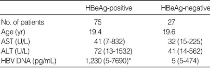
The demographics, liver biochemistry, and HBV DNA levels of all the patients are listed in Table 1. HBeAg-positive patients had a higher median HBV DNA level compared to HBeAg-negative patients (p=0.001). Among 27 patients without HBeAg, inactive carrier (ALT within normal range) was 15 (56%) and HBeAg negative hepatitis (increased ALT) was 12 (44%).
Sixty-nine of 75 (92.0%) patients with HBeAg and 16 of 27 (59%) patients without HBeAg had significant difference for intrahepatic HBcAg staining (p=0.000). The HBcAg sta- ining pattern of 69 patients with HBeAg was as follows (Table 2): 20 nuclear (30%), 47 both nuclear and cytoplasmic (68%) and 2 cytoplasmic alone (2%). The HBcAg staining pattern of 16 patients without HBeAg was as follows: 9 nuclear (56%), 4 both nuclear and cytoplasmic (25%) and 3 cytoplasmic alone (19%). Patients with HBeAg had significantly higher expression of mixed pattern of nuclei of intrahepatic HBcAg than HBeAg-negative patients (p<0.001) (Table 2).
Continous variables are expressed in median (range). Undetectable HBV DNA level (<5 pg/mL) was accepted as 5 pg/mL. ALT, alanine ami- notransferase; AST, aspartate aminotransferase.
*p=0.001.
HBeAg-positive HBeAg-negative
No. of patients 75 27
Age (yr) 19.4 19.6
AST (U/L) 41 (7-832) 32 (15-225)
ALT (U/L) 72 (13-1532) 41 (14-562)
HBV DNA (pg/mL) 1,230 (5-7690)* 5 (5-474) Table 1.Clinical and laboratory data of the patients studied
Data presented as number (percentage). Chi-square test, *p<0.0001.
Distribution pattern of Intrahepatic HBcAg
Nuclear pattern 9 (56) 20 (30)
Cytoplasmic pattern 3 (19) 2 (3)
Mixed pattern 4 (25) 47 (67)*
Table 2.The distribution pattern of HBcAg in hepatocyte accord- ing to HBeAg status
HBeAg
Negative (%) Positive (%)
Relationship between the degree of expression of HBcAg in hepatocyte nucleus and cytoplasm with the histologic activity of liver disease
For HBeAg-positive patients, there was a highly significant correlation between the degrees of expression of HBcAg in the hepatocyte cytoplasm and lobular activities, and portal/
periportal activities (Spearman rank correlation coefficient r=0.314, p=0.006 and r=0.283, p=0.01, respectively). There was no significant correlation between fibrosis and the degree of expression of HBcAg in the nucleus (r=0.023, p=0.84).
On the other hand, there was a negative correlation between the degree of expression of HBcAg in the nucleus and lobu- lar, and portal activities (r=-0.293, p=0.01 and r=-0.229, p=
0.04, respectively).
For HBeAg-negative patients, the degrees of expression of HBcAg in the hepatocyte cytoplasm correlated positively with the lobular activities (r=0.512, p=0.006, respectively), but did not correlate with the portal activities and fibrosis.
On the other hand, no significant correlation between the de- grees of expression of HBcAg in the hepatocyte nucleus and histologic activities, and fibrosis was noted.
Relationship between the level of HBV DNA in serum and the degrees of expression of HBcAg in the hepato- cyte nucleus and cytoplasm
For HBeAg-positive patients, there was a highly significant correlation between the levels of HBV DNA in serum and the degree of expression of HBcAg in the nucleus (r=0.507, p=0.000). On the other hand, no significant correlation bet- ween the level of HBV DNA in serum and the degree of expression of HBcAg in the cytoplasm was noted (r=0.069, p>0.5).
For HBeAg-negative patients, there was a highly significant correlation between the levels of HBV DNA in serum and the degrees of expression of HBcAg in the nucleus and cyto-
plasm (r=0.392, p=0.043 and r=0.502, p=0.008, respectively).
Relationship between HBV DNA levels and with the histologic activity of liver disease
For HBeAg-positive patients, there were negative correla- tions between HBV DNA levels and histological activities (lobular activity and portal/periportal activity, p=0.05 and 0.02, respectively). For HBeAg-negative patients, HBV DNA levels positively correlated with lobular activity (p=0.028) while HBV DNA levels did not correlate with portal activi- ty and fibrosis.
DISCUSSION
In the previous results, the expression pattern of HBcAg in hepatocytes was found to be related to the activity of liver disease in chronic HBV infection (11) especially when HBcAg was located in the cytoplasm of the hepatocyte (12). It also was found that nuclear but not cytoplasmic expression of HB- cAg is associated with high HBV replication and low activi- ty of liver disease in the chronic B viral infection (13). In this study, all these factors were assessed in the young age popu- lation which enabled us to compare the effects of these factors on the degree of HBcAg expression. The results suggested that only the extent of nuclear HBcAg expression correlated with HBV replication and also the extent of cytoplasmic HB- cAg expression correlated with histological activity of liver disease in chronic HBV infection. The limitation of this study was that the HBV DNA levels were only measured at a sin- gle time point and most of patients had mild or moderate histologic activities of liver disease.
The interesting and significant finding was that there was a positive correlation between the histologic activity of liver
Statistical results was determined by Spearman rank correlation.
*p<0.05.
HBcAg HBcAg HBV Lobular Portal Fibro- nucleus cytoplasm DNA activity activity sis HBcAg r 1.000 0.114 0.392 0.770 0.323 0.237
nucleus p 0.570 0.043* 0.059 0.100 0.236 HBcAg r 0.114 1.000 0.502 0.512 0.235 0.155 cytoplasm p 0.570 0.008* 0.006* 0.237 0.443 HBV DNA r 0.392 0.502 1.000 0.424 0.286 0.164 p 0.043* 0.008* 0.028* 0.148 0.412 Lobular r 0.770 0.512 0.424 1.000 0.571 0.472 activity p 0.059 0.006* 0.028* 0.002* 0.013 Portal r 0.323 0.235 0.286 0.571 1.000 0.472 activity p 0.100 0.237 0.148 0.002* 0.739 Fibrosis r 0.237 0.155 0.164 0.472 0.739 1.000
p 0.236 0.443 0.412 0.013 0.000* Table 4.Relation of Histologic scores, HBV DNA and the deg- rees of intrahepatic HBcAg expression in the HBeAg-negative patients
Statistical results was determined by Spearman rank correlation.
*p<0.05.
HBcAg HBcAg HBV Lobular Portal Fibro- nucleus cytoplasm DNA activity activity sis HBcAg r 1.000 0.069 0.507 -0.293 -0.229 0.023
nucleus p 0.554 0.000* 0.011* 0.048* 0.846 HBcAg r 0.069 1.000 0.066 0.314 0.066 0.137 cytoplasm p 0.554 0.571 0.006* 0.571 0.242 HBV DNA r 0.507 0.066 1.000 -0.227 -0.280 -0.123 p 0.000* 0.571 0.050* 0.015* 0.293 Lobular r -0.293 0.314 -0.227 1.000 0.486 0.345 activity p 0.011* 0.006* 0.050* 0.000* 0.002* Portal r -0.229 0.283 -0.280 0.486 1.000 0.458
activity p 0.048* 0.014* 0.015* 0.000* 0.000* Fibrosis r 0.23 0.137 -0.123 0.345 0.458 1.000
p 0.846 0.242 0.293 0.002* 0.000* Table 3.Clinical and laboratory data of the patients studied
disease and the degrees of expression of HBcAg in the hepa- tocyte cytoplasm in both HBeAg-positive and -negative pa- tients. In the HBeAg-positive patients, there was a inverse correlation between the degree of expression of HBcAg in the hepatocyte nucleus and the histological activity of liver disease. This finding supported the importance of hepatocyte injury in determination of HBcAg expression pattern. To our knowledge, this could be the first study that the degree of expression of HBcAg in the hepatocyte cytoplasm as assayed by immunohistochemical techniques is helpful for estimat- ing histological activity of liver disease in the young patients with chronic HBV infection. This inverse relation could sug- gest that the lysis of HBV infected hepatocytes was followed not only with the decreasing of serum viral load but also the degree of expression of HBcAg in the hepatocyte nucleus.
In 1987, Hsu et al. (7) showed that HBcAg was expressed at a relatively higher level on the nucleus than on the cyto- plasm during the immune tolerance phase, in which there was little or no inflammatory activity in the liver, whereas expression of HBcAg in the nuclei decreased with a concomi- tant increase in the expression of HBcAg in cytoplasm dur- ing the immune clearance phase, in which there is active and ongoing hepatitis. Both studies postulated that expression of HBcAg on the cell membrane is the important event that triggers cytotoxic T cells with HBcAg receptors, resulting in lysis of HBV-infected hepatocytes. Viral peptides, rather than the whole viral antigen, are presented by the human lymphocytes antigen (HLA) class I molecules to the cytotoxic T cells in conjunction with adhesion molecules (14). The exact mechanism by which viral peptides are being incorporated into the HLA molecules was fully understood. The positive relationship between the degree of expression of HBcAg in the hepatocyte cytoplasm and histologic activity of liver dis- ease supported that high level expression of HBcAg in the cytoplasm could reflect an increase in the availability of HB- cAg for intracellular processing into the HLA molecules, and presentation as immune target to the cytotoxic T cells. In 1995, Chu et al. (12) denied the above-mentioned postulation and concluded that the cytoplasmic localization of HBcAg in patients with active hepatitis may be secondary to liver damage and regeneration.
In the present study, the interesting other finding was that there was a highly significant positive correlation between the levels of HBV DNA in serum and the degrees of expres- sion of HBcAg in the hepatocyte nucleus, but there was no correlation between the degree of expression of HBcAg in the hepatocyte cytoplasm and the level of viral replication in the HBeAg-positive patients. Our result confirmed the relation- ship between nuclear HBcAg expression and viral replication.
It has been shown that HBcAg was localized in the nucleus in quiescent cells but diminished in proliferating cells, and the replication of HBV was enhanced in quiescent cells but diminished in the proliferating cells (12). Expression of HB- cAg in the hepatocyte nucleus was likely to be important in
viral replication.
However, the mechanism for a significant correlation bet- ween the level of HBV DNA in serum and the degrees of expression of HBcAg in the hepatocyte nucleus rather than in the cytoplasm has remained unclear. HBcAg was a trans- lational product of the pre-core/core gene of HBV and the carboxyl-terminal arginine-rich region of HBcAg has a nucle- ar localization signal (15). After entry into hepatocytes and transportation of the uncoated virus into the nucleus, viral DNA exists as a covalently closed circular (ccc) DNA, which serves as the template for transcription of pregenomic RNA (16). Some pregenomic RNAs was transported to the cytosol where formation of the viral nucleocapsid occurs along with the polymerase into core particles (17). Cytoplasmic core-pac- ked viral DNA could also reenter the nucleus to amplify nu- clear ccc DNA which in turn could provide more viral DNA for the production of new virions (18, 19). Our finding of a significant correlation between the degrees of HBcAg expres- sion in the hepatocyte nucleus and the level of viral replica- tion in chronic HBV infection supports this re-entry ampli- fying mechanism.
Though HBeAg seroconversion was commonly taken as a therapeutic endpoint in the past, increasing evidence showed that disease progression can continue in a significant portion of patients after HBeAg seroconversion, especially in the Asia and Mediterranean population (20, 21). We found that there was correlation between HBV DNA levels with lobular activi- ty of liver disease in the HBeAg-negative which is in agree- ment with other previous studies (22, 23). This finding con- firms that active viral replication is still present in a certain proportion of HBeAg-negative patients, and increased HBV DNA might be used as a marker to identify HBeAg-nega- tive patients who have a higher risk of active liver disease.
Some HBeAg-negative patients who have the inflammatory response and viral clearance might have precore mutation or/
and core promoter mutations to escape immune clearance (24-27). In contrast, one reported that core promotor muta- tions were not associated with the enhanced viral replication (28). These mutations and unknown other factors may be related to the localization of HBcAg in the hepatocyte, there- fore the virus might gradually replicate and develop inflam- matory response in the liver. More studies are required to doc- ument whether core promotor mutations have any effects on the chronic hepatitis B disease.
Our study had limitations similar to those of other cross- sectional studies (8, 13, 28). As hepatic inflammation may fluctuate with time, we can not be absolutely sure of the final outcome of these patients. This issue could only be satisfactori- ly addressed by a longitudinal study with paired liver biopsies.
In conclusion, the degrees of cytoplasmic HBcAg expres- sion correlated with histological activity of liver and also the degrees of nuclear HBcAg expression correlated with HBV replication in the young patients with chronic HBV infection.
These finding leads to the proposition that the nuclear local-
ization of HBcAg may amplify ccc DNA in the replication period of HBV and cytoplasmic localization may induce inter- action between T cell and infected hepatocyte.
REFERENCES
1. Lee WM. Hepatitis B virus infection. N Engl J Med 1997; 337: 1733-45.
2. Okada K, Kamiyama I, Inomata M, Imai M, Miyakawa Y. e antigen and anti-e in the serum of asymptomatic carrier mothers as indica- tors of positive and negative transmission of hepatitis B virus to their infants. N Engl J Med 1976; 294: 746-9.
3. Stevens CE, Neurath RA, Beasley RP, Szmuness W. HBeAg and anti-HBe detection by radioimmunoassay: Correlation with vertical transmission of hepatitis B virus in Taiwan. J Med Virol 1979; 3:
237-41.
4. Hadziyannis SJ, Lieberman HM, Karvountzis GG, Shafritz DA. Anal- ysis of liver disease, nuclear HBcAg, viral replication, and hepatitis B virus DNA in liver and serum of HBeAg Vs. anti-HBe positive car- riers of hepatitis B virus. Hepatology 1983; 3: 656-62.
5. Chu CM, Liaw YF. Membrane staining for hepatitis B surface anti- gen on hepatocytes: a sensitive and specific marker of active viral replication in hepatitis B. J Clin Pathol 1995; 48: 470-3.
6. Chu CM, Liaw YF. Intrahepatic distribution of hepatitis B surface and core antigens in chronic hepatitis B virus infection. Hepatocyte with cytoplasmic/membranous hepatitis B core antigen as a possible target for immune hepatocytolysis. Gastroenterology 1987; 92: 220-5.
7. Hsu HC, Su IJ, Lai MY, Chen DS, Chang MH, Chang SM, Sung JL.
Biologic and prognostic significance of hepatocyte hepatitis B core antigen expressions in the natural course of chronic hepatitis B virus infection. J Hepatol 1987; 5: 45-50.
8. ter Borg F, ten Kate FJ, Cuypers HT, Leentvaar-Kuijpers A, Oosting J, Wertheim-van Dillen PM, Honkoop P, Rasch MC, de Man RA, van Hattum J, Chamuleau RA, Reesink HW, Jones EA. Relation bet- ween laboratory test results and histological hepatitis activity in in- dividuals positive for hepatitis B surface antigen and antibodies to hepatitis B e antigen. Lancet 1998 27; 351: 1914-8.
9. Chan HL, Hui Y, Leung NW, Ching JY, Chan FK, Sung JJ. Risk fac- tors for active liver disease in HBeAg-negative chronic hepatitis B virus-infected patients. Am J Gastroenterol 2000; 95: 3547-51.
10. Desmet VJ, Gerber M, Hoofnagle JH, Manns M, Scheuer PJ. Clas- sification of chronic hepatitis: diagnosis, grading and staging. Hep- atology 1994; 19: 1513-20.
11. Mangia A, Chung YH, Hoofnagle JH, Birkenmeyer L, Mushahwar I, Di Bisceglie AM. Pathogenesis of chronic liver disease in patients with chronic hepatitis B virus infection without serum HBeAg. Dig Dis Sci 1996; 41: 2447-52.
12. Chu CM, Yeh CT, Sheen IS, Liaw YF. Subcellular localization of hepatitis B core antigen in relation to hepatocyte regeneration in chronic hepatitis B. Gastroenterology 1995; 109: 1926-32.
13. Chu CM, Yeh CT, Chien RN, Sheen IS, Liaw YF. The degrees of hepatocyte nuclear but not cytoplasmic expression of hepatitis B core antigen reflects the level of viral replication in chronic hepatitis B
virus infection. J Clin Microbiol 1997; 35: 102-5.
14. Thomas HC, Carman WF. The host immune response may be respon- sible for selection of envelope and precore/core variants of HBV. Prog Liver Dis 1992; 10: 239-51.
15. Yeh CT, Liaw YF, Ou JH. The arginine-rich domain of hepatitis B virus precore and core proteins contains a signal for nuclear trans- port. J Virol 1990; 64: 6141-7.
16. Bock CT, Schranz P, Schroder CH, Zentgraf H. Hepatitis B virus genome is organized into nucleosomes in the nucleus of the infected cell. Virus Genes 1994; 8: 215-29.
17. Hirsch RC, Loeb DD, Pollack JR, Ganem, D. cis -Acting sequences required for encapsidation of duck hepatitis B virus pregenomic RNA.
J Virol 1991; 65: 3309-16.
18. Tuttleman JS, Pourcel C, Summers J. Formation of the pool of cova- lently closed circular viral DNA in hepadnavirus-infected cells. Cell 1986; 47: 451-60.
19. Wu TT, Coates L, Aldrich CE, Summers J, Mason WS. In hepato- cytes infected with duck hepatitis B virus, the template for viral RNA synthesis is amplified by an intracellular pathway. Virology 1990;
175: 255-61.
20. Hsu YS, Chien RN, Yeh CT, Sheen IS, Chiou HY, Chu CM, Liaw YF. Long-term outcome after spontaneous HBeAg seroconversion in patients with chronic hepatitis B. Hepatology 2002; 35: 1522-7.
21. Yuen MF, Yuan HJ, Hui CK, Wong DK, Wong WM, Chan AO, Wong BC, Lai CL. A large population study of spontaneous HBeAg seroconversion and acute exacerbation of chronic hepatitis B infec- tion: implications for antiviral therapy. Gut 2003; 52: 416-9.
22. Lindh M, Horal P, Dhillon AP, Norkrans G. Hepatitis B virus DNA levels, precore mutations, genotypes and histological activity in chron- ic hepatitis B. J Viral Hepat 2000; 7: 258-67.
23. Karayiannis P, Fowler MJ, Lok AS, Greenfield C, Monjardino J, Thomas HC. Detection of serum HBV-DNA by molecular hybridis- ation. Correlation with HBeAg/anti-HBe status, racial origin, liver histology and hepatocellular carcinoma. J Hepatol 1985; 1: 99-106.
24. Lindh M, Gustavson C, Mardberg K, Norkrans G, Dhillon AP, Horal P. Mutation of nucleotide 1762 in the core promoter region during hepatitis B e seroconversion and its relation to liver damage in hep- atitis B e antigen carriers. J Med Virol 1998; 55: 185-90.
25. Kidd-Ljunggren K, Oberg M, Kidd AH. Hepatitis B virus X gene 1751-1764 mutations: implications for HBeAg status and disease. J Gen Virol 1997; 78: 1469-78.
26. Li J, Buckwold VE, Hon MW, Ou JH. Mechanism of suppression of hepatitis B virus precore RNA transcription by a frequent double mu- tation. J Virol 1999; 73: 1239-44.
27. Buckwold VE, Xu Z, Chen M, Yen TS, Ou JH. Effects of a natural- ly occurring mutation in the hepatitis B virus basal core promoter on precore gene expression and viral replication. J Virol 1996; 70:
5845-51.
28. Wu PC, Lau JY, Lau TK, Lau SK, Lai CL. Relationship between intrahepatic expression of hepatitis B viral antigens and histology in Chinese patients with chronic hepatitis B virus infection. Am J Clin Pathol 1993; 100: 648-53.