INTRODUCTION
The occurrence of extrapulmonary small cell carcinoma has been reported in a variety of organs (1). Small cell carci- noma of the genitourinary tract is rarely encountered in the renal parenchyma (2), renal pelvis (3), ureter (4, 5), urinary bladder (6), urethra (7), and prostate (8). Especially, small cell carcinoma of the ureter itself is extremely rare, except for that of the renal pelvis including ureteropelvic junction (9) and only two cases have been reported in the previous literature (4, 5). The histogenesis of this tumor remains uncer- tain, although several hypotheses have been suggested. More- over, due to its rarity, the clinical behavior of small cell car- cinoma of the ureter has not been well established.
In this paper, we report the clinical characteristics, patho- logical features, and immunohistochemical results in a case of small cell carcinoma combined with squamous cell and transitional cell carcinomatous components which was asso- ciated with ureteral stone.
CASE REPORT
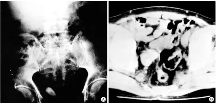
A 60-yr-old man without significant prior medical histo- ry presented himself with a 2-weeks history of right flank pain in September 1998. At admission, he complained of urinary frequency, hesitancy, and dysuria. However, he had no history of gross hematuria. After admission, his flank pain partially subsided, but the urinary frequency, hesitancy, dysuria, and sense of residual urine persisted. He had a smok- ing history of about or less than 0.5 pack per day, and he denied any respiratory symptoms. Physical examination was unremarkable except for a soft, nontender mass palpated in his right upper abdomen. All laboratory investigations, in- cluding complete blood cell count, biochemical parameters, and urinalysis, yielded values within normal limits. 99mTc- labeled dimercaptosuccinic acid scan revealed no uptake in the right kidney. Chest radiography and plain radiography of the kidney, ureter, and urinary bladder (KUB) revealed no remarkable findings except for a radiopaque density in the right pelvic cavity (Fig. 1A). Abdominal computed
Tae Sook Kim, Do Hwan Seong*, Jae Y. Ro�
Departments of Pathology and Urology*, Inha University College of Medicine, Inchon;
Department of Diagnostic Pathology�, University of Ulsan College of Medicine, Asan Medical Center, Seoul, Korea
Address for correspondence Tae Sook Kim, M.D.
Department of Pathology, Inha University College of Medicine, 7-241, 3rd St., Shinheung-dong, Choong-gu, Inchon 400-103, Korea Tel : +82.32-890-0943, Fax : +82.32-890-0944 E-mail : [email protected]
*This work was partially supported by Inha Univer- sity Research Grant (1998) (INHA-14382).
796 J Korean Med Sci 2001; 16: 796-800
ISSN 1011-8934
Copyright � The Korean Academy of Medical Sciences
Small Cell Carcinoma of the Ureter with Squamous Cell and
Transitional Cell Carcinomatous Components associated with Ureteral Stone
We report a case of primary small cell carcinoma of the ureter with squamous cell and transitional cell carcinomatous components associated with ureteral stone, which is unique in that the patient has remained free of tumor recurrence for 36 months after the surgery without adjuvant chemotherapy or radiotherapy.
A 60-yr-old man presented himself with a right flank pain. Computed tomogra- phy revealed an ill-defined mass and a stone in the lower one third of the right ureter, and hydronephroureterosis above the stone-impacted site. The patient underwent right nephroureterectomy and stone removal. Upon gross examina- tion, a 3.8×1.8×1.2 cm white and partly yellow mass was noted in the anterior part of the ureter, resulting in indentation of the ureteral lumen on the posterior side. Light microscopic examination revealed that the mass was mainly composed of small cell carcinoma, and partly squamous cell and transitional cell carcino- matous components. The overlying ureteral mucosa and renal pelvis also con- tained multifocal dysplastic transitional epithelium and transitional cell carcinoma in situ. There was no vascular invasion, and the surgical margins were free of tumor. The small cell carcinomatous component was positive for chromogranin, neuron specific enolase, synaptophysin, and pancytokeratin but negative for high molecular-weight cytokeratin (K-903) by immunohistochemistry.
Key Words : Carcinoma, Small Cell; Carcinoma, Squamous Cell; Carcinoma, Transitional Cell; Ureter- al Calculi
Received : 9 November 2000 Accepted : 15 February 2001
tomography (CT) revealed right hydronephroureterosis and a stone in the right distal ureter with an irregularly thick- ened ureteral wall just above the stone-impacted site (Fig.
1B). However, neither a definite ureteral mass nor enlarged pelvic lymph nodes were identified on CT scans. Based on the imaging findings, right nephroureterectomy with stone removal was performed on the patient. A palpable mass, which was found in the distal anterior ureter just above the stone-impacted site during the operation, was sent for frozen section evaluation. The pathologic findings of frozen and permanent sections are described below. The patient refused either adjuvant chemotherapy or radiotherapy. He has been well without evidence of tumor recurrence or metastasis for 36 months after surgery.
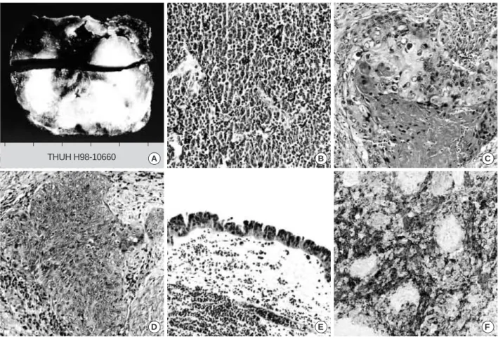
Gross examination of the right nephroureterectomy spec- imen revealed a hydronephrotic kidney with a dilated renal pelvis and calyces with renal cortical thinning, and ureteral dilation. A segment of the right distal ureter contained an ill-defined, ovoid mass (3.8×1.8×1.2 cm) that protruded into the ureteral lumen and caused partial obliteration of the ureter. The cut surface of the mass was grayish-white with patchy yellow areas and small hemorrhagic foci (Fig.
2A). The mass in the anterior side of the ureter indented the ureteral lumen on the posterior side. Upon light microscop- ic examination of the mass, various malignant epithelial components including small cell carcinoma, squamous cell carcinoma, and transitional cell carcinoma were observed.
The ureteral lumen was almost totally occluded by the small cell carcinomatous component, located mainly in the anteri- or side of the ureter (Fig. 2B). The squamous cell and tran-
sitional cell carcinomatous components were seen especially on the posterior side of the ureter (Fig. 2C, D). Three tumor components were intermingled on the lateral and posterior side of the ureter and a gradual transition of small cell carci- noma to transitional cell and squamous cell carcinoma was noted. The tumor invaded throughout the proper muscle of the ureter, but resection margins were free of tumor. There was no vascular invasion. The overlying ureteral mucosa of the tumor was almost entirely denuded, and several foci of dysplastic transitional epithelium and transitional cell carcino- ma in situ were found (Fig. 2E). The renal pelvis also showed focal transitional cell carcinoma in situ. The overlying ureter- al lumen of the tumor was focally covered with squamous cell carcinoma but the transition from transitional cell carci- noma in situ was not found due to ureteral mucosal denuda- tion. The renal parenchyma had interstitial inflammation and fibrosis with glomerular sclerosis but no evidence of tumor. Immunohistochemical staining for chromogranin showed strong reactivity in the small cell carcinomatous component (Fig. 2F), but not in the squamous cell and tran- sitional cell carcinomatous components. Immunohistochem- ical staining for another neuroendocrine markers including neuron specific enolase and synaptophysin was also positive in small cell carcinomatous component. High molecular- weight cytokeratin (K-903) staining revealed strong reac- tivity in the cytoplasm of the transitional cell carcinoma- tous component, weak reactivity in the squamous cell carci- nomatous component, but no reactivity in the small cell carcinomatous component. Pancytokeratin was present in all three tumorous components.
A B
Fig. 1.Simple KUB shows a radiopaque stone in right distal ureter (A). Computed tomographic scan shows an ill-defined, low-density mass in the right distal ureter resulting in irregular thickening of the ureteral wall and obliteration of the ureteral lumen just above the stone-impacted site (B).
DISCUSSION
The majority of small cell carcinomas of the genitourinary tract occur in combination with other tumorous components, such as transitional cell carcinoma, squamous cell carcino- ma, and adenocarcinoma. Its variable histologic feature sup- ports the notion that small cell carcinoma in any sites of the urinary tract, including renal pelvis (3), urinary bladder (6), and ureter (4, 5), originates from multipotential stem cells.
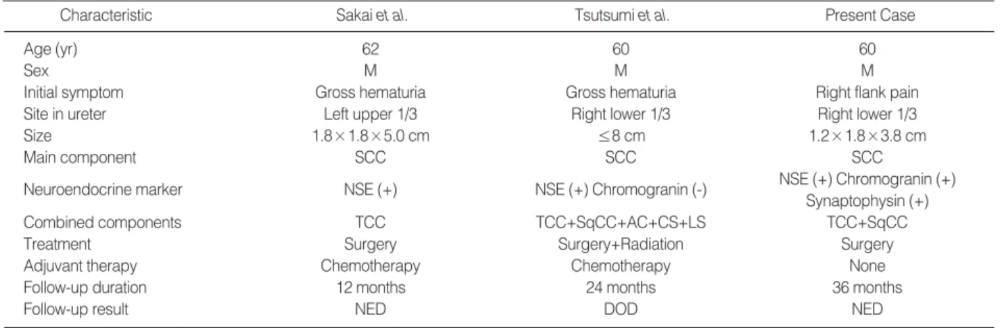
Also in this case, small cell carcinoma appeared with squa- mous cell and transitional cell carcinomatous components, which is in agreement with the hypothesis that this tumor probably originates from multipotential stem cells of the ureter. Two previously reported cases of small cell carcinoma of the ureter also had other tumorous components; one had transitional cell carcinoma (4), and the other leiomyosarco- matous and chondrosarcomatous elements, in addition to transitional cell and squamous cell carcinomatous, and adeno- carcinomatous components (5) (Table 1). The other hypothe- sis is that small cell carcinoma originates from intrinsic neuro-
endocrine cells within the normal genitourinary tract, derived from the neural crest during the embryogenesis. It was reported that a few number of neuroendocrine cells have been noted in the normal urinary tract including the renal pelvis (11), ureter (11), urinary bladder (12), urethra (13), and prostate (14), which are derived from mesonephros.
However, this theory does not support two previously report- ed cases, which showed combined histologic features of other carcinomatous components without exception (4, 5).
It has also been reported that no neuroendocrine cells were noted in the normal renal parenchyma (15), which is derived from metanephros.
The most common symptom of small cell carcinoma of the ureter is gross hematuria, which is also seen in small cell carcinomas at other sites of the urinary tract (6). In the pre- vious report (4, 5), asymptomatic gross hematuria was the initial symptom of small cell carcinoma of the ureter with- out exception. Our patient presented with flank pain with- out gross hematuria. An ill-defined ureteral mass seen on radiological findings was initially interpreted as a periureter-
798 T.S. Kim, D.H. Seong, J.Y. Ro
D E F
A B C
Fig. 2.On gross examination, cut surface of the right ureter shows the luminal narrowing caused by intramural infiltrative mass, which mainly involved the anterior half (A). On light microscopic examination, the tumor is mainly composed of small cell carcinomatous component, which shows densely packed small round tumor cells with hyperchromatic nuclei, scanty cytoplasms with delicate stro- ma (B). The squamous cell (C) and transitional cell (D) carcinomatous components are intermingled. The overlying ureteral mucosa of the tumor shows multifocal dysplastic transitional epithelium and transitonal carcinoma in situ (E) with almost entire denudation. Im- munohistochemial staining for chromogranin shows positive reactivity only in the small cell carcinomatous component (F) (B-F,×200).
THUH H98-10660
al inflammatory lesion, because the ureteral stone impacted upon the ureteral lumen. It is of note that our patient sought treatment earlier due to the presence of an ureteral stone than the patients in previously reported cases, which may explain why the outcome for this patient was favorable. There have been few reports that carcinomas of the urinary tract at the stone-impacted sites during long-term follow-up (16).
It has been also proposed that chronic mechanical irritation by stone may induce morphological changes of the transi- tional epithelium including dysplastic changes might lead to carcinoma of the urinary tract (17). Primary squamous cell carcinoma of the ureter associated with ureteral stone has been already reported (18).
It has also been suggested that smoking is related to the carcinogenesis of small cell carcinoma in extrapulmonary areas, expecially the genitourinary tract (19, 20). It has been reported that small cell carcinomas of the urinary tract includ- ing kidney (21), renal pelvis (3), urinary bladder (22), and prostate (21) have developed in heavy smokers. Our patient had a smoking history of less than 0.5 pack per day for 30 yr and it has also been reported that cases of small cell carci- noma occurs in any site of the genitourinary tract in non- smokers (8, 21, 22). Therefore, the role of cigarette smok- ing in the development of small cell carcinoma of the ureter and other extrapulmonary sites remains undetermined. In this case, the patient had neither respiratory symptoms nor remarkable findings by chest radiography. In addition, only one case of metastatic small cell carcinoma of the lung to the ureter has been reported (23). Therefore, the possibility of metastatic small cell carcinoma of the lung to the ureter was not taken into consideration in our case.
The treatment and prognosis of small cell carcinoma of the ureter are not well established (9) because of its rarity (4, 5). Many clinicians have proposed that multimodality therapy which includes surgery, radiation, and chemothera- py is essential for patients with small cell carcinoma of the
urinary tract (3, 6, 9). It has been reported that combined chemotherapy with methotrexate, vinblastine, doxorubicin, and cisplatin (M-VAC) is effective against small cell carci- nomas of the genitourinary tract (24). However, it is diffi- cult to determine the most effective therapeutic modality for small cell carcinoma in the ureter because of the small number of reported cases (9).
The majority of small cell carcinomas of the genitourinary tract progress rapidly to the regional lymph nodes, liver, and other organs (21). So the majority of patients with small cell carcinoma die of the disease within a year (7). However, some cases of small cell carcinomas in the renal pelvis and urinary bladder showed relatively slow growth in the primary sites (10).
In summary, we report upon an unique case of small cell carcinoma of the ureter combined with squamous cell and transitional cell carcinomatous components, associated with ureteral stone. The patient is getting well 36 months after the surgery without any adjuvant therapy.
ACKNOWLEDGMENTS
The authors would like to thank Dr. Nasakazu Tsutsumi, Department of Urology, Hitachi General Hospital, Ibaraki, Japan, for providing information about a previous case of small cell carcinoma of the ureter, and Dr. John R. Mackey, Department of Medical and Experimental Oncology, Cross Cancer Institute, Edmonton, Canada, who kindly provided data upon previously reported small cell carcinoma cases.
REFERENCES
1. Ibrahim NB, Briggs JC, Corbishley CM. Extrapulmonary oat cell carcinoma. Cancer 1984; 54: 1645-61.
Sakai et al.
Characteristic Tsutsumi et al. Present Case
Age (yr) 62 60 60
Sex M M M
Initial symptom Gross hematuria Gross hematuria Right flank pain
Site in ureter Left upper 1/3 Right lower 1/3 Right lower 1/3
Size 1.8×1.8×5.0 cm ≤8 cm 1.2×1.8×3.8 cm
Main component SCC SCC SCC
Neuroendocrine marker NSE (+) NSE (+) Chromogranin (-) NSE (+) Chromogranin (+)
Synaptophysin (+)
Combined components TCC TCC+SqCC+AC+CS+LS TCC+SqCC
Treatment Surgery Surgery+Radiation Surgery
Adjuvant therapy Chemotherapy Chemotherapy None
Follow-up duration 12 months 24 months 36 months
Follow-up result NED DOD NED
Table 1.The characteristics of reported cases of small cell carcinoma of the ureter
SCC, small cell carcinoma; NSE, neuron specific enolase; TCC, transitional cell carcinoma; SqCC, squamous cell carcinoma; AC, adenocarcinoma;
CS, chondrosarcoma; LS, leiomyosarcoma; NED, no evidence of disease; DOD, dead of disease (bone and liver metastasis).
2. T tu B, Ro JY, Ayala AG, Ordonez NG, Johnson DE. Small cell carcinoma of the kidney: A clinicopathologic, immunohistochemi- cal, and ultrastructural study. Cancer 1987; 60: 1809-14.
3. Guillou L, Duvoisin B, Chobaz C, Chapuis G, Costa J. Combined small cell and transitional cell carcinoma of the renal pelvis: A light microscopic, immunohistochemical, and ultrastructural study of a case with literature review. Arch Pathol Lab Med 1993; 117: 239- 43.
4. Sakai N, Ogawa T, Ishibashi Y, Fukuoka H, Sakanishi S. A case of small cell carcinoma of the ureter. Hinyokika Kiyo 1990; 36: 1455- 8 (in Japanese).
5. Tsutsumi M, Kamiya M, Sakamoto M, Tobisu K, Kakizoe T. A ureteral small cell carcinoma mixed with malignant mesodermal and ectodermal elements: A clinicopathological, morphological, and immunohistochemical study. Jpn J Clin Oncol 1993; 23: 325-9.
6. Grignon DJ, Ro JY, Ayala AG, Shum DT, Ordonez NG, Logothetis CJ, Johnson DE, Mackey B. Small cell carcinoma of the urinary bladder. A clinicopathologic analysis of 22 cases. Cancer 1992; 69:
527-36.
7. Fukuda T, Kamishima T, Saito T, Itoh S, Suzuki T. Small cell car- cinoma arising from the outer urethral orifice: A case report exam- ined by histologic, ultrastructural, and immunohistochemical meth- ods. Pathol Int 1997; 47: 497-501.
8. T tu B, Ro JY, Ayala AG, Johnson DE, Logothetis CJ, Ordonez NG.
Small cell carcinoma of the prostate Part I: A clinicopathological study of 20 cases. Cancer 1987; 59: 1803-9.
9. Mackey JR, Au H, Hugh J, Venner P. Genitourinary small cell car- cinoma: Determination of clinical and therapeutic factors associat- ed with survival. J Urol 1998; 159: 1624-9.
10. Ordonez NG, Khorsand J, Ayala AG, Sneige N. Oat cell carcinoma of the urinary tract: An immunohistochemical and electron micro- scopic study. Cancer 1986; 58: 2519-30.
11. Fetissof F, Dubois MP, Lanson Y, Jobard P. Endocrine cells in renal pelvis and ureter: An immunohistochemical analysis. J Urol 1986;
135: 420-1.
12. Gosling JA, Dixon JS, Humpherson JR. Functional anatomy of the urinary tract. An integrated text and color atlas. New York: Gower, 1983.
13. Zavia i M, Brozman M, Zaji kova M, Blazekova J, Oberu ova J.
The adult human female urethra: Enzyme-histochemical study. Acta
Histochem 1985; 77: 165-75.
14. Di Sant’Agnese PA. Neuroendocrine cells of the prostate and neu- roendocrine differentiation in prostatic carcinoma: A review of mor- phologic aspects. Urology 1998; 51: 121-4.
15. Guy L, Begin LR, Oligny LL, Brock GB, Chevalier S, Aprikian AG.
Searching for an intrinsic neuroendocrine cell in the kidney: An immunohistochemical study of the fetal, infantile and adult kidney.
Pathol Res Pract 1999; 195: 25-30.
16. Beyer-Boon ME, Cuypers LH, de Voogt HJ, Brussee JA. Cytologi- cal changes due to urinary calculi: A consideration of the relation- ship between calculi and the development of urothelial carcinoma.
Br J Urol 1978; 50: 81-9.
17. Harzmann R, Schubert GE, Gericke D, Altenahr E, Bichler KH.
Morphology of the urinary bladder following long-term experimen- tal irritation of the urothelium. Urol Int 1983; 38: 166-72.
18. Koh E, Hamada S, Saiki S, Kinouchi T, Kuroda M, Miki T, Kiyohara H, Usami M, Kotake T. A case of primary ureteral squamous cell carcinoma associated with calculus. Hinyokika Kiyo 1989; 35: 105- 9 (in Japanese).
19. Auerbach O, Garfinkel L. Histologic changes in the urinary bladder in relation to cigarette smoking and use of artificial sweeteners.
Cancer 1989; 64: 983-7.
20. McLaughlin JK, Blot WJ, Mandel JS, Schuman LM, Mehl ES, Frau- meni JF Jr. Etiology of cancer of the renal pelvis. J Natl Cancer Inst 1983; 71: 287-91.
21. Christopher ME, Seftel AD, Sorenson K, Resnick MI. Small cell carcinoma of the genitourinary tract: An immunohistochemical, elec- tron microscopic, and clinicopatholocial study. J Urol 1991; 146:
382-8.
22. Mills SE, Wolfe JT 3rd, Weiss MA, Swanson PE, Wick MR, Fowler JE Jr, Young RH. Small cell undifferentiated carcinoma of the uri- nary bladder: A light microscopic, immunocytochemical, and ultra- structural study of 12 cases. Am J Surg Pathol 1987; 11: 606-17.
23. DiPietro M, Zeman RK, Keohane M, Rosenfield AT. Oat cell car- cinoma metastatic to ureter. Urology 1983; 22: 419-20.
24. Oesterling JE, Brendler CB, Burgers JK, Marshall FF, Epstein JI.
Advanced small cell carcinoma of the bladder: Successful treatment with combined radical cystoprostatectomy and adjuvant methotrex- ate, vinblastine, doxorubicin, and cisplatin chemotherapy. Cancer 1990; 65: 1928-36.
800 T.S. Kim, D.H. Seong, J.Y. Ro
~
~
~
、
、
、
、
、
、
、~