INTRODUCTION
Mucocele-like tumor (MLT) of the breast was first report- ed by Rosen (1) in 1986 as benign neoplasia analogous to mucocele of the minor salivary gland. This lesion is charac- terized by mucus-filled cysts lined by flattened epithelium with only a focal tendency to papillary hyperplasia. Another constant finding is an extrusion of mucin into the surround- ing stroma. Subsequent reports identified MLT associated with ductal hyperplasia and carcinoma (2-4). More recently, MLT has been considered as a spectrum of pathologic lesions, including benign tumor, atypical ductal hyperpla- sia, ductal carcinoma in situ, and mucinous carcinoma (5).
In Korea only a few cases of MLT have been reported (6-8).
We describe a case of MLT associated with ductal carcinoma in situ and mucinous carcinoma in a 34-yr-old female.
CASE REPORT
A 34-yr-old female presented with a palpable mass in the right breast. The mammograph showed an ill-defined and lobulated mass with tiny microcalcifications. Excisional
biopsy was done. The specimen measured 2.6×2.0×1.5 cm and had a lobulated surface. The cut section revealed multiple aggregated cysts containing gelatinous materials.
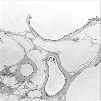
Histological examination showed multiple cysts of varying size (Fig. 1). The cysts contained an amorphous mucinous secretion. Extrusion of mucinous material into the sur- rounding stroma was also observed. The mucinous content in the cysts and in the stroma was positive for periodic acid- Schiff with diastase and mucicarmine. The lining of the cysts in most areas were of flat or cuboidal epithelium and devoid of cellular atypia (Fig. 2). The lining epithelium showed proliferative change ranging from atypical ductal hyperplasia to ductal carcinoma in situ, micropapillary type (Fig. 3). A microscopic focus of mucinous carcinoma within MLT was also noted (Fig. 4). None of the neoplastic lesions were positive for S-100 protein (Zymed, San Francisco, CA, U.S.A., predilute), carcinoembryonic antigen (Zymed, predilute), and p53 protein (Zymed, dilution 1:50).
There was no residual MLT or mucinous carcinoma in the subsequent modified radical mastectomy specimen. Axil- lary lymph nodes were free of tumor metastasis. The patient is alive and well without evidence of disease 54 months after operation.
Ji Shin Lee, Hyung Seok Kim, Jong Jae Jung, Min Cheol Lee*
Department of Pathology, Seonam University, College of Medicine, Namwon; Department of Pathology*, Chonnam National University Medical School and Medical Center, Kwangju, Korea
Received : 1 June 2000 Accepted : 17 August 2000
Address for correspondence Ji Shin Lee, M.D.
Department of Pathology, Seonam University, College of Medicine, 720 Kwangchi-dong, Namwon 590-170, Korea
Tel: +82.63-620-0352, Fax: +82.63-620-0355 E-mail: [email protected]
516 J Korean Med Sci 2001; 16: 516-8
ISSN 1011-8934
Copyright � The Korean Academy of Medical Sciences
Mucocele-Like Tumor of the Breast Associated with Ductal Carcinoma In Situ and Mucinous Carcinoma
: A Case Report
Mucocele-like tumor (MLT) of the breast is a rare neoplasm. Although this lesion was considered benign when first described, the concept of a pathologic contin- uum with mucinous carcinoma was evident in subsequent reports. Only a few cases of MLT have been reported in Korea. We describe a case of MLT associ- ated with ductal carcinoma in situ and mucinous carcinoma in a 34-yr-old female. Histological examination showed multiple mucus-filled cysts of varying size. Extravasated mucin was present in the surrounding stroma. The lining of the cysts in most areas were of flat or cuboidal epithelium and devoid of cellular atypia. The lining epithelium showed proliferative change ranging from atypical ductal hyperplasia to ductal carcinoma in situ, micropapillary type. A microscop- ic focus of mucinous carcinoma within MLT was also noted. None of the lesions exhibited epithelial reactivity for p53 protein. The patient is alive and well without evidence of disease 54 months after initial treatment. This case supports the concept that MLT encompasses a spectrum of pathologic lesions including benign tumor, atypical ductal hyperplasia, ductal carcinoma in situ, and muci- nous carcinoma.
Key Words : Breast; Mucocele; Adenocarcinoma, Mucinous
Mucocele-Like Tumor of the Breast 517
DISCUSSION
The term MLT of the breast was used by Rosen (1) to describe mucus-filled cysts lined by flattened epithelium with focal areas of hyperplasia often producing a papillary pattern. The extruded mucinous material is commonly pre- sent within the stroma. Rosen distinguished MLT from
mucinous carcinoma. In contrast to the lack of association with malignancy in Rosen’s cases, Ro et al. (2) reported seven cases of MLT associated with atypical ductal hyper- plasia or microscopic foci of mucinous carcinoma. The mucin of MLT was identical with that of mucinous carcino- ma, and they suggested that some MLTs may be the early form of mucinous carcinoma of the breast. Subsequent
Fig. 1.Mucoele-like tumor with mucin-filled epithelial lined cysts and extravasated mucin in the stroma (H&E, ×40).
Fig. 2.The lining cells of cysts are usually flat (H&E, ×400).
Fig. 3.Mucocele-like tumor with intraductal carcinoma. Ductal carcinoma in situ of micropapillary type is present (H&E, ×400).
Fig. 4.A microscopic focus of mucinous carcinoma (arrow) is adjacent to the mucocele-like tumor (H&E, ×100).
518 J.S. Lee, H.S. Kim, J.J. Jung, et al.
reports identified MLT associated with ductal hyperplasia or mucinous carcinoma (3, 4). In this case all these morpho- logical features of MLT were identified. In addition, ductal carcinoma in situ and mucious carcinoma were also observed.
Weaver et al. (9) suggested that MLT and mucinous car- cinoma of the breast may represent the two ends of patho- logical spectrum of mucinous lesions of the breast. More recently, MLT was considered as a spectrum of pathologic lesions including benign tumor, atypical ductal hyperplasia, ductal carcinoma in situ, and mucinous carcinoma (5). It is important, therefore, to exclude the possibility of carcinoma by examining adequate tissue samples when MLT is found in a breast biopsy.
Hamele-Bena et al. (10) compared the clinical features of benign and malignant MLTs. Malignant MLT had ductal carcinoma in situ or mucinous carcinoma. There were no appreciable differences in age, tumor size, or laterality bet- ween patients with benign MLT or malignant one, although MLT with carcinoma had coarse calcification more often than benign MLT. All of the patients were alive without evidence of disease. In this case tiny microcalcification was mammographically detected. The neoplastic epithelium of benign and malignant lesion was negative for p53 protein.
The patient is alive and well after the follow-up of 54 months. Our case supports the concept that MLT encom- passes a spectrum of pathologic lesions including benign tumor, atypical ductal hyperplasia, ductal carcinoma in situ, and mucinous carcinoma and MLT with mucinous carcino- ma is a low-grade neoplasm of the breast.
The pathogenesis of MLT of the breast is uncertain, but excess production of mucinous secretion or ductal obstruc- tion may be the contributing factors (1, 2).
The differential diagnosis of MLT includes cystic hyper- secretory hyperplasia and cystic hypersecretory duct carcino- ma of the breast (11). However, these lesions show cystically dilated ducts containing a homogeneous secretion. But these lesions are not associated with extravasated mucinous
material into the stroma, which is a typical feature of MLT.
REFERENCES
1. Rosen PP. Mucocele-like tumors of the breast. Am J Surg Pathol 1986; 10: 464-9.
2. Ro JY, Sneige N, Sahin AA, Silva EG, del Junco GW, Ayala AG.
Mucocelelike tumor of the breast associated with atypical ductal hyperplasia or mucinous carcinoma. Arch Pathol Lab Med 1991;
115: 137-40.
3. Fisher CJ, Millis RR. A mucocele-like tumour of the breast associ- ated with both atypical ductal hyperplasia and mucoid carcinoma.
Histopathology 1992; 21: 69-71.
4. Kulka J, Davies JD. Mucocele-like tumours: more associations and possibly ductal carcinoma in situ? Histopathology 1993; 22: 511-2.
5. Yeoh GP, Cheung PS, Chan KW. Fine-needle aspiration cytology of mucocelelike tumors of the breast. Am J Surg Pathol 1999; 23:
552-9.
6. Kim HS, Park JM, Ji EK, Gong GY, Ahn SH. Mucocele-like tumor of the breast: a case report. J Korean Radiol Soc 1999; 41: 607-9.
7. Kim DS, Lee SJ, Hwang MS, Bae YK, Lee JK, Kwun KB. Muco- cele-like tumor. In: Disease of the breast: case reviews. 1st ed.
Taegu: Sae Han Press, 2000; 46-9.
8. Jang KY, Park HS, Chung MJ, Moon WS, Kang MJ, Lee DG. Muco- celelike tumor of the breast associated with atypical ductal hyper- plasia and invasive lobular carcinoma (abstract). Korean J Pathol 1999; 33: 10: 788.
9. Weaver MG, Abdul-Karim FW, Al-Kaisi N. Mucinous lesions of the breast. A pathologic continuum. Pathol Res Proc 1993; 189:
873-6.
10. Hamele-Bena D, Cranor ML, Rosen PP. Mammary mucocele-like lesions: benign and malignant. Am J Surg Pathol 1996; 20: 1081-5.
11. Colandrea JM, Shmookler BM, O’Dowd GJ, Cohen MH. Cystic hypersecretory duct carcinoma of the breast: report of a case with fine-needle aspiration. Arch Pathol Lab Med 1988; 112: 560-3.