INTRODUCTION
In vitro fertilization and embryo transfer (IVF-ET) was developed to overcome mechanical obstruction attributable to tubal diseases. However, several recent reports have demon- strated significantly lower pregnancy rates and higher risk of pregnancy loss with IVF-ET in the presence of a unilateral or bilateral hydrosalpinx (1-3). The possibility has been raised of a connection between the hydrosalpinx and the uterine cavity allowing a direct flow of hydrosalpinx fluid (HSF) into the uterus, thereby exposing the endometrium and embryo to HSF (4). It is postulated that the fluid in damaged tubes contains microorganisms, debris, lymphocytes, and other toxic agents which may exert potentially a detrimental effect on the developing embryos (5). Schenk et al. (6) proposed that hydrosalpinx contents are directly toxic to embryos, and this embryotoxic effects are evident both before and after im- plantation leading to decreased pregnancy rates and increased miscarriage rates. However, the mechanisms of the embryo- toxic effect of hydrosalpinx are still controversial.
Numerous studies have demonstrated the beneficial effects of cellular monolayer of various somatic cell types on mam- malian embryonic development (7-10). Many different so- matic cell types have been used for co-culture, including hu-
man oviductal epithelial cells, human endometrial fibroblasts, human granulosa and cumulus, bovine oviductal epithelial cells, fetal bovine endometrial fibroblasts, and African green monkey kidney epithelial cells. However, the use of human or animal cell lines for the co-culture of embryos pose many problems such as viral infections.
Vero cells, derived from African green monkey kidney, share a common embryonic origin (mesoderm) with cells from the genital tract (7). In addition, they are potentially safe to use since they are highly controlled for viruses and other contam- inants. Therefore, coculture using Vero cell have been wide- ly utilized to enhance embryo viability and development.
The aims of this study were to investigate the detrimen- tal effects of HSF on the development of mouse embryos in vitro and to demonstrate whether Vero cells overcome these adverse effects.
MATERIAL AND METHODS HSF collection
Fluid was collected laparoscopically from three women with bilateral hydrosalpinx who had a history of chlamydial expo-
Yong Bong Kim, Sung Ho Ahn, Doo Young Chang, Kyung Nam Chung, Jae Whoan Koh
Department of Obstetrics and Gynecology, College of Medicine, InJe University, Seoul Paik Hospital, Seoul, Korea
Address for correspondence Yong Bong Kim, M.D.
Department of Obstetrics and Gynecology, College of Medicine, InJe University, Seoul Paik Hospital, 85, 2-ka, Jur-dong, Chung-gu, Seoul 100-032, Korea
Tel : +82.2-2270-0053, Fax : +82.2-2270-0055 E-mail : [email protected]
217 J Korean Med Sci 2002; 17: 217-9
ISSN 1011-8934
Copyright � The Korean Academy of Medical Sciences
Vero Cell Co-culture Counteracts the Detrimental Effects of
Hydrosalpinx Fluid on the Development of Mouse Embryos in vitro
Recent studies have suggested that the hydrosalpinx has a negative effect on pregnancy outcome, with markedly diminished implantation and increased early pregnancy loss. Fluid from the hydrosalpinx may leak into and accumulate in the uterine cavity. It is not clear, however if this creates a hostile local environment in the uterus for embryo implantation or exerts a direct embryotoxic effect. This study was conducted to investigate the detrimental effects of hydrosalpinx fluid (HSF) on the development of mouse embryos in vitro and to demonstrate whether Vero cells overcome these adverse effects. HSF was collected from three women with bilateral hydrosalpinx at the time of laparoscopic surgery. Collected fluid was centrifuged and the supernatant was frozen at -20°C. For co-culture, Vero cells were commercially obtained in a frozen state and cultured using Ham's F10 medi- um. Single-cell mouse embryos (B6CBAF1) were cultured for 5 days in 0, 0.4, 0.8, and 1.2% of HSF in media with and without Vero cells and examined daily to record the number of embryos reaching expanded blastocyst and hatching stage. Co-culture of mouse embryos with Vero cells at 0.8% HSF concentration significantly enhanced embryo development, but not at 1.2% hydrosalpinx fluid concentration. These results suggest that HSF is highly embryotoxic and Vero cells are likely to overcome these detrimental effects to some degree.
Key Words : Hydrosalpinx; Mouse Embryo Culture; Embryotoxicity; Vero Cell, Co-culture
Received : 19 November 2001 Accepted : 7 January 2002
218 Y.B. Kim, S.H. Ahn, D.Y. Chang, et al.
sure and long-term infertility. On collection, HSF was cen- trifuged to remove any cellular debris, filtered through a 0.22 m filter (Millipore, Molsheim, France) and frozen at -20℃ until use. At each time of the experiment, HSF was thawed and its osmolarity and pH were measured.
Maintenance of Vero cell monolayer
Vero cells were commercially obtained in a frozen state.
From the frozen cells, flasks are seeded at 2 to 3×10⁶cells, reaching confluency within 4 days. After trypsinization, the cell suspension was divided into three parts. One third was used to seed again in a new flask, another one third was frozen, and the remaining portion was used to seed wells at a concen- tration of 105cells per well. Confluency was reached in wells within 3 days. The passaged cells, once stressed by trypsiniza- tion, may release nonspecific stress proteins that may provide beneficial effects for embryo viability. The cells must not be passaged repeatedly because the growth was significantly re- duced after four subpassages.
Embryo collection
Embryos were obtained by superovulating B6CBAF1 fe- male hybrid mice at 4 to 5 weeks of age. Mice were injected intraperitoneally with 5 IU pregnant mare's serum gonado- tropins (PMSG; Sigma Chemical Co., St. Louis, MO, U.S.A.).
Ovulation was induced 16 to 18 hr thereafter by adminis- tering 5 IU human chorionic gonadotropins (hCG; Sigma Chemical Co., St. Louis, MO, U.S.A.). The mice were then placed overnight with 15-week-old males, and mating was confirmed by the presence of a vaginal plug the next morn-
ing. Females were killed, and cumulus-enclosed single-cell embryos were recovered from the oviducts and immediated- ly transferred to Ham's F-10 medium (Gibco BRL, N.Y., U.S.A.) containing 0.4% bovine serum albumin. One exper- iment was performed to demonstrate the detrimental effects of HSF on the mouse embryo development. Single-cell em- bryos were washed and transferred into 40 L drops of culture medium containing only Ham's F-10 (controls) or 0.4%, 0.8%, or 1.2% HSF (study group). Similarily, the other experiment was performed whether Vero cells overcome this detrimental effect of HSF. Single-cell embryos were washed and trans- ferred into 80 L drops of culture medium containing Ham's F-10 or 0.8%, or 1.2% HSF without Vero cells (controls) or with Vero cells (study group) respectively. Embryos were examined 96 hr later to record the number of those reaching blastocyst and hatching or hatched blastocyst stage. Compari- sons were made between the control and the study groups using the Fisher's exact test to detect any difference in the blastulation and hatching rates.
RESULTS
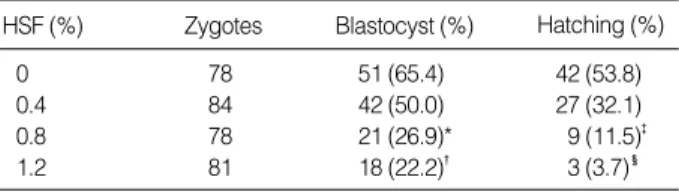
The pH and osmolarity of the thawed HSF were in the phys- iologic range. Table 1 demonstrates a significant decline in blastulation and hatching or hatched rates at 0.8% and 1.2%
HSF concentrations, compared with those at 0%. Table 2 demonstrates that the co-culture of mouse embryos with Vero cells at 0.8% HSF concentration significantly enhanced em- bryo development to blastocyst, but not at 1.2% HSF con- centration. In hatching process, however, the beneficial effects of Vero cells were significantly noted both at 0.8% and 1.2%
HSF concentrations.
DISCUSSION
In spite of intensive research, there is no clear explanation for the detrimental effect of hydrosalpinx on pregnancy rate.
Several authors have suggested that HSF contains embryotox- ic and lipophilic factors which are detrimental to the normal development of embryos (5, 11). Cultured embryos in the HSF have lower cell numbers and more fragmentation dur- ing cleavage than conventionally cultured ones, resulting in decreased pregnancy rate. Our study demonstrated a signifi- cant decline in the mouse embryo blastulation and hatching or hatched rates between 0.8% and 1.2% HSF concentration, suggesting that the hydrosalpinx is involved in the pathogen- esis of embryotoxicity. HSF has been reported to be similar to that of serum with respect to sodium, potassium, chloride, and bicarbonate, but lower for calcium, phosphate, glucose, total protein, and osmolality in its chemical analysis (12).
Because hydrosalpinx usually occurs after infectious process, this fluid may contain cytokines, leukotrienes, and prosta-
0 78 51 (65.4) 42 (53.8)
0.4 84 42 (50.0) 27 (32.1)
0.8 78 21 (26.9)* 9 (11.5)�
1.2 81 18 (22.2)� 3 (3.7)�
Table 1. The effects of hydrosalpinx fluid (HSF) on the devel- opment of mouse embryos in vitro
HSF (%) Zygotes Blastocyst (%) Hatching (%)
*p<0.05 compared with 0 (control), using Fisher's exact test.
�,�,�p<0.01 compared with 0 (control), using Fisher's exact test.
0.8 without Vero cell 78 21 (26.9) 9 (11.5) with Vero cell 66 60 (60.6)* 45 (45.5)� 1.2 without Vero cell 81 18 (22.2) 3 (3.7)
with Vero cell 66 39 (39.4) 27 (27.1)� Table 2. The effects of Vero cells in the presence of hydro- salpinx fluid (HSF)
HSF (%) Zygotes Blastocyst (%) Hatching (%)
*,�,�p<0.05 compared with controls (without Vero cells), using Fisher's exact test.
Hydrosalpinx Fluid and Vero Cells in Mouse Embryo Culture 219
glandins that may be detrimental to the developing embryos.
It has been repeatedly demonstrated that mouse embryo developmental arrest can be overcome by co-culture with Vero cells. Co-cultured embryos on the Vero cell monolayer have higher cell numbers and less fragmentation during cleavage than conventionally cultured ones (9, 13). In addition, Vero cells have been shown to provide significant improvements in morphology and cleavage of embryos (14). The resulting embryos, therefore, are likely to implant and develop progres- sively. Vero cells, although their physiological roles are yet to be determined, may provide beneficial effects on the devel- opment of mouse embryos in vitro through the removal of toxic compounds from the culture medium such as heavy metal divalent cations and metabolic inhibitors (8, 15), or through the secretion of various soluble factors including mitogenic factors (16).
We could observe that the co-culture of mouse embryos with Vero cells at 0.8% HSF concentration significantly en- hanced embryo development to blastocyst, but not at 1.2%
HSF concentration. This finding suggests that Vero cells are likely to overcome the detrimental effects of HSF to some degree through the removal of potential toxins from the cul- ture medium. In hatching process, however, the beneficial effects of Vero cells were significantly noted both at 0.8% and 1.2% HSF concentrations. It is suggested that Vero cells assist the hatching process either by reducing substances which are inhibitory to the hatching process or by thinning the zona pellucida, which may be due to the physical expansion of blastocyst or release of zona digestive substances, as demon- strated by Wiemer et al. (17). Thus, certain factors in the coculture system seem to overcome the zona hardening pro- cess, which may be triggered by a detrimental environment.
In conclusion, we have demonstrated that HSF is embryo- toxic and Vero cells are likely to overcome these detrimental effects to some degree. It is suggested that Vero cells provide beneficial effects on mouse embryo development by creating an in vitro environment favorable for the developing embryos.
REFERENCES
1. Kassabji M, Sims JA, Butler L, Muasher SJ. Reduced pregnancy out- come in patients with unilateral or bilateral hydrosalpinx after in vitro fertilization. Eur J Obstet Gynecol Reprod Biol 1994; 56: 129- 32.
2. Strandell A, Waldenstrom U, Nilssen L, Hamberger L. Hydrosalpinx reduces in-vitro fertilization/embryo transfer pregnancy rates. Hum Reprod 1994; 9: 861-3.
3. Vandromme J, Chasse E, Lejune B, Van Rysselberge M, Delvigne A, Leroy F. Hydrosalpinges in in-vitro fertilization: an unfavorable prognostic feature. Hum Reprod 1995; 10: 576-9.
4. Monsour RT, Aboulghar MA, Sevour B, Sarov GI, Riad R. Fluid accumulation of the uterine cavity before embryo transfer: a possi- ble hindrance for implantation. J In Vitro Fert Embryo Transfer 1991; 8: 157-9.
5. Ben-Rafael Z, Orvieto R. Cytokines: involvement in reproduction Fertil Steril 1992; 58: 1093-9.
6. Schenk LM, Ramey JW, Taylor SL, Swanson RJ, Morshedi MS, Muasher SJ. Embryotoxicity of hydrosalpinx fluid. Presented at the 43rd annual meeting of the Society of Gynecologic Investigators 1996. J Soc Gynecol Invest 1996; 88A.
7. Menezo YJR, Guerin JF, Czyba JC. Improvement of human early embryo development in vitro by co-culture on monolayers of Vero cells. Biol Reprod 1990; 42: 301-6.
8. Ouhibi N, Hamidi J, Guillaud J, Menezo Y. Co-culture of 1-cell mouse embryos on different cell supports. Hum Reprod 1990; 5: 737-43.
9. Sakkas D, Trounson AO, Kola I. In-vitro cleavage rates and viability obtained for early cleavage mouse embryos in co-culture with oviduct cells. Reprod Fertil Dev 1989; 1: 127-36.
10. Yeung WSB, Ho PC, Lau EYC, Chan STH. Improved development of human embryo in vitro by a human oviductal cell co-culture sys- tem. Hum Reprod 1992; 7: 1144-9.
11. Mukherjee T, Copperman AB, McCaffrey C, Cook CA, Bustillo M, Obasaju MF. Hydrosalpinx fluid has embryotoxic effects on murine embryogenesis: a case for prophylactic salpingectomy. Fertil Steril 1996; 66: 851-3.
12. David A, Garcia CR, Czernobilsky B. Human hydrosalpinx: histo- logic study and chemical composition of fluid. Am J Obstet Gynecol 1969; 105: 400-11.
13. Goodeaux LL, Voekel SA, Anzalone CA, Menezo Y, Graves KH.
The effect of Rhesus uterine epithelial cell monolayers on in-vitro growth of Rhesus embryos. Theriogenology 1989; 39: 197.
14. Van Blerkom J. Development of human embryos to the hatched blas- tocyst stage in the presence or absense of a monolayer of Vero cells.
Hum Reprod 1993; 8: 1525-39.
15. Bongso A, Fong CY, Ng SC, Ratnam S. The search for improved in-vitro systems should not be ignored: embryo co-culture may be one of them. Hum Reprod 1993; 8: 1155-62.
16. Gandolfi F, Brevini TAL, Moor RM. Effect of oviductal environ- ment on embryonic development. J Reprod Fertil Suppl 1989; 38:
107-15.
17. Wiemer KE, Cohen J, Amborski GF, Wright G, Wiker S, Munyakazi L, Godke RA. In-vitro development and implantation of human embryos following culture on fetal bovine uterine fibroblast cells.
Hum Reprod 1989; 4: 595-600.