414
©The Korean Society of Food Science and Technology
다이오스민(diosmin)에 의한 병원성 대장균 세포부착 및 생물막 형성 억제
김현정
1·김승민
2,*
1한국식품연구원 소비안전연구단, 2한국방송통신대학교 생활과학과
Inhibition of adhesion and biofilm formation in
Escherichia coli O157:H7 by diosmin
Hyun Jung Kim1 and Seung Min Kim2,*
1Research Group of Consumer Safety, Korea Food Research Institute 2Department of Human Ecology, Korea National Open University
Abstract Escherichia coli O157:H7 is one of the most common foodborne pathogens responsible for outbreaks of hemorrhagic colitis, which can lead to the life-threatening hemolytic-uremic syndrome. In this study, we identified phytochemicals that specifically inhibit the expression of LEE operon in E. coli O157:H7. Among phytochemicals, diosmin decreased the adherence of E. coli O157:H7 towards Caco-2 cells in vitro (p<0.01) and its biofilm formation activity (p<0.05). Quantitative RT-PCR analysis revealed that the transcripts of Ler-regulated genes and genes related to curli production were significantly reduced in the presence of diosmin. However, diosmin does not affect bacterial viability, indicating that the resistance rate to diosmin was remarkably low. Overall, these results provide significant insights into the development of a novel anti-infective agent that is different from conventional antibiotics.
Keywords: E. coli O157:H7, adhesion, biofilm, diosmin, antibiotic resistance
서
론
병원성 대장균은 그람음성의 식품유래 병원성 세균으로서 최 근 5년 동안 우리나라에서 발생된 총 31,216명의 식중독 환자 중 32.5%에 이르는 10,158명이 병원성 대장균에 의해 발생되어 가 장 많은 식중독 환자를 발생시키는 원인물질이라 알려져 있다 (MFDS, 2018). 이 균이 식중독을 일으키는 첫 번째 단계는 소화 기계 세포에 부착이며 이후 인체로 침입하게 된다. 이 때 숙주 상피세포 부착에 필수적인 인티민(intimin)을 암호화하는 eae라는 유전자가 작용하여 사람 장 상피세포에 부착-소멸적 손상(attaching and effacing, A/E) 병변을 형성한다고 알려져 있으며 eae 유전자 는 LEE (locus of enterocyte effacement)로 알려져 있는 35.5 kb 의 염색체상 병원성섬(pathogenicity island, PAI)에 위치하고 있다. 이외에도 LEE 오페론에는 제3형 분비 시스템, 숙주세포로 effector molecule을 이동시켜 숙주 세포골격을 파괴시키는 분비 단백질 (Esp), 인티민 수용체(Tir) 등을 암호화하고 있다. 이 LEE 오페론 은 LEE operon master regulator (Ler)에 의해 전사가 활성화 된 다고 알려져 있으므로 Ler은 병원성 대장균 O157:H7의 식중독 유발에 중요한 역할을 한다고 할 수 있다(Kim, 2014; Tarr 등, 2005).병원성 대장균 O157:H7의 LEE 오페론에 암호화되어 있는 EspA 는 장 상피세포 부착을 도와 생물막을 형성하는 데에 중요한 역 할을 한다고 보고되어 있다(Moreira 등, 2006; Sharma 등, 2016). 병원성 대장균은 장 상피세포 뿐만 아니라 식물체, 유리, 스테인 리스강 등 다양한 표면에 생물막을 형성한다(Patel 등, 2011; Ryu 와 Beuchat, 2005). 이처럼 생물막은 미생물이 다양한 환경의 표 면에 부착함으로써 형성되며 항생제나 숙주의 방어 메커니즘, 외 부 스트레스와 같은 외부의 가혹한 환경 조건에 대해 물리적 보 호작용을 하고 유전적 전이를 용이하게 하는 등 일반적으로 미 생물 생육에 이로운 영향을 미친다고 알려져 있다(Watnick과 Kolter, 2000). 이에 따라 병원성 대장균 O157:H7과 같은 식중독 균들이 생물막을 형성하면 쉽게 제거되지 않아 식중독을 일으킬 수 있으므로 생물막 제거를 위한 다양한 시도가 이루어지고 있 다. 일반적으로는 경제적인 측면과 사용 편리성 등을 고려하여 식품첨가물, 살균제와 살균소독제를 이용하는 화학적 방법이 가 장 많이 사용되고 있다(Kim 등, 2009). 하지만 화학적 제어 방법 은 미생물의 저항성과 돌연변이 혹은 유전자 전이에 의한 내성 화 등의 문제점을 가져올 수 있고 소비자에 있어서는 장기 손상, 면역 과민 반응 등 부작용이 발생될 우려가 있어 식물화학물질 과 같은 자연 항균물질에 대한 요구도가 높아지고 있다(Srey 등, 2013). 자연물 유래 식물화학물질은 항미생물 효과를 비롯한 다양한 생리 활성을 가지는 것으로 보고되고 있다(Cowan, 1999). 따라서 식물화학물질은 병원균의 독성과 발병 메커니즘을 타깃으로 하 는 새로운 항미생물제재로서 뛰어난 원천이라 할 수 있다. 자몽 리모노이드(Vikram 등, 2010), 귤 껍질 유래 베타 시토스테롤 (Vikram 등, 2013), 계피나무껍질 유래 유제놀(Kim 등, 2015) 등 다양한 식물 유래 화학물질들이 이미 미생물 제어에 있어 광범 *Corresponding author: Seung Min Kim, Ph. D., Department of
Human Ecology, Korea National Open University, Seoul 03087, Korea
Tel: +82-2-3668-4531 Fax: +82-2-2088-4306 E-mail: [email protected]
Received July 18, 2018; revised August 10, 2018; accepted August 13, 2018
위한 활성을 나타냄이 증명되었다. 그러나 각 식물화학물질의 작 용 메커니즘은 완전히 알려지지 않았다. 의료계뿐만 아니라 식품 산업계에서도 항생제 내성 문제는 심 각한 문제로 대두되고 있어 새로운 항미생물제재를 시급히 개발 해야 하는 시점에 놓여 있다(Clatworthy 등, 2007). 항생제 내성 을 가지는 균주들의 출현을 극복하기 위해서는 기존의 미생물 세 포벽 생합성, 단백질 합성, DNA 복제와 복구 등의 억제를 통해 생장을 억제시키는 전통적인 방법에서 벗어나 새로운 타깃이 지 속적으로 발굴되어야 한다. 따라서 전세계적으로는 미생물의 제 3형 분비 시스템(Kline 등, 2012), 쿼럼 센싱(Brackman과 Coenye, 2015), 숙주-병원체간 상호작용(Eichinger, 2011) 등과 같은 병원성 에 관련된 유전자를 타깃으로 삼아 새로운 항미생물제재를 개발 하고자 하는 연구들이 수행되고 있다. 따라서 본 연구에서는 Ler에 의해 전사가 조절되는 LEE 오페 론을 타깃으로 삼아 병원성 대장균 O157:H7의 생장은 억제하지 않으면서 LEE 오페론 발현을 감소시키는 식물화학물질을 스크리 닝하여 장 상피세포 부착과 생물막 형성을 억제할 수 있는 천연 물을 탐색함으로써 새로운 병원성 대장균 O157:H7 제어제로서의 가능성을 타진하고자 하였다.
재료 및 방법
균주 배양 조건 및 식물화학물질 본 실험에 사용된 균주와 플라스미드는 Table 1에 제시되었다. 병원성 대장균 O157:H7 EDL933 및 reporter strain은 Luria-Ber-tani (LB, Difco Inc., Detroit, MI, USA) 배지에서 37oC의 호기적 조건으로 배양하였고 생물막 형성 시에는 Brain Heart Infusion (BHI, Difco Inc.) 배지에서 정치배양하였다. 식물화학물질은 fla-vones, flavanones, flavonols, isoflafla-vones, flavanonols, catechins 계열이 포함된 총 32종류가 사용되었으며 Extrasynthese (Genay, France)에서 구매하여 다이메틸설폭사이드(DMSO, Gibco, Grand Island, NY, USA)에 녹여서 20 mM 농도의 용액을 만들어 희석 한 후 사용하였다.병원성 대장균 O157:H7 억제제를 찾기위한 high-throughput screen
Kim과 Kim의 논문(2017)에서 사용된 reporter strain KS02가 병 원성 대장균 O157:H7 억제제를 찾기위한 스크리닝 시스템에 사 용되었다. KS02를 하룻밤 배양한 후 적절한 항생제(앰피실린과 클로람페니콜)가 포함된 새로운 LB 배지에 100분의 1로 희석해 접종했다. 600 nm에서의 흡광도가 0.5가 될 때까지 37oC에서 배
양한 후, 0.0001% L-(+)아라비노스(Sigma-Aldrich, St. Louis, MO, USA)와 각 식물화학물질(최종농도 100 μM)이 들어있거나 혹은
대조군으로서 0.0001% L-(+)아라비노스와 DMSO (최종농도 1%) 가 들어있는 96-well microtiter plates에 200 μL씩 분주했다. 세포 내 발광정도는 발광측정기(Bio-Tek Instruments, Inc., Winooski, VT, USA)로 측정되었고 Jeong 등(2010)이 설명한 바와 같이 relative light units (RLUs)로 표현되었다(Jeong 등, 2010). Percent inhibition은 다음 식에 의해 계산되었다.
100×
(
1− RLU in the presence of compound−RLU of the negative control)
RLU of the positive control−RLU of the negative control 병원성 대장균 O157:H7의 사람 결장 상피세포 부착능에 대 한 식물화학물질 효과 측정3.0 cm 직경의 조직 배양 플레이트 바닥에 덮개 유리를 놓고 Caco-2 사람 결장 상피세포를 2×10⁶ cell/well 로 dividing 한 후 5% 이산화탄소 농도 하에 37oC에서 overnight로 부착시켰다. 부 착된 세포는 적절한 농도의 DMSO나 식물화학물질 100 μM이 포 함된 병원성 대장균 O157:H7에 의해 MOI 10으로 2시간 동안 감 염되었다. 부착되지 않은 병원성 대장균 O157:H7을 제거하기 위 해 단층 배양된 세포는 미리 데워진 PBS로 두 번 씻었다. 세포 에 부착된 병원성 대장균 O157:H7은 0.1% Triton X-100 (Sigma-Aldrich)로 5분 동안 Caco-2 세포를 용해시킨 후 LB agar plate에 서 회복된 세균 수를 세어 집락형성단위(colony forming unit, CFU)로 나타내었다(Jeong 등, 2009). 병원성 대장균 O157:H7의 생물막 형성에 대한 식물화학물질 효과 측정 병원성 대장균 O157:H7에 대한 식물화학물질의 생물막 형성 억제 효과는 Kim 등(2013)의 방법으로 확인하였다. 병원성 대장 균 O157:H7은 BHI 배지에서 16시간 배양 후 1/100 희석하여 준 비하였다. DMSO나 식물화학물질 100 μM가 분주된 96-well plate (Nunc, Roskilde, Denmark)에 희석된 병원성 대장균 O157:H7을 접종하여 37oC에서 24시간 배양 후 균액을 제거하고 PBS buffer
로 3번 씻어 내었다. 플레이트 벽면에 형성된 생물막은 1.0% 크 리스탈 바이올렛을 이용하여 15분간 염색하였다. 염색에 이용된 잔여 크리스탈 바이올렛을 제거하기 위하여 증류수로 3번 씻어 내었고, 형성된 생물막은 95% 에탄올로 10분 동안 용해 후 UV/ VIS 분광광도계(Thermo Fisher Scientific, Waltham, MA, USA)를 이용하여 570 nm에서 측정하여 정량하였다.
RNA 순수분리 및 전사물 분석
전사물 분석을 위해 병원성 대장균 O157:H7을 식물화학물질 이 있는 상태, 없는 상태에서 각각 8시간 동안 배양한 후, RNeasy minikit (QIAGEN, Valencia, CA, USA)를 사용하여 병원성 대장 Table 1. Strains and plasmids used in this study
Strain or plasmid Relevant characteristicsa Reference or source Strains
E. coli
EDL933 Wild-type O157:H7 Riley et al. (1983)
DH5α λ−Φ80dlacZΔM15 Δ(lacZYA-argF)U169 recA1 endA1 hsdR17(rK− mK−) supE44 thi-1 gyrA relA1 Laboratory collection KS02 DH5α containing pKS1303 and pKS1304; Apr, Cmr Kim and Kim (2017) Plasmids
pKS1303 pKS1101 with 390-bp ler; Apr Kim and Kim (2017)
pKS1304 pBBR-lux with 420-bp fragment of LEE3 upstream region; Cmr Kim and Kim (2017) aApr, ampicillin resistant; Cmr, chloramphenicol resistant
균 O157:H7으로부터 전체 RNA를 순수분리했다. 정량적 real-time PCR (qRT-PCR)을 위해 maxima first strand cDNA synthesis kit (Thermo Fisher Scientific)로 cDNA가 합성되었고, PikoReal 24 RT-PCR (Thermo Fisher Scientific)을 사용하여 cDNA가 증폭되었 으며, PikoReal software 2.2 (Thermo Fisher Scientific)로 분석되 었다. cDNA의 증폭에 사용된 특이적 프라이머는 Table 2에 제시 되었다. 전사물의 상대적 발현 정도는 Allen 등(2008)의 이전 연 구에서 기술된 바와 같이 tufA 항존유전자의 발현을 참고하여 보 정되었다. 시간에 따른, 식물화학물질 존재에 따른 tufA 항존유전 자의 발현에는 차이가 없었다. 모든 샘플은 세 번 반복 실험하였 고 Livac과 Schmittgen(2001)이 보고한 바에 따라 2−ΔΔCq방법으로 분석되었다. 병원성 대장균 O157:H7에 대한 식물화학물질의 생장 억제 효 과 측정 식물화학물질이 없을 때와 있을 때 계대 배양된 균주의 600 nm에서 흡광도 값을 37oC에서 통기배양하면서 마이크로플레이트 판독기(Bio-Tek Instruments, Inc.)로 측정했다.
통계분석
모든 실험 단계는 3반복 실시하였으며 결과의 통계분석은 GraphPad software version 5.02 (Graphpad Software, La Jolla, CA, USA) 프로그램을 통하여 일원배치 분산분석(ANOVA) 방법 을 이용하여 p<0.05 유의 수준에서 통계분석하였다.
결과 및 고찰
식물화학물질의 high-throughput screen
병원성 대장균 O157:H7에 의한 감염을 예방할 수 있는 항부 착 물질을 찾기 위하여 식물 유래의 화학물질 32종에 대한 LEE 오페론 발현 저해능을 스크리닝했다. LEE3 promoter가 promoter-less luxCDABE 오페론에 결합된 reporter 플라스미드 pKS1304와 아라비노스에 의해 야생형 ler 유전자 발현이 유도되는 pKS1303 플라스미드가 E. coli DH5에 함께 형질전환된 KS02 균주를 스 크리닝에 사용했다. 따라서 식물화학물질 처리 시 KS02 발광 정 도가 감소하면 그 물질은 Ler 억제제로 간주했다. 이 때 제 3형 분비 시스템에 포함되는 eae 유전자의 발현을 유의미하게 억제 시켰다고 보고된 브로콜리 성분의 미리세틴(myricetin)은 양성대 조군으로 사용되었고(Lee 등, 2011), LEE 오페론 발현에 큰 영향 을 미치지 않았다는 Vikram 등(2010)의 연구결과에 따라 자몽 성 분인 노밀린(nomilin)은 음성대조군으로 사용되었다. 100 μM의 농 도에서 50% 이상의 발광 억제 효과를 나타낸 물질은 아카세틴 (acacetin), 다이오스민, 아이소람네틴(isorhamnetin) 세 종으로 각 각 57.3, 64.7, 50.2%의 억제 효과를 나타내어, LEE 오페론 발현 양을 두 배 이상(p<0.05) 감소시킴을 확인했다(Fig. 1). 56.5%의 억제 효과를 보인 양성대조군보다 더 높은 저해 효과를 나타낸 아카세틴은 국화(Chrysanthemum lavandulifolium)에 들어있는 성 분으로서 황색포도알세균에 대한 항균활성이 보고된 바 있으며 (Komape 등, 2014) 다이오스민은 자몽, 레몬과 같은 감귤류 과일 Table 2. Oligonucleotides used for quantitative real-time PCR
Oligonucleotide Oligonucleotide sequence, 5'-3'
a
Forward Reverse
tufA Housekeeping gene TCCGGAATGTAGGAATCCAG TGGTTGATGACGAAGAGCTG
escJ GCCATCACAAGAAAATGCAA TTAACCTCTGGCGAGCTGAT
sepZ AGCCGGATTAGCACTTACCA GGCATATTTCATCGCTAATGC
escV CGGTGAAAGCGAAAAAGAAG GCAAACCGCCAGAAATAAGA
escN TCGTGTTGGAGACGCACTAC GCCCTGACGCCAAGTATAAA
tir Translocated intimin receptor ACTTCCAGCCTTCGTTCAGA TTCTGGAACGCTTCTTTCGT
eae Intimin CAACATGACCGATGACAAGG GATTAACCTCTGCCGTTCCA
espA EspA protein CGGCACAAAAGATGGCTAAT ACCAGCGCTTAAATCACCAC
espD EspD protein ACGAACGGTATTCGTTCTGC TAACTCGCTTGCCGCTTTAT csgA Curli subunit, major curli subunit AGATGTTGGTCAGGGCTCAG CGTTGTTACCAAAGCCAACC csgB Curli nucleator protein curlin CsgB ATCAGGCAGCCATAATTGGT CCATAAGCACCTTGCGAAAT csgD Transcriptional activator for csgBA CCGCTTGTGTCCGGTTTT GAGATCGCTCGTTCGTTGTTC csgF Protein required for curli nucleation by CsgB TTGAAACACCCTCAGCGTTA CTGGATGGTCGAGGTTTGTC csgG Pore-forming lipoprotein for curli subunit secretion CCGGGTAGGGTTTAAATTTG GCAGCGCTTATTTCTTTTGG aThe oligonucleotides were designed using the E. coli O157:H7 genome sequence (GenBank accession numbers AE005174.2).
Fig. 1. High-throughput screening assay using a reporter strain KS02 for identifying E. coli O157:H7 inhibitors. The reporter plasmid pKS1304 contains the LEE3 promoter fused to promoterless luxCDABE operon. pKS1304 was co-transformed with pKS1303 which includes an arabinose-inducible wild-type ler gene into E. coli DH5 cells. Cellular luminescence was measured in an arabinose concentration-dependent manner. *p<0.05; **p<0.01.
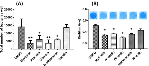
에 존재하는 성분으로서 정맥질환(Jantet, 2002), 당뇨(Vinayagam 과 Xu, 2015) 등에 효능을 발휘한다고 알려져 있으나 식중독균 저해에 대한 효과는 현재까지 알려진 바 없다. 아이소람네틴은 비브리오 콜레라의 세포벽에 침투하여 산화적 스트레스를 야기 시킴으로써 항미생물 효과를 나타내는 것으로 보고된 바 있다 (Bhattacharya 등, 2018). 하지만 아카세틴, 다이오스민, 아이소람 네틴 모두 병원성 대장균의 독성 인자 발현에 미친 영향에 대해 보고된 바는 없었다. 한편 음성대조군으로 사용한 노밀린을 처리 했을 시에는 DMSO 처리 때와 유사한 수준의 RLU 값을 나타 냈다. 식물화학물질 처리에 따른 병원성 대장균 O157:H7의 부착능 저해 KS02를 이용한 high-throughput screen을 통해 선정된 식물화학 물질들이 실제 병원성 대장균 O157:H7에서도 효과를 나타내는지 확인해 보고자 사람 결장 상피세포에 대한 부착 억제 활성을 평 가했다. Caco-2 세포에 DMSO, 미리세틴, 아카세틴, 다이오스민, 아이소람네틴, 노밀린을 각각 처리한 병원성 대장균 O157:H7을 MOI 10으로 감염시켜 2시간 후 Caco-2 세포에 부착된 박테리아 수를 세어 비교하였다. 미리세틴, 아카세틴, 다이오스민, 아이소 람네틴 처리 시, 병원성 대장균 O157:H7의 Caco-2 세포 부착 정 도가 DMSO 처리 때에 비하여 각각 3.63, 2.23, 3.62, 2.34배 감 소하여, 다이오스민은 미리세틴과 유사한 수준의 세포 부착 억제 능을 나타냈다(Fig. 2A). 그러나 노밀린 처리 시에는 1.03배 감소 하는 것으로 나타나 DMSO 처리 때와 유사함을 확인할 수 있었 다. 한편 병원성 대장균 O157:H7없이 미리세틴, 아카세틴, 다이 오스민, 아이소람네틴만 처리했을 경우에는 Caco-2 세포의 형태 및 증식에 아무런 영향을 미치지 않은 것으로 보아 식물화학물 질 자체가 사람 결장 상피세포에 독성을 나타내는 것은 아님을 알 수 있었다.
Fig. 2. Effects of selected phytochemicals on cell adhesion activity and biofilm formation. (A) Caco-2 cells were cultured on glass coverslips and infected at an MOI of 10. After incubation with or without phytochemicals, adherent bacteria were quantified and expressed as the number of bacteria per well in tissue culture. (B) Biofilms with or without phytochemicals were grown on microtiter plate walls and quantitated using crystal violet staining. *p<0.05; **p<0.01. Error bars represent the SEM. Means and SEM values were calculated from three independent experiments.
Fig. 3. Effects of myricetin and diosmin on the relative expression levels of LEE operon and genes related to biofilm formation. qRT-PCR analyses were used to determine the effects of myricetin (A) and diosmin (B). Each column represents the mRNA expression level in the presence of phytochemicals relative to that in the absence of phytochemicals. Gene names are based on the database of the E. coli O157:H7 genome, which was retrieved from GenBank (accession numbers AE005174.2). The data are the means plus standard errors of the means (SEM) of results from three independent experiments.
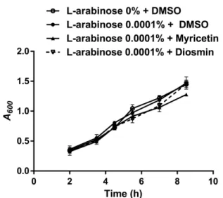
식물화학물질 처리에 따른 병원성 대장균 O157:H7의 생물막 형성 저해 사람 결장 상피세포에 대한 부착 억제능을 보였던 식물화학물 질들이 생물막 형성 억제능 역시 보이는지 확인하였다. 미리세틴, 아카세틴, 다이오스민, 아이소람네틴 처리 시, 병원성 대장균 O157:H7의 생물막 형성 정도가 DMSO 처리 때에 비하여 각각 18.1, 17.0, 25.6, 17.3% 감소하여 유의적 차이가 확인되었다(Fig. 2B). 이 중 다이오스민은 이미 보고된 미리세틴보다 뛰어난 활성 을 보여 새로운 생물막 형성 억제제로서의 가능성을 보였다. 따 라서 본 연구를 통해 효과가 검증된 flavone 유도체 다이오스민 은 선행연구들에서 보고된 바와 같이 인간에게 안전할 뿐만 아 니라 생물막 형성 저해 효과도 새로이 증명되었으므로 식중독 예 방 및 제어에 이용될 수 있으리라 판단된다. 식물화학물질에 의한 LEE 오페론 및 생물막 형성 관련 유전 자 발현의 영향 양성대조군으로 사용된 미리세틴과 비교하여 저해 효과가 좋 았던 다이오스민에 대해 검증실험을 실행하였다. 병원성 대장균 O157:H7의 LEE 오페론 및 생물막 형성 관련 유전자 발현 정도 가 미리세틴과 다이오스민 처리에 의해 어떻게 바뀌는지 알아보 고자 qRT-PCR을 이용하였다. LEE 오페론에 속하는 유전자로는 escJ, sepZ, escV, escN, tir, eae, espA, espD의 전사물을 분석했다. 병원성 대장균 O157:H7의 생물막 형성에 있어서는 curli fimbriae 생성이 중요한 역할을 한다고 알려져 있으므로(Arita-Morioka 등, 2018), 생물막 형성에 관련하여서는 curli 유전자인 csgABDFG의 발현 변화를 보았다. DMSO 처리 때에 비해 미리세틴과 다이오 스민 처리 시 LEE 오페론에 속하는 유전자와 curli 유전자의 발 현은 현저하게 줄어들었다(Fig. 3). 또한 다이오스민 처리 시 curli 유전자의 발현이 미리세틴 처리 때보다 1.57-2.60배 유의미하게 감소하는 것으로 나타나 Fig. 2B의 표현형 결과와 일치함을 확 인했다. 한편 미리세틴과 다이오스민 모두 tufA 항존유전자의 발 현에는 전혀 영향을 미치지 않았다. 식물화학물질의 병원성 대장균 O157:H7 생장율에 대한 효과 각 식물화학물질이 병원성 대장균 O157:H7의 생장에 영향을 미치는지 알아보기 위해 미리세틴과 다이오스민 유무에 따른 병 원성 대장균 O157:H7의 생장능을 측정했다. Fig. 4에서와 같이 실험조건에서 병원성 대장균 O157:H7의 생장에 아무런 영향을 미치지 않음을 확인할 수 있었다. 따라서 본 연구에 활용된 식물 화학물질 중 특히 효과가 좋았던 다이오스민은 내성 발생률이 저 감화된 새로운 항미생물제재로서 사용 가능하리라 판단된다.
요
약
병원성 대장균 O157:H7은 식중독 사고를 일으키는 주된 원인 균으로서 항생제 내성 문제를 극복하기 위해서는 병원성 대장균 O157:H7을 제어하기 위한 새로운 방법이 개발되어야 한다. 따라 서 본 연구에서는 식물화학물질을 스크리닝하여 병원성 대장균 O157:H7의 부착에 주된 역할을 하는 LEE 오페론의 발현을 감소 시키는 물질을 찾고자 하였다. 스크리닝을 통해 선발된 식물화학 물질 중 다이오스민은 사람 결장 상피세포 부착능을 3.62배 감소 시킴으로써(p<0.01) 양성대조군으로 사용된 미리세틴과 유사한 정 도의 효과를 나타냈다. 생물막 형성에 있어서는 다이오스민 처리 시 표현형이 25.6% 감소하여 유의적 차이가 확인되었고(p<0.05), curli 유전자의 발현 역시 미리세틴 처리 때보다 1.57-2.60배 더 유의미하게 감소하는 것으로 나타나 양성대조군보다 더 좋은 효 과를 보였다. 한편 다이오스민은 병원성 대장균 O157:H7의 생장 에는 아무런 영향을 미치지 않는 것으로 나타나 내성 발생률이 저감화된 새로운 항미생물제재로서 사용 가능하리라 판단된다.감사의 글
이 연구는 한국식품연구원 사업연구비의 지원을 받아 수행한 연구결과로 이에 감사드립니다.References
Allen KJ, Lepp D, McKellar RC, Griffiths MW. Examination of stress and virulence gene expression in Escherichia coli O157:H7 using targeted microarray analysis. Foodborne Pathog. Dis. 5: 437e447 (2008)
Arita-Morioka KI, Yamanaka K, Mizunoe Y, Tanaka Y, Ogura T, Sugimoto S. Inhibitory effects of Myricetin derivatives on curli-dependent biofilm formation in Escherichia coli. Sci. Rep. 8: 8452 (2018)
Bhattacharya D, Ghosh D, Bhattacharya S, Sarkar S, Karmakar P, Koley H, Gachhui R. Antibacterial activity of polyphenolic frac-tion of Kombucha against Vibrio cholerae: targeting cell mem-brane. Lett. Appl. Microbiol. 66: 145-152 (2018)
Brackman G, Coenye T. Quorum sensing inhibitors as anti-biofilm agents. Curr. Pharm. Des. 21: 5-11 (2015)
Clatworthy AE, Pierson E, Hung DT. Targeting virulence: a new par-adigm for antimicrobial therapy. Nat. Chem. Biol. 3: 541-548 (2007)
Cowan MM. Plant products as antimicrobial agents. Clin. Microbiol. Rev. 12: 564-582 (1999)
Eichinger L. Model organisms to study host-pathogen interaction: prerequisites for the identification of novel drug targets. Curr. Drug Targets. 12: 934-5 (2011)
Jantet G. Chronic venous insufficiency: Worldwide results of the RELIEF study. Reflux assessment and quality of life improve-ment with micronized flavonoids. Angiology. 53: 245-56 (2002) Jeong HS, Kim SM, Lim MS, Kim KS, Choi SH. Direct interaction
between quorum-sensing regulator SmcR and RNA polymerase is mediated by integration host factor to activate vvpE encoding elastase in Vibrio vulnificus. J. Biol. Chem. 285: e9357-e9366 (2010)
Fig. 4. Effects of myricetin and diosmin on the growth of E. coli O157:H7. Growth curves in LB at 37oC for E. coli O157:H7 in the absence or presence of phytochemicals. A600, optical density measured at 600 nm.
Jeong HG, Oh MH, Kim BS, Lee MY, Han HJ, Choi SH. The capa-bility of catabolic utilization of N-acetylneuraminic acid, a sialic acid, is essential for Vibrio vulnificus pathogenesis. Infect. Immun. 77: e3209-e3217 (2009)
Kim SM. Characterization and regulation of Escherichia coli O157:H7. Safe Food. 9: 35-40 (2014)
Kim IJ, Ha JH, Kim YS, Kim HI, Choi HC, Jeon DH, Lee YJ, Kim AJ, Bae DH, Kim KS, Lee C, Ha SD. Evaluation for efficacies of commercial sanitizers and disinfectants against Bacillus cereus strains. Food Sci. Biotechnol. 18: 537-540 (2009)
Kim SM, Kim HJ. Development of an HTS system to identify natu-ral chemicals that specifically inhibit Escherichia coli O157:H7 adhesion to host cells. Food Control. 73: 185-192 (2017)
Kim YG, Lee JH, Kim SI, Baek KH, Lee J. Cinnamon bark oil and its components inhibit biofilm formation and toxin production. Int. J. Food Microbiol. 195: 30-39 (2015)
Kim SM, Park JH, Lee HS, Kim WB, Ryu JM, Han HJ, Choi SH. LuxR homologue SmcR is essential for Vibrio vulnificus patho-genesis and biofilm detachment, and its expression is induced by host cells. Infect. Immun. 81: 3721-3730 (2013)
Kline T, Felise HB, Sanowar S, Miller SI. The type III secretion sys-tem as a source of novel antibacterial drug targets. Curr. Drug Targets. 13: 338-351 (2012)
Komape NP, Aderogba M, Bagla VP, Masoko P, Eloff JN. Anti-bac-terial and anti-oxidant activities of leaf extracts of Combretum vendae (Combretecacea) and the isolation of an anti-bacterial compound. Afr. J. Tradit. Complement. Altern. Med. 11: 73-77 (2014)
Lee KM, Lim J, Nam S, Yoon MY, Kwon YK, Jung BY, Park Y, Park S, Yoon SS. Inhibitory effects of broccoli extract on Escher-ichia coli O157:H7 quorum sensing and in vivo virulence. FEMS Microbiol. Lett. 321: 67-74 (2011)
Livak KJ, Schmittgen TD. Analysis of relative gene expression data using real-time quantitative PCR and the 2(-Delta Delta C(T)) method. Methods. 25: e402-e408 (2001)
Ministry of Food and Drug Safety (MFDS). Food Poisoning
Statis-tics. Available from: http://www.foodsafetykorea.go.kr/. Accessed July 6, 2018.
Moreira CG, Palmer K, Whiteley M, Sircili MP, Trabulsi LR, Castro AF, Sperandio V. Bundle-forming pili and EspA are involved in biofilm formation by enteropathogenic Escherichia coli. J. Bacte-riol. 188: 3952-3961 (2006)
Patel J, Sharma M, Ravishakar S. Effect of curli expression and hydrophobicity of Escherichia coli O157:H7 on attachment to fresh produce surfaces. J. Appl. Microbiol. 110: 737-745 (2011) Riley LW, Remis RS, Helgerson SD, McGee HB, Wells JG, Davis
BR., Hebert RJ, Olcott ES, Johnson LM, Hargrett NT, Blake PA, Cohen ML. Hemorrhagic colitis associated with a rare Escheri-chia coli serotype. N. Engl. J. Med. 308: 681-685 (1983)
Ryu JH, Beuchat LR. Biofilm formation by Escherichia coli O157:H7 on stainless steel: effect of exopolysaccharide and curli production on its resistance to chlorine. Appl. Environ. Microbiol. 71: 247-254 (2005)
Sharma VK, Kudva IT, Bearson BL, Stasko JA. Contributions of EspA filaments and curli fimbriae in cellular adherence and bio-film formation of enterohemorrhagic Escherichia coli O157:H7. PLoS One. 11: e0149745 (2016)
Srey S, Jahid IK, Ha SD. Biofilm formation in food industries: A food safety concern. Food Control. 31: 572-585 (2013)
Tarr PI, Gordon CA, Chandler WL. Shiga-toxin-producing Escheri-chia coli and haemolytic uraemic syndrome. Lancet. 365: 1073-1086 (2005)
Vikram A, Jayaprakasha GK, Uckoo RM, Patil BS. Inhibition of Escherichia coli O157:H7 motility and biofilm by β-sitosterol glucoside. BBA-Gen Subjects. 1830: 5219-5228 (2013)
Vikram A, Jesudhasan PR, Jayaprakasha GK, Pillai BS, Patil BS. Grapefruit bioactive limonoids modulate E. coli O157:H7 TTSS and biofilm. Int. J. Food Microbiol. 140: 109-116 (2010)
Vinayagam R, Xu B. Antidiabetic properties of dietary flavonoids: a cellular mechanism review. Nutr. Metab (Lond). 12: 60 (2015) Watnick P, Kolter R. Biofilm, city of microbes. J. Bacteriol. 182: