467
"J. Korean Soc. Radiol., Vol. 11, No. 6, November 2017"
A Study of the Characteristics of the Human External Auditory Canal Using 3-Dimensional Medical Imaging
Hyeong-Gyun Kim
Department of Radiological Science, Far East University
Received: November 13, 2017. Revised: November 23, 2017. Accepted: November 25, 2017
ABSTRACT
Using Digital Imaging and Communications in Medicine(DICOM) and a 3D medical imaging program, the characteristics of the external auditory canal(EAC) were compared. Using images of the ears of 63 different male and female subjects of varying age, this study measured and compared EAC transverse axis lengths, internal diameter circumferences, and upper and lower curvature angles. The findings of the study indicated differences in EAC shapes according not only to age and sex but also to the left and right of the same subject. A comparison between the sexes of the subjects (35 males and 28 females) indicated that, on average, the length of the EAC was 4.75mm longer in males. Based on the lower curvature angle, the interior side of the diameter circumference of the EAC was found to be reduced on average by 37.2% compared to the exterior side. Although the upper curvature angle was on average 25.7° larger than the lower curvature angle, 4 subjects showed a larger lower curvature angle and large differences between the upper and lower curvature angles were observed in 8 subjects of the younger age group (4~14 years old). This indicated changes in EAC curvature shapes during growth. This study presents a method to raise safety and precision by comparing direct measurements taken through physical means and indirect measurements acquired from existing ear samples. This was possible due to technological developments in which 3D medical image representation technology creates images close to reality, and, through further development, this method is expected to be used for standardization research of EAC shapes.
Keywords: Digital Imaging and Communications in Medicine(DICOM), External Auditory Canal(EAC), 3-Dimensional Image, Age, Sex
Ⅰ. INTRODUCTION
인체의 청각 기관으로 소리가 전달되는 방법은 반향체로 작용하는 측두부의 골전도와 이개강 (concha cavity)과 외이도(external auditory canal)로 고막(tympanic membrane)까지 전달되는 방법이 있 다.[1] 이러한 청각기관은 소리를 중추로 전달하면서 매질의 변화로 발생하는 에너지 손실을 보상하기 위해 인체의 해부학적 모양과 형태에 따라 공명을 통해 음압을 증강시킨다.[2] 이러한 이유로 음향학에 서는 청각의 외부 및 내부에 대한 형태, 크기비율에 대한 연구들이 많이 진행되었다.[3,4] 이는 귓본
(impression)을 제작하거나 물리적 측정 도구를 직.
간접으로 측정하는 방법이며, 이러한 방법들은 고 막 손상과 채취된 귓본의 정밀성으로 여러 문제를 일으킬 수 있다. 따라서 본 연구에서는 청각기관 중 외부에 노출된 이개와 이개강을 제외한 외이도에서 고막까지의 3차원적인 형태를 의료용 디지털 영상 및 통신(Digital Imaging and Communications in Medicine; DICOM) 표준 영상을 이용하여 외이도 길 이, 직경 둘레, 굴곡 각도의 특징들을 분류하고자 하였다. 이러한 특징을 구분하는 목적은 청각학적 으로 외이도 길이나 용적에 따라 소리가 전달되는 공명주파수가 유의하게 낮아지는 현상이 있어 청 각 음향학적으로 유익한 정보가 될 수 있다.[5,6] 즉,
* Corresponding Author: Hyeong-Gyun Kim E-mail: [email protected] Tel: +82-10-3526-7818
Address: Dept. of Radiological Science, Far East University, 76-32 Daehakgil, Gamgok-myeon, umseong-gun, Chungbuk-do, https://doi.org/10.7742/jksr.2017.11.6.467
소리가 사람의 고막을 통해 청각기관으로 들어오 는 물리량들이 외이도의 형태에 따라 크게 또는 작 게 인식될 수 있는 차이를 비교할 수 있기 때문이 다.[7] 이와 같이 본 연구는 인체의 다양한 3차원 (demension; D) 형태를 의료영상 기반으로 동일하 게 표현 할 수 있는 2D 및 3D 영상 재구성 기술의
발전으로[8,9] 연령과 성별에 따른 외이도의 길이, 굴
곡도, 내경의 변화를 DICOM 영상의 후향적 영상 평가를 통해 분류하고자 하였다.
Ⅱ. MATERIAL AND METHODS
뇌(Brain) 측두부(Temporal) 전산화단층촬영(Computed Tomography; CT)으로 검사된 0.5mm DICOM 볼륨데이터를 이용하여 63귀(남자 35귀, 여자 28귀)의 2D 영상과 3D 영 상을 시각화 하여 측정하였다.[10-12] DICOM 영상은 연령, 성별만 제공된 영상으로 교통사고 및 수술 특이점이 있는 영상들이 배제된 후향적 평가로 분석하였다.
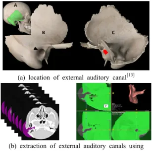
(a) location of external auditory canal[13]
(b) extraction of external auditory canals using DICOM images
Fig. 1. Location of external auditory canal and method of extracting external auditory canals using DICOM
images.
Fig. 1. (a)는 인체의 머리 측두부(A-green color)와 연관된 외이도의 외측부(B-arrow head)와 내측부 (C-red arrow)를 나타내고 있다. Fig. 1. (b)는 DICOM 볼륨영상의 외이도 영역을(violet color) 나 타내고 있으며 이러한 외이도의 영상분석 모델링 은 Terarecon(AquariusNET, TERARECON, USA) 프
로그램을 사용하였다. Fig. 2 는 본 연구에서 2D 및 3D 모델링된 외이도의 측정 기준을 정의하였다. 외 이도의 전체길이는 A1, A3에 나타내고 있으며 이 개에서 상부측 외이도가 시작되는 선분(a)을 기준 으로 고막(A)까지로 하였다. 외이도 내경 둘레는 A1, A2에 나타내고 있으며 각도가 급격한 하부 굴 곡각(F)을 기준으로 외측(B)과 내측(C)의 직경으로 비교하였다. 외측 내경 둘레(B)는 외이도 상부의 접선부(b), 내측 내경 둘레는(C) 외이도 하부의 접 선부(c)를 기준으로, 외이도 내부와 직각으로 이어 지는 선분에서 직경이 제일 큰 지점으로 하였다.
외이도 굴곡 각도는 B1에 나타내고 있으며 인체 해부학적 구조에서 앞쪽(frontal)을 상부 굴곡 각도 (D), 뒤쪽(posterior)을 하부 굴곡 각도(F)로 정의하 고 측정하였다. 측정된 기초 자료는 외이도의 횡축 길이, 직경 둘레 외측과 내측의 크기, 상하부 굴곡 각도로 연령과 성별로 분류하여 비교하였다.
Fig. 2. Analysis of external auditory canal measurements using 2D and 3D images.
Ⅲ.RESULT
1. 성별의 위치에 따른 분류
Table 1의 성별, 연령대 분포를 바탕으로 외이도 횡축 길이, 직경 둘레, 굴곡 각도의 평균값을 좌우 위치별로 분류하였다.
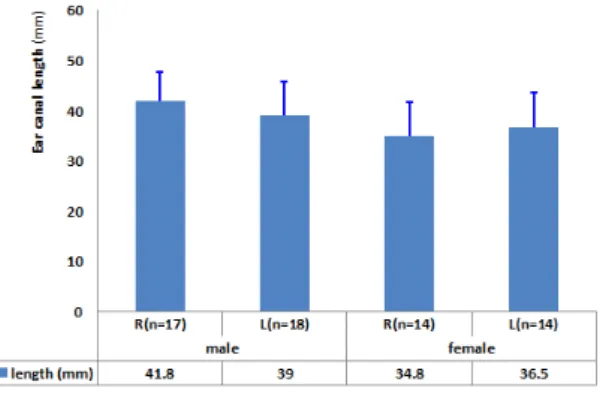
1.1 외이도 횡축 길이
남자 오른쪽 귀의 평균 길이는 41.8 ±5.96mm, 왼 쪽 39.0 ±6.80mm, 여자 오른쪽 34.8 ±6.77mm, 왼쪽 36.5 ±6.99mm로 나타나 남자의 외이도 횡축 길이 는 오른쪽에서 2.8mm, 여자는 왼쪽에서 1.7mm 더
"J. Korean Soc. Radiol., Vol. 11, No. 6, November 2017"
길게 나타났다. 또한 남자의 외이도 횡축 길이는 여 자에 비해 오른쪽 7.0mm(20.1%), 왼쪽 2.5mm(6.8%) 평균적으로 길게 나타났다.
Fig. 3. Comparison of average external auditory canal’s transverse axis lengths and standard diviations.
1.2 외이도 직경 둘레
외이도에서 첫 번째 굴곡 된 하부 굴곡 각도를 중심 으로 내측(고막 방향)과 외측(이개 방향)의 내경 둘레 를 비교하였다. 남자의 외측 오른쪽은 40.4 ±4.77mm2, 왼쪽은 39.3 ±4.96mm2, 내측은 25.7 ±3.63mm2, 24.1
±3.96mm2, 여자의 외측 오른쪽은 38.2 ±4.83mm2, 왼쪽 은 39.0 ±4.84mm2, 내측은 24.1 ±3.87mm2, 24.7
±3.85mm2 로 나타나 남자는 오른쪽에서 외측 1.1mm2, 내측 1.6mm2 크게, 여자는 왼쪽에서 외측 0.8mm2, 내측 0.6mm2 로 크게 나타났다. 이는 외이도 내경이 하부 굴 곡 각도를 기준으로 남자는 오른쪽에서 36.4%, 왼쪽에 서 38.7%, 여자는 오른쪽에서 36.9%, 왼쪽에서 38.7%로 고막 방향으로 갈수록 좁아졌다.
Fig. 4. Comparison of average external auditory canal’s diameter circumference and standard diviations.
1.3 외이도 굴곡 각도
외이도 내에서 급격하게 굽어진 상하부 굴곡 각 도를 측정한 결과 남자의 오른쪽 상부(top)는 137.9
±11.21°, 하부(bottom)는 116.6 ±14.81°, 왼쪽 상부는 142.1 ±11.03°, 하부는 113.8 ±16.56°, 여자의 오른쪽 상 부는 141.1 ±10.74°, 하부는 114.7 ±15.98°, 왼쪽 상부는 139.7 ±10.74°, 하부는 113.0 ±16.11°로 나타나 남자의 상부는 왼쪽에서 4.2°, 하부는 오른쪽에서 2.8° 크게 나 타났다. 여자는 상하부 모두 1.4°, 1.7° 차이로 오른쪽에 서 크게 나타났다. 또한 상하부 굴곡 각도 차이는 남자 24.8°, 여자 26.6° 로 상부 각도 에서 모두 크게 나타났 다.
Fig. 5. Comparison of average external auditory canal’s upper and lower curvature angles and standard
diviations.
2. 연령에 따른 분류
실험한 63귀의 외이도 횡축 길이, 직경 둘레, 굴곡 각도의 개인별 측정값들을 나이별로 구분하여 Fig.
6과 같이 그래프로 나타내었다. 가로축은 실험한 63 귀의 연령이 높은 순으로 성별, 좌우가 구분되어 나 타냈다. 세로축은 실험에서 측정된 결과 값으로 파 란(bule)색은 외이도 횡축 길이(mm), 빨강(violet)과 녹색(green)은 외·내측 직경둘레(mm2), 자주(violet)와 청록(turquoise)은 상하부 굴곡 각도(°)의 값들을 그 래프로 나타내고 있다. 그래프 분포에서 63귀에 대 한 연령별 외이도 횡축 길이는 5세 이하의 4귀에서 좀 낮아지는 경향이 보였으나 외이도 직경둘레는 전 체적으로 특징적인 구분이 없었다. 외이도 상하부 굴곡 각도 차이는 8귀(4~14세, 점선타원)에서 다른 연령층에 비해 간격이 넓게 나타났다. 그 변화율은
전체 평균 18%에 비해 본 구간에서는 38.4%로 나 타났다. 또한 외이도 상부 굴곡 각도는 하부보다 대부분 크게 나타났지만 67세 왼쪽, 21세 오른쪽,
19세에서 왼쪽 두 곳(빨간 화살촉)은 하부 각도가 더 크게 나타나는 개별적 특징을 보였다.
Fig. 6. Overall comparison graph regarding external auditory canal measurement values by age(transverse axis length, diameter circumference, angle)
Table 1. No. of ears of subjects (breakdown by sex and age)
sex / age 0 - 10 11 - 20 21 - 30 31 - 40 41 - 50 51 - 60 61 - 70 71 - 80 81 - 90 total
male 4 10 2 4 4 6 4 1 0 35
female 2 0 4 2 4 6 6 2 2 28
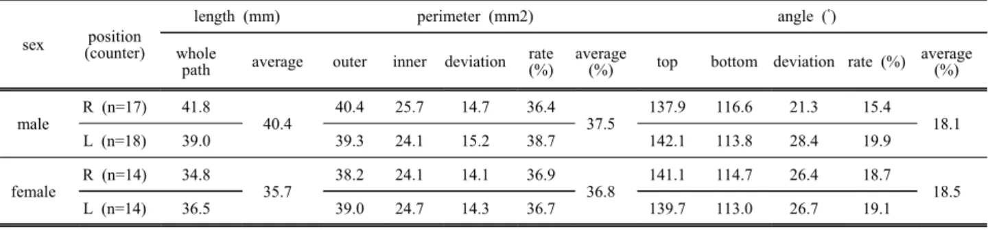
Table 2. Average transverse axis length, diameter circumference, and curvature angles found through the analysis of external auditory canal characteristics
sex position (counter)
length (mm) perimeter (mm2) angle (˚)
whole
path average outer inner deviation rate
(%) average
(%) top bottom deviation rate (%) average (%)
male R (n=17) 41.8
40.4 40.4 25.7 14.7 36.4
37.5 137.9 116.6 21.3 15.4
18.1
L (n=18) 39.0 39.3 24.1 15.2 38.7 142.1 113.8 28.4 19.9
female R (n=14) 34.8
35.7 38.2 24.1 14.1 36.9
36.8 141.1 114.7 26.4 18.7
L (n=14) 36.5 39.0 24.7 14.3 36.7 139.7 113.0 26.7 19.1 18.5
"J. Korean Soc. Radiol., Vol. 11, No. 6, November 2017"
Ⅳ. DISCUSSION
의료영상을 이용하여 특징을 분류하는 방법 중 표준 연령을 판단하는 근거로 손목뼈, 대퇴골, 치아 등의 생성이나 크기를 분류하는 기준이 있다.[14-16]
이와 같이 개인마다 다른 특이점을 분류하기 위해 본 연구에서는 외이도의 횡축 길이, 내경 둘레, 굴 곡 각도를 측정하여 분류하고자 하였다.
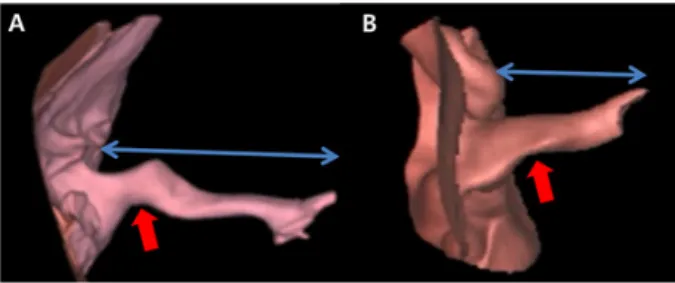
Fig. 7. 3D external auditory canals extracted from DICOM images
실험 결과 Fig. 6과 같이 외이도의 횡축 길이, 내 부 직경 둘레, 상하부 굴곡 각도는 개인마다 조금 씩 다른 특징으로 나타났다. 특히 외이도 상하부 굴곡 각도 차이는 전체 연령층과 비교하여 8귀 (4~14세)에서 뚜렷한 특징을 보였다. 이러한 변화는 상하부 굴곡 각도가 줄어드는 형태로 나타나 성장 하면서 외이도의 굴곡 모양이 변화됨을 의미했다.
이 결과는 실험 모수의 부족으로 그 변화를 특정하 기에는 한계가 있지만 전체 평균 18% 와 비교하여 38.4% 로 높게 나타난 값이다. 이는 하부(bottom)의 굴곡 각도가 낮은 것이 직접적인 원인이었으며 얼 굴뼈의 하악뼈 악관절 돌기가 연결되는 해부학적 위치이다. 실험에 이용된 63귀는 Fig. 7과 같이 외 이도 형태가 개별적 특징으로 나타났으며 동일인 이라도 좌. 우가 다르게 나타났다. 외이도 횡축 길 이와 직경 둘레는 여자에 비해 남자에서 측정값이 높았으며, 좌. 우 길이가 긴 쪽에서 내경크기가 크 게 나타났다. 상하부 굴곡각도는 외이도 길이와 내 경이 높은 곳에서 모두 낮게 나타났다. 이러한 외 이도 특징과 공명주파수와의 상관관계에[2] 대해 외 이도 길이를 측정하는 방법은 오래전부터 많이 사 용되었다. johansen 등[17] 은 사체에서 귀본 (impression)을 얻어 외이도의 부피, 길이를 구하였
으며, Egolf 등[18] 은 시상 및 횡단면의 3차원 방사 선영상을 얻어 외이도를 측정하였다. 또한 jeon 등
[2] 은 17명을 대상으로 고막의 하연까지 눈금이 표 시된 실라스틱 관을 주입하여 길이를 측정하고 정 확성을 알아보기 위해 컴퓨터 단층촬영 하였다. 이 와 같이 외이도의 길이를 측정하는 방법으로 침습 적, 비침습적 방법이 오래전부터 사용 되었지만 인 체에 주입하여 측정하는 방법은 감염과 고막손상 의 위험이 있다. 또한 Egolf 와 jeon 등 이 사용했든 컴퓨터 단층촬영 방법은 1.0mm 간격으로 획득된 영상으로 본 연구의 0.5mm 간격으로 획득된 영상 으로 측정된 결과와는 차이가 있을 수 있다. 본 연 구에서 외이도 특징을 분류하기 위한 63귀 실험은 한국인의 외이도 표준화 실험을 위한 기초단계 실 험으로, 향후 좀 더 많은 사례의 특징을 신뢰적인 방법으로 분류 해 보고자 한다.
Ⅴ. CONCLUSION
의료 영상을 이용한 63귀의 외이도의 특징을 분 석하고 다음과 같은 결론을 도출하였다.
첫째, 외이도 횡축 길이는 좌. 우 모두에서 여자 에 비해 남자가 더 길었으며, 남자는 오른쪽, 여자 는 왼쪽에서 더 길게 나타났다. 또한 동일인이라도 좌우 외이도 횡축 길이는 일치하지 않았다.
둘째, 외이도 직경 둘레는 내측이 외측에 비해 오른쪽은 36%, 왼쪽은 38% 내경 감소가 일어나 외 부의 공명된 소리가 내부로 전달되는 형태의 특징 들을 알 수 있었다.
셋째, 외이도 굴곡 각도는 하부에 비해 상부가 평균 25° 정도 높았으나 4귀에서는 하부 각도가 크 게 나타나는 개별적 특징이 있었다. 또한 8귀의 낮 은 연령(4~14세)에서 전체 연령층에 비해 상하부 굴곡 각도 차이가 크게 나타나 성장 변화에 따른 굴곡 각도가 하부 각도에서 주로 변화됨을 알 수 있었다.
Acknowledgement
본 연구는 2017년도 극동대학교 교내연구비의 지원에 의해 수행되었다(FEU2017R28).
Reference
[1] Shaw EAG, “The external ear: new knowledge,” Scan dinavian audiology, Vol. 5, pp. 24-50, 1975.
[2] S. D. Heo, J. H. Lee, S. M. Jeon, I. H. Kim, "Reso nance in the Concha Cavity is Associated with the S ize of the Pinna," Korean Journal of Communication Disorders, Vol. 15, pp. 107-113, 2010.
[3] H. J. Kang, K. S. Hu, W. C. Song, H. J. Kwon, D.
K. Park, H. J. Sohn, K. S. Koh, S. H. Han, D. J. P ark, R. H. Chung, H. J. Kim, "Physical Anthropologi c Characteristics of the Auricle through the Metric an d Non-metric Analysis in Korean Young Adults," Th e Korean Journal of Physical Anthropology, Vol. 19, No. 4, pp. 255-265, 2006.
[4] S. O. Jang, B. Y. Choi, "Surgery of ear deformity a nd auricle," Department of Otorhinolaryngology, Seoul National University College of Medicine, Otolaryngol ogy Head and Neck Surgery Seoul Symposium, Vol.
8, pp. 1-23, 2003.
[5] H. S. Jeong, H. E. Koo, S. M. Lee, S. Koo, S. H.
Lee, T. H. Yu, "Changes in Resonance Frequency an d Length of External Auditory Canal Related to the Age," Korean J Otolaryngol, Vol. 44, pp. 144-147, 2 001.
[6] A. H. Choi, M. S. Lee, A. R. Choi, S. D. Heo, "Re sonance Changes in the External Auditory Canal Ass ociated with the Ear Canal Volume," Speech Science s, Vol. 1, No. 3, pp. 151-154, 2009.
[7] S. J. Oh, Introduction to Hearing Aids, Sigma Press, pp. 3-26, 2015.
[8] Y. M. Shin, “3D Printing Medical Application," Proc eedings of the Medical Society of fusion for 3D prin ting 2017, pp. 50-52, 2017.
[9] Ferreira. A, F. Gentil, J. M. R. S. Tavares, "Segment ation algorithms for ear image data towards biomecha nical studies," Computer Methods in Biomechanics an d Biomedical Engineering, Vol. 17, No. 8, pp. 888–9 04, 2014.
[10] J. K. Lee, Y. M. Kim, D. Y. Kim, "Segmentation a nd Visualization of Human Anatomy using Medical Imagery," Journal of Korea Institute of Information, Electronics, and Communication Technology, Vol. 8, No. 1, pp. 191-197, 2013.
[11] S. E. Umbaugh, "Computer Vision and Image Proces sing," Prentice-Hall, pp. 84-86, 1998.
[12] J. R. Parker, "Algorithms for Image Processing and Computer Vision," John-Wiley & Sons, pp. 116-128, 1997.
[13] http://applications.3d4medical.com/eula
[14] J. W. Yoo, J. M. Lee, W. Y. Kim, "A Bone Age Assessment Method Based on Normalized Shape Mo del," Journal of Korea Multimedia Society, Vol. 12, No. 3, pp. 383-396, 2009.
[15] E. Pietka, L. Kaabi, "Feature Extraction in Carpal-B one Analysis," IEEE Transactions on Medical Imagin g, Vol. 12, No. 1, pp. 44-49, 1993.
[16] J. S. Lee, "The Application of TW3 method for Pre diction about Bone Age in Hand AP Image of Chil dren," Journal of the Korean Society of Radiology, Vol. 9, No. 6, pp. 349-356, 2015.
[17] Johansen PA, "Measurement of the human ear canal,
" Acoustica, Vol. 33, pp. 349-351, 1975.
[18] D. P. Egolf, D. K. Nelson, H. C. Howell, V. D Lar son, "Quantifying earcanal geometry with multiple c omputer-assisted tomographic scans," J Acoust Soc Am, Vol. 93, pp. 2809-2819, 1993.
"J. Korean Soc. Radiol., Vol. 11, No. 6, November 2017"
3차원 의료영상을 이용한 인체 외이도 특징에 관한 연구
김형균 극동대학교 방사선학과
요 약
의료용 디지털 영상 및 통신 표준과 3차원 의료 영상 프로그램을 이용하여 외이도의 해부학적 특징들을 비교하였다. 실험은 연령과 성별이 다른 63귀의 영상을 이용하여 외이도의 횡축 길이, 내부 직경 둘레, 상 하부 굴곡 각도를 측정한 후 비교하였다. 실험 결과 외이도 형태는 연령과 성별 뿐 만 아니라 동일인의 좌.
우 에서도 다르게 나타났다. 성별 비교에서 남자 35귀, 여자 28귀에 대한 평균 길이는 남자가 4.75mm 길게 나타났다. 외이도 직경 둘레는 하부 굴곡 각도를 중심으로 내측이 외측 보다 평균 37.2% 감소된 형태로 나 타났다. 상하부 굴곡 각도는 상부가 평균적으로 25.7° 높았으나 4귀에서 하부 각도가 높고, 연령이 낮은(4~
14세) 8귀에서 상하부 굴곡 각도 차이가 크게 나타났다. 이는 성장하면서 외이도 굴곡 모양이 변화됨을 나 타냈다. 본 연구는 기존 귀본 채취를 통한 간접 측정과 물리적 방법에 의한 직접 측정과 비교하여 안전성 과 정밀성을 높일 수 있는 방법이다. 이는 3차원 의료 영상 표현 기술이 실제에 가깝게 표현할 수 있는 기 술 향상 때문이며 더 발전하여 외이도 형태의 표준화 연구에도 활용될 수 있는 측정 방법이라 판단된다.
중심단어: 의료용 디지털 영상 및 통신 표준, 외이도, 3차원 영상, 연령, 성별