논문 2016-53-9-18
흉부 디지털 단층영상합성 시스템의 영상 재구성 알고리즘 개발 및 선량과 화질 평가
( Development of Image Reconstruction Algorithm for Chest Digital Tomosynthesis System (CDT) and
Evaluation of Dose and Image Quality )
김 민 경*, 곽 형 주*, 김 종 훈*, 최 원 호*, 하 연 경*, 이 소 정*, 김 대 호*, 이 용 구**, 이 영 진*
( Min Kyoung Kim*, Hyeng Ju Kwak*, Jong Hun Kim*, Yun Kyung Ha*, So Jung Lee*, Dae Ho Kim*, Yong-Gu Lee**, and Youngjin Lee*ⓒ)
요 약
최근 디지털 단층영상합성 시스템 (digital tomosynthesis system, DTS)은 일반 X-ray의 영상 중첩현상과 전산화단층촬영장 치 (computed tomography, CT)의 높은 선량의 문제점을 해결하기 위하여 개발되었다. 본 연구의 목적은 흉부 촬영용 디지털 단층영상합성 시스템 (chest digital tomosynthesis, CDT)의 재구성 알고리즘 개발 및 화질과 선량 평가를 수행하는 것이다. 영 상의 재구성은 필터 후 역투영 (filtered back-projection, FBP)을 모델링하였고, 팬텀 영상을 획득하기 위한 X-선과 검출기 사 이의 각도를 ±10°, ±15°, ±20°, 그리고 ±30°로 구성하였다. 영상의 화질 평가는 평균 제곱근 편차 (root mean square error, RMSE)와 신호대 잡음 변화율 (signal difference-to-noise ratio, SDNR)로 수행하였고, 선량 평가는 ±20°의 범위에서 유효선량 으로 수행하였다. 결과적으로, 모든 각도에서 Slice thickness 필터를 적용한 팬텀영상이 가장 우수한 RMSE와 SDNR 결과를 나타내었고, 최종 유효 선량은 0.166 mSv로 측정되었다. 결론적으로, 개발한 CDT 재구성 알고리즘의 유용성을 증명하였고, 최 종 유효 선량을 측정하여 CDT의 기초 실험 데이터를 구축할 수 있었다.
Abstract
Recently, digital tomosynthesis system (DTS) has been developed to reduce overlap using conventional X-ray and to overcome high patient dose problem using computed tomography (CT). The purpose of this study was to develop image reconstruction algorithm and to evaluate image characteristics and dose with chest digital tomosynthesis (CDT) system.
Image reconstruction was used for filtered back-projection (FBP) methods and system geometry was constructed ±10°,
±15°, ±20°, and ±30° angular range for acquiring phantom images. Image characteristics carried out root mean square error (RMSE) and signal difference-to-noise ratio (SDNR), and dose is evaluated effective dose with ±20° angular range.
According to the results, the phantom image with slice thickness filter has superb RMSE and SDNR, and effective dose was 0.166 mSv. In conclusion, we demonstrated usefulness of developed CDT image reconstruction algorithm and we constructed CDT basic output data with measuring effective dose.
Keywords : Digital tomosynthesis system (DTS), Chest digital tomosynthesis (CDT), Imgae reconstruction algorithm, Image characteristics and dose evaluation
*학생회원, 을지대학교 방사선학과 (Dept. of Radiological Science, Eulji University)
**정회원, 한림성심대학교 방사선과 (Dept. of Radiological Science, Hallym Polytechnic University)
ⓒCorresponding Author (E-mail : [email protected])
Received ; July 11, 2016 Revised ; July 18, 2016 Accepted ; August 29, 2016
Ⅰ. Introduction
X-ray imaging system provides various informations for patient and has advantage for improvement of diagnostic accuracy.[1~2] Among X-ray imaging systems, conventional planar X-ray and computed tomography (CT) systems are widely used in the field of diagnostic imaging.[1~3] However, these systems have major disadvantages : (1) anatomical structures overlap in conventional planar X-ray system and (2) high patient dose in CT system.[2] To address these disadvantages, a new diagnostic device, digital tomosynthesis system (DTS), has been introduced.[3~5] DTS has moderate ability between conventional planar X-ray and CT systems with limited exposure angle. less than 360°.
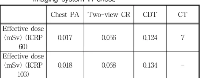
In particle, chest digital tomosynthesis (CDT) has greatly been paid attention in the DTS imaging because of improvement for lung nodule detection and acquiring low patient dose. In ICRP 60 report, the effective dose using CDT was lower than that by a chest X-ray imaging using CT. Table 1 shows the acquired effective dose with respect to the imaging system.[6] In addition, we can acquire improved depth resolution and structure visibility in the chest by using a CDT.[7]
In this study, we modelled CDT system with various angular range and acquired projection data with MATLAB simulation. Then, we developed image reconstruction algorithm with filtered back-projection (FBP) using different filters and evaluated image characteristics with two parameters, such as root mean square error (RMSE) and signal difference-to-noise ratio (SDNR) with simulation condition. Finally, we acquired effective dose by using PCXMC 1.5 software.
표 1. 흉부에서 획득된 다양한 영상 시스템별 유효 선량
Table1. The acquired effective dose as function of imaging system in chest.
Chest PA Two-view CR CDT CT Effective dose
(mSv) (ICRP 60)
0.017 0.056 0.124 7
Effective dose (mSv) (ICRP
103)
0.018 0.068 0.134 -
※ CR: computed radiography
Ⅱ. Materials and Methods
1. CDT system modelling and FBP image reconstruction
We performed simulation study using MATLAB.
The angular range was ±10°, ±15°, ±20°, and ±30°
with 180 cm source-to-detector distance (SDD). Fig.
1 shows schematic diagrams of CDT geometry.
Image reconstruction was carried out FBP, which is widely used in the CT system with many projection data acquired at greater than 360°, using various filter combinations. Fig. 2 shows the schematic diagram of FBP reconstruction process.
The number of projections generally ranges from few hundred to approximately one thousand. In both CT and DTS, the Fourier central slice theorem is general theory with X-ray beam in the Fourier space.[5]
Especially, the Fourier data are sampled for relative exposure angle and this data is mapped into the Fourier domain.
그림 1. CDT 시스템 기하학적 구조
Fig. 1. A schematic illustration of the CDT geometry.
그림 2. 기본적인 FBP 영상 재구성 과정
Fig. 2. A schematic diagram of basic FBP image reconstruction process.
In this study, we used four filters for FBP image reconstruction: (1) Filter 1 : none, (2) Filger 2 : Ram- Lak, (3) Filger 3 : Ram-Lak × Spectral Apodizing, and (4) Filter 4 : Ram-Lak ×Spectral Apodizing×Slice Thickness.
3. Image characteristics evaluation
To evaluate image characteristics, we used the RMSE and SDNR as function of angular range. The RMSE and SDNR are calculated as follows:
RMSE N
i N
fi f (1)
SDNR =
(2)
where is the number of evaluations, is the intensity of each pixel, is the intensity from referenced pixel, is the standard deviation in the background image, and and
are the average pixel values in the ROI and background image, respectively.
4. Effective dose evaluation
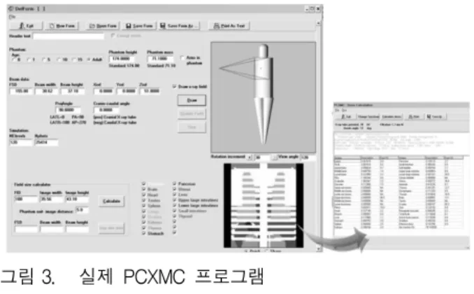
We used PCXMC program for evaluation of effective dose. This program is based on Monte Carlo and is modelled by attenuation of the radiation when passing through patient body.[6] Also, the mathematical phantom was designed in PCXMC program and the patient size can be selected in both weight and height.
The results from this program was estimated using a weighting factors of ICRP 60 or ICRP 103.
Fig. 3 shows the PCXMC program.
그림 3. 실제 PCXMC 프로그램 Fig. 3. Photo of PCXMC program.
Ⅲ. Results and Discussion
The recent introduction of DTS system into routine clinical imaging has possible for the acquisition of 3-dimension image within a standard chest X-ray radiographic examination. When we compared between conventional X-ray imaging system and CDT, more improved depth information was obtained with CDT. Also, patient dose with CDT has lower than that of CT system. However, CDT can acquire tomographic image by using a limited angle so development of appropriate image reconstruction algorithm is essential. Thus, we performed evaluation of image characteristics with FBP image reconstruction and evaluation of patient dose using a PCXMC program.
Fig. 4 shows the reconstructed phantom images with different filter combinations as function of angular range.
그림 4. 영상 획득 각도별 FBP 방법을 통해 재구성된 팬텀영상: (a) Filter 1, (b) Filter 2, (c) Filter 3, and (d) Filter 4.
Fig. 4. Reconstructed phantom images using FBP method with respect to acquiring angular range: (a) Filter 1, (b) Filter 2, (c) Filter 3, and (d) Filter 4.
Figs. 5 and 6 show the RMSE and SDNR results with different filter combinations as function of angular range, respectively. In evaluated RMSEs, differences with respect to the angular range are very similar in all reconstruction filter combinations. Also, the RMSE result goes from Filter 4, via Filter 3, and Filter 2, to Filter 1 in increasing order. In evaluated SDNRs, Filter 4 is superb result (approximately 22 at
±30°). According to the results, increasing the angular range has the effect of both only improving the depth resolution and increases the blurring of out of plane for object description.
그림 5. 영상 획득 각도별 필터 조합에 따른 RMSE 결과 Fig. 5. The RMSE result with filter combinations according
to the total angular range.
그림 6. 영상 획득 각도별 필터 조합에 따른 SDNR 결과 Fig. 6. The SDNR result with filter combinations according
to the total angular range.
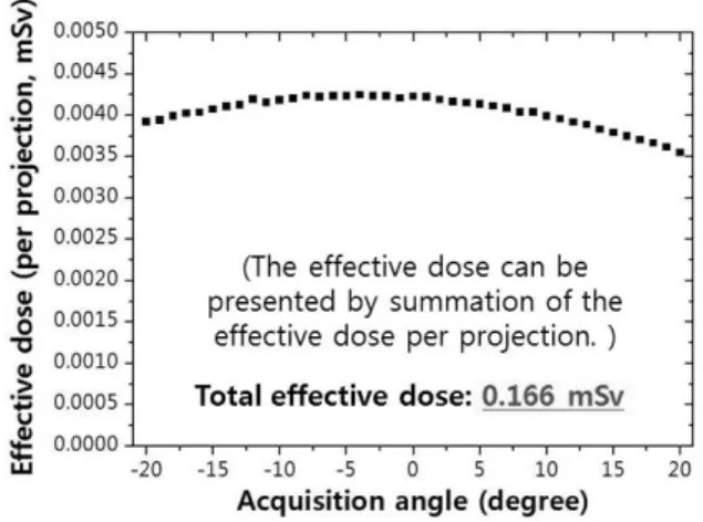
Fig. 7 shows the effective dose results as function of angular range (±20°). Total effective dose was 0.166 mSv that is similar to the ICRP 60 (0.124 mSv) and ICRP 103 (0.134 mSv). Especially, this effective dose is approximately twice that of a two-view chest examination. Also, the CDT represents less than 2%
of the effective dose in comparison with Ct imaging system.[6]
그림 7. 영상 획득 각도에 따른 유효선량 결과
Fig. 7. The effective dose result according to the total angular range.
Ⅳ. Conclusion
CDT was demonstrated to outperform conventional X-ray or CT imaging system. Also, CDT may be considered the high potential device for lung nodule detection with improvement of depth resolution. In this study, we developed FBP reconstruction algorithm with various filter combinations and evaluated image characteristics and patient dose. In conclusion, our results demonstrated that CDT with FBP reconstruction method is possible in the chest diagnostic imaging.
Acknowledgment
This paper was supported by Eulji University in 2016.
REFERENCES
[1] Y. Lee, S. Lee and H. Kim, “Comparison of spectral CT imaging methods based on a photon-counting detector: Experimental study,”
Nuclear Instruments and Methods in Physics Research A, vol. 815, pp. 68-74, 2016.
[2] Y. Lee, J. Shin, K. Seo, Y. Choi, S. Lee, Y. Lee and H. Kim, “The study on optimal acquisition condition and image processing,” Journal of the
Institute of Electronics and Information Engineers, vol. 51, pp. 897-902, 2014.
[3] Y. Lee and S. Lee, “Preliminary study on a chest digital tomosynthesis: development and evaluation,” Journal of Instrumentation, doi:10.1088/1748-0221/10/10/P10035, 2015.
[4] I. Sechopoulos, “A review of breast tomosynthesis.
Part 1. The image acquisition process”, Medical Physics, vol. 40, pp. 014301-1-12, 2013.
[5] J. T. Dobbins III and D. J. Godfrey, “Digital X-ray tomosynthesis: current state of the art and clinical potential,” Physics in Medicine and Biology, vol. 48, pp. R65-R106, 2003.
[6] J. M. Sabol, “A Monte Carlo estimation of effective dose in chest tomosynthesis,” Medical Physics, vol 36, pp. 5480-5487, 2009.
[7] S. H. Park , J. M. Goo and C. H. Jo, “Receiver Operating Characteristic (ROC) Curve: Practical Review for Radiologists,” orean Journal of Radiology, vol. 5, no. 1, pp. 11-18, 2004.
저 자 소 개 김 민 경(학생회원)
2008년 연세대학교 방사선학과 학 사 졸업.
2016년 을지대학교 방사선학과 석 사 재학 중.
<주관심분야 : 의학영상, 영상처리>
김 종 훈(학생회원)
2016년 을지대학교 방사선학과 학 사 재학 중.
<주관심분야 : 의학영상, 영상처리>
하 연 경(학생회원)
2016년 을지대학교 방사선학과 학 사 재학 중.
<주관심분야 : 의학영상, 영상처리>
김 대 호(학생회원)
2016년 을지대학교 방사선학과 석 사 재학 중.
<주관심분야 : 의학영상, 영상처리>
이 영 진(학생회원)
2007년 연세대학교 방사선학과 학 사 졸업.
2015년 연세대학교 방사선학과 박 사 졸업.
<주관심분야 : 의학영상, 의료공학, 영상처리, 신호처리>
곽 형 주(학생회원)
2016년 을지대학교 방사선학과 학 사 재학 중.
<주관심분야 : 의학영상, 영상처리>
최 원 호(학생회원)
2016년 을지대학교 방사선학과 학 사 재학 중.
<주관심분야 : 의학영상, 영상처리>
이 소 정(학생회원)
2016년 을지대학교 방사선학과 학 사 재학 중.
<주관심분야 : 의학영상, 영상처리>
이 용 구(정회원) 대한전자공학회 논문지 제 51권 4호 (2014년) 참조